Mood Stabilizer Toxicities

Written by: Justine Ko, MD (NUEM PGY-4) Edited by: Sarah Dhake, MD (NUEM ‘19) Expert Commentary by: Patrick Lank, MD, MS

Written by: Justine Ko, MD (NUEM PGY-4) Edited by: Sarah Dhake, MD (NUEM ‘19) Expert Commentary by: Patrick Lank, MD, MS


mood-stabilizer_36306943.png

Expert Commentary


This is a great summary of the causes, symptoms, work-up, and treatment of two relatively common medications that cause toxicity. In fact, these (along with carbamazepine) are levels I routinely recommend checking in patients with a history of bipolar disorder who come to the emergency department with altered mental status even if they do not report a history of being on these medications. That is because these three medications are very commonly used in the treatment of bipolar disorder and all have quite different treatment courses. In addition to the great summary above, below are some of my usual teaching points about these medications in overdose.

Let's tackle them separately as they are quite different toxicities.

First let's talk about lithium. In almost all medical texts, the tissue distribution of lithium is appropriately identified as being "complex." The easiest way I communicate that with patients, families, and medical learners is that in chronic therapy, lithium forms "stores" of drug in the body and intracellularly. Clinically that is relevant because after performing hemodialysis (HD) for lithium toxicity, you will reliably see an initial drop in lithium concentration followed by elevation approaching pre-dialysis levels if routine HD is performed. Although that could make one feel ambivalent about routinely recommending HD for lithium toxicity, there is suggestion of an alternate advantage of performing HD for lithium toxicity.  Vodovar et al published a study in 2016 showing that patients who met their institutional criteria for HD and had HD performed had significantly fewer neurologic side effects from their toxicity than those who met criteria but did not have HD. So even though it did not impact usual measurements that we would expect HD to influence -- mortality and ICU length of stay -- its performance in this study seems to have been clinically beneficial.

 The other big thing to discuss with lithium toxicity is that there are many known medication interactions with lithium. In short, any medications that impair renal function should not be used in someone on chronic lithium therapy. The main list of those medications includes NSAIDs, ACE inhibitors, ARBs, and thiazide diuretics.

 Most of the unique aspects of valproate toxicity focus on its diagnosis and treatment. In the setting of acute intentional overdose of valproate, one of the most important things for emergency physicians to be aware of is that there can be a delay of peak valproic acid level. There are cases of patients presenting to an emergency department with stated valproate ingestion, initial negative level, then repeat level hours later being in the toxic range. So I recommend serial valproate levels until both down-trending and non-toxic. For treatment, there is a great summary of recommendations by the Extracorporeal Treatments in Poisoning Workgroup (EXTRIP) published online (https://www.extrip-workgroup.org/valproic-acid). In short, consult nephrology for dialysis if the patient is super sick.

 As always, I recommend you consult your regional poison center when you are worried your patient is experiencing medication toxicity. But I hope this infographic and some of my comments helps you understand their recommendations.

 References

  1. Vodovar V, et al. Lithium poisoning in the intensive care unit: predictive factors of severity and indications for extracorporeal toxin removal to improve outcome. Clin Tox (Phila) 2016 Sep; 54(8): 615-23.

  2.  Lank P and Bryant S. "Valproic Acid" In: Wolfson A, Hendey G, Ling L, Rosen C, Schaider J, Cloutier R (eds.): Harwood-Nuss’ Clinical Practice of Emergency Medicine, 6th edition. Lippincott Williams & Wilkins 2014.

Patrick_Lank-04.jpg
 

Patrick Lank, MD, MS

Assistant Professor of Emergency Medicine

Medical Toxicologist

Department of Emergency Medicine


 How To Cite This Post

[Peer-Reviewed, Web Publication] Ko J, Dhake S. (2020, July 13). Mood Stabilizer Toxicities [NUEM Blog. Expert Commentary by Lank P. Retrieved from http://www.nuemblog.com/blog/mood-stabilizer-tox


Other Posts You May Enjoy

Posted on July 13, 2020 and filed under Toxicology.

Management of Environmental Heat Injury in the ED

heat injury image.png

Written by: Sean Watts, MD (NUEM PGY-3) Edited by: Phil Jackson, MD (NUEM ‘20) Expert Commentary by: George Chiampas, DO, CAQSM, FACEP


Heat related illness has become an increasing source of morbidity and mortality due to environmental injuries from rising global temperatures and increased interest in outdoor activities. The National Oceanic and Atmospheric Administration reported that 2016 was the hottest year on record, and that temperatures were on average 3.2 F° higher than the 20th century averages.[1] Increasing temperatures have manifested in fatal heat waves such as one claiming the lives of 70,000 individuals living in Europe during 2003.[1] The population most subject to these heat waves include the extremes of age and athletes.

Human body temperature is normally set at 37 ° C, and is maintained via the preoptic nucleus of the anterior hypothalamus.[1,2] Hyperthermia results from exposure to an exogenous heat source without altering the hypothalamic set point. As core temperatures elevate during exertion and with exposure to heat, the posterior hypothalamic nucleus signals sympathetic pathways that result in vasodilation of peripheral vascular beds and shunting blood away from gastrointestinal vasculature in order to maximize heat dissipation. Additionally, eccrine sweat glands are cholinergically activated resulting in an evaporative cooling effect. When the duration and magnitude of heat exposure outpace these physiologic mechanisms, the symptoms of heat-related illness become evident and vary from mild heat cramps to severe heat stroke and death.[2]

Heat cramps result from both potassium wasting from persistent utilization of aldosterone in order to maintain a euvolemic state and sodium loss through sweat. Edema can result from increased hydrostatic pressure of the peripheral vasculature. Additionally, syncope and hypotension can manifest due to dehydration, orthostatic pooling of blood, peripheral vasodilation, and a subsequent decrease in cardiac output. Without appropriate treatment, heat exhaustion and the more extreme heat stroke can present.

Heat exhaustion is defined as a core temperature between 37 ° C and 40 ° C with signs and symptoms including intense thirst, weakness, discomfort, anxiety and dizziness.[1,2,6,8] Heat stroke, on the other hand, is defined as a core temperature greater than 40 C° with signs of central nervous system dysfunction. Heat stroke can be further categorized into exertional and non-exertional.[4] The demographic of exertional heat stroke includes athletes, military personal, or young individuals participating in prolonged exercise.[4,8] Non-exertional heat stroke includes the elderly, young children, or individuals with metabolic or cardiac comorbidities that engage in brisk to minor activity at elevated temperatures.[1,4] When the body reaches 40 C° denaturation of proteins, release of pro-inflammatory mediators, and direct activation of the coagulation cascade occurs.[1,2] This can ultimately result in disseminated intravascular coagulation, which is a common complication of heat stroke.[1,4,5] Disruption of the liver and the cerebellum from tissue ischemia, hypoxia, vascular dysfunction, secondary cascade inflammation manifest with elevated liver function tests and ataxia dysmetria, and coma.[1,6]

Summary of the Pathophysiology of Heat Stroke [1]

Summary of the Pathophysiology of Heat Stroke [1]

Treatment of heat related illness in the emergency department rests on appropriate recognition of the severity of disease. For heat syncope and heat cramps, isotonic or hypotonic electrolyte solutions may be administered in addition to actively flexing leg muscles to prevent peripheral pooling of blood.[7,8] Ice packs or cold towels around the neck, axillae and groin can also be used for comfort measures 6. In general, these heat illnesses are self-limiting.

For heat exhaustion and heat stroke, treatments become more aggressive and should be initiated within 30 minutes of recognition of the signs/symptoms.[1,4] These patients often present critically ill and rapid assessment of the patient’s airway, breathing, and circulation is paramount. Caregivers should obtain good IV access, as well as intubate the patient if they are obtunded or in danger of loss of airway protection.[1,6] Broad spectrum critical care labs should be obtained, as well as a CK to assess for evidence of rhabdomyolysis.[5]  Additionally, obtaining an accurate core body temperature is a crucial first step to determine the severity of illness.[1,2,4,5,6] This is best performed through continuous rectal probe monitoring. Rehydration should then be performed, preferably with 1 to 2 L of isotonic fluids.[1,4] Care should be taken to not over-correct hypovolemia as the aforementioned pathophysiology makes this population vulnerable to pulmonary edema.[1] Additionally, care should be taken not to over bolus hypotonic or isotonic solutions as this population, especially those involved in long distance endurance sports like triathlons or marathons, are particularly prone to hyponatremia.[9] If these patients are given too much of these solutions, this can actually exacerbate the hyponatremia. Patients with profound hyponatremia will actually require IV hypertonic solutions or salt tabs.[9]

 Clinicians should next focus on cooling core body temperature. The best treatment for exertional heat stroke is cold-water immersion therapy—where the patient gets placed in a cold body of water.[5,7] This method takes advantage of the high thermal conductivity of water and is most effective when the patient’s clothing is removed. Studies have demonstrated that immersion in an ice-water slurry at 2°C generated cooling rates of 0.35°C/min.[4] Comparatively, allowing hyperthermic subjects to rest in air-conditioned or temperature-controlled rooms only resulted in cooling rates of only 0.03°–0.06°C/min.[4] Evidence regarding an optimal temperature to halt cooling is still under debate, but is thought to be somewhere between 38°C to 39°C, with the fear that overcooling may result in cardiac arrhythmias, especially in the elderly suffering from non-exertional heat stroke.[1,4]

Subject in a cold water-immersion bath after heat- stroke [4]

Subject in a cold water-immersion bath after heat- stroke [4]

The use of cold-water immersion therapy in non-exertional heat stroke is still under debate, but the limited evidence shows that evaporative and convective cooling by a combination of cool water spray with continual airflow over the body may be superior, especially in the elderly suffering from non-exertional heat stroke.[4] In many emergency departments, complete cold water immersion therapy may not be readily  available and limited by the placement of cardiac leads, intubation, and IV access, so evaporative and convective cooling methods become first-line for both exertional and non-exertional heat stroke in the emergency department setting should cold water immersion be unavailable.[1,4,5] Should shivering become problematic, benzodiazepines are considered first line therapy.[1,6] In severe or refractory cases the patient may benefit from ECMO.[6]

Evaporative and conductive cooling methods--note the placement of ice packs in axilla, groin as well as the cooling fan overhead [4]

Evaporative and conductive cooling methods--note the placement of ice packs in axilla, groin as well as the cooling fan overhead [4]

With the rapid increase in heat-related injuries, and projected increase in global warming, researchers are continually seeking new and efficacious treatments. For example, recombinant activated protein C is currently being explored to manage the disseminated intravascular coagulation that may result from heat stroke.[2] Additionally, application of cold packs versus other methods of rapid cooling has been explored. An experimental study published in the journal of Wilderness and Environmental Medicine found that the use of ice packs provided a significantly higher enthalpy change over cold packs—suggesting that ice packs are more efficacious than cold packs when managing heat-injury.[3] Additionally, the study found that application of cold packs or ice packs to locations high in AV anastomoses provided superior cooling rates.[3] Evaporative plus convective cooling units are also under study as an alternative means to cold water immersion for the treatment of non-exertional heat stroke.[5]


Key Points and Summary

  • Heat Injury continues to be a major cause of environmental morbidity and mortality, and will likely increase due to rising global temperatures

  • Heat Injury exists on a continuum, with heat cramps/syncope on one end and heat exhaustion/stroke on the other end

  • Obtain a rectal temperature if you suspect heat exhaustion/stroke, assess ABC’s, get good IV access, and be careful not to over bolus isotonic/hypotonic solutions due to the risk of worsening hyponatremia in athletes

  • If feasible, cold water immersion is superior for exertional heat stroke, in the ED setting evaporative and conductive cooling with ice packs can be used

  • In severe or resistant cases cardiopulmonary bypass can be effective

table of cooling methods [6]

table of cooling methods [6]


Expert Commentary

A great review and reminders of what is a preventable death especially in exertional heatstroke. Unfortunately, still in the United States there are still approximately fifteen to twenty heat-related deaths in athletes annually, mostly seen in august. While there is a spectrum of illness, preventative measures, a high index of concern and management can all mitigate negative outcomes.

In non-exertional heat illness, removal from the environment, addressing the medical condition and or removing any contributing factors is key. Cooling methods and the aggressiveness of cooling are determined by the patient’s mental status and stability. As highlighted, Heat exhaustion presents with headaches, nausea, dizziness, and weakness. Using cooling blankets and cold packs to the groin axilla and circulating fans all are measures in passive cooling. One key element to address as typically a patient presents undifferentiated is to obtain a rectal temperature in a timely fashion as highlighted. Temperatures and glucose in altered mental status patients are critical for efficient management and positive outcomes. There are key studies that highlight that time and duration above 42C lead to higher morbidity including death. 

In athletics, the death of Minnesota football player Korey Stringer in August of 2001 shed greater light on the risks of exertional heatstroke. Since his death, more work and research has been done including best practices in sport to mitigate these outcomes. Across many sports, including Marathons, best practices as outlined in the blog are being implemented pre-hospital. These measures are comparable to the recent out of hospital cardiac arrest best practices of on-sight CPR and utilization of an AED and transport second mantra. In heatstroke “cooling” on sight with ice tub submersion is the current thread being communicated. This messaging is evidenced by a recent EMS consensus paper  that highlights to first-responders the importance of recognizing but also cooling on-sight prior to transport. The delay of cooling and transport times to delay of recognition and cooling in emergency departments may lead to not initiating life-saving rapid cooling beyond the thirty minutes highlighted in the blog.

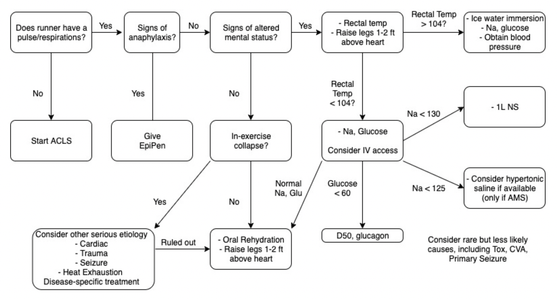
As you accurately highlighted patients can present differently, however, the key is altered mental status (AMS). Based on experience this can have the forms of patients collapse and obtunded, seizing, irritable and combative to just being confused. Rapid assessments in the right environment with excluding other AMS possibilities will allow the practitioner to respond and manage in a timely fashion. At Northwestern, both Dr. Malik and Dr. Chiampas have published the attached “collapse algorithm” (below) which allows for a quick assessment and possible differential diagnoses. Lastly obtaining a rectal temperature, which at times may be challenging with the combative patient, allows the staff in the Emergency room to objectively determine when to cease cooling. I will share that some of these patients based on their presentation would traditionally be intubated upon arrival. I would caution and remind the practitioner that if you have prepared in advance and can rapidly cool the symptoms are reversible within 10-15 minutes of ice submersion.

Lastly for emergency departments, where out-door events (sporting, festivals or concerts) with the possibility of stimulant use, preparedness is key. At Northwestern, we have secured 100-gallon ice tubs, implemented the collapse algorithm in our trauma bay and on when high-risk events take place to trigger necessary resources. For the Chicago Marathon, Triathlon or major concerts such as Lolla Palooza we order ice to the ER, towels, and prep the tub while educating our staff of the likelihood of these conditions. As we head towards the summer ahead with all of the environmental concerns of climate change and increased temperatures, this blog provides key reminders of the emergency department’s role.

Collapse_Algorithm[1] (1) (1).png
chiampas.png
 

George Chiampas DO CAQSM

Assistant Professor Northwestern University, Feinberg School of Medicine

Departments of Emergency and Orthopedic Surgery

Chief Medical Officer U.S. Soccer

Chief Medical and Safety Officer Bank of America Chicago Marathon

Team Physician Chicago Blackhawks


How To Cite This Post

[Peer-Reviewed, Web Publication] Watts S, Jackson P. (2020, July 6). Management of Environmental Heat Injury in the ED [NUEM Blog. Expert Commentary by Chiampas G. Retrieved from http://www.nuemblog.com/blog/environmental-heat-injury


Other Posts You May Enjoy


References 

  1. Heat-Related Illness. Walter F. Atha, MD. Emerg Med Clin N Am 31 (2013) 1097–1108. http://dx.doi.org/10.1016/j.emc.2013.07.012

  2. Wilderness Medical Society Practice Guidelines for the Prevention and Treatment of Heat-Related Illness: 2014 Update. Grant S. Lipman, MD; Kurt P. Eifling, MD; Mark A. Ellis, MD; Flavio G. Gaudio, MD; Edward M. Otten, MD; Colin K. Grissom, MD. WILDERNESS & ENVIRONMENTAL MEDICINE, 25, S55–S65 (2014)

  3. Chemical Cold Packs May Provide Insufficient Enthalpy Change for Treatment of Hyperthermia. Samson Phan, MS; John Lissoway, MD; Grant S. Lipman, MD. WILDERNESS & ENVIRONMENTAL MEDICINE, 24, 37–41 (2013)

  4. Cooling Methods in Heat Stroke Flavio G.Gaudio MD∗Colin K.Grissom MD†The Journal of Emergency Medicine, Volume 50, Issue 4, April 2016, Pages 607-616

  5. Heat Stroke. Alan N. Peiris, MD, PhD, FRCP(London); Sarah Jaroudi, BS; Rabiya Noor, BS. JAMA. 2017;318(24):2503. doi:10.1001/jama.2017.18780

  6. Tintinalli’s Emergency Medicine: A Comprehensive Study Guide, 8e. Judith E. Tintinalli, J. Stephan Stapczynski, O. John Ma, Donald M. Yealy, Garth D. Meckler, David M. Cline. Section 16, Chapter 210: Heat Emergencies. http://accessmedicine.mhmedical.com.ezproxy.galter.northwestern.edu/content.aspx?bookid=1658&sectionid=109384117. Accessed June 10, 2019.

  7. Heat-Related Illness in Athletes Allyson S. Howe MD, Barry P. Boden, MD First Published August 1, 2007 https://doi-org.ezproxy.galter.northwestern.edu/10.1177/0363546507305013

  8. Heat-Related Illnesses. ROBERT GAUER, MD, Womack Army Medical Center, Fort Bragg, North Carolina. BRYCE K. MEYERS, DO, MPH, 82nd Airborne Division, Fort Bragg, North Carolina. Am Fam Physician. 2019 Apr 15;99(8):482-489.

  9. Hyponatremia among runners in the Boston Marathon. Almond CS, Shin AY, Fortescue EB, Mannix RC, Wypij D, Binstadt BA, Duncan CN, Olson DP, Salerno AE, Newburger JW, Greenes DS. N Engl J Med. 2005 Apr 14;352(15):1550-6.


Posted on July 6, 2020 and filed under Environmental.

Chief Complaint: Sexual Assault

OB_Gyn-17 (1).png

Written by: Logan Wedel, MD (NUEM PGY-3) Edited by: Jason Chodakowski, MD (NUEM ‘20) Expert Commentary by: Erin Lareau, MD


ED track board reads: 24 F *****, CC: SA

Unfortunately, this is not an uncommon complaint we see in the ED

Stay engaged, and prepare for a prolonged patient stay

Sexual Assault has reached Epidemic Proportions in the United States and Globally

19.3% of women and 1.7% of men are raped at some point in their lifetime
Of female rape victims, 78.2% have their first experience of rape before the age of 25 Recent data suggests estimated cost is $122,461 per rape victim .

In one study it was found that alcohol/substance abuse was involved in over 50% of cases.

Screen Shot 2020-06-28 at 12.57.36 PM.png


Screen Shot 2020-06-28 at 1.00.07 PM.png

Only a small proportion of victims present to the emergency department

When they do the we play a vital role in treating injuries, providing prophylaxis, and collecting evidence that can be used to apprehend the attacker

It is not our role to judge the validity of the patient's accusations, to identify the attacker, nor file a police report. The latter is at the patient's discretion.

The process will be time consuming but these patients deserve our full attention: minimize distractions, sit down, provide deep empathy, and give them the space to tell their full story.

Perhaps most importantly you must provide patients with what was violently take from them: a sense of control and safety.

Step 1: Obtain History

  1. Time and Location

    • Exact details if able to remember

  2. Identity of the Attacker (if known) Number of individuals

    • Possible identifying information

  3. Specific Encounter Details - Use Patient Quotes Penetration (vaginal, anal, oral)

    • Ejaculation?

    • Condom use?

    • Use of other foreign bodies?

    • Licking, kissing, biting?

  4. Post Assault Activities

    • Shower, urination, defecation?

    • Did they change their tampon, diaphragm, or clothing? Any oral intake or vomiting?

  5. Patient's Medical History

    • HIV and Hepatitis B status and vaccination

    • Recent consensual sexual encounters

Step 2: Physical Exam

  1. General Physical Exam

    • Immediate and acute interventions always take precedent

  2. Pelvic and GU exam

    • Can be done with Evidence Collection Kit If patient consents

  3. Detailed skin and soft tissue exam

    • Again can be conducted with Evidence Collection

Step 3: Medical Management

  1. Always tend to trauma first

    • Primary and Secondary surveys

    • Workup traumatic injuries (XR / CT / FAST)

  2. Baseline Labs and Blood draws

    • CBC, CMP, LFT's, UA, Urine Pregnancy, HIV

  3. Offer Medical Advice and Inform patient of Risks, and Potential Prophylaxis Options

    1. High Risk (By Prevalence) --> Empiric Treatment

      • Chlamydia: 528.8 per 100,000 -> Azithromycin 1g PO

      • Gonorrhea:171.9 per100,000 -> Ceftriaxone 250mg IM

      • Trichomonas: 3.1% -> Metronidazole 2g PO

      • Bacterial V.: 29.2% -> Metronidazole 2g PO

    2. Lower Risk--> PEP options

      • HIV: 0.1% vaginal / 2.0% Anal ->

        • Emtricitabine/Tenofovir 200/300mg PO: 1 tab QD

        • Raltegravir 400mg PO: 1 tab BID

      • Hep B: <1% ->

        • Hep B vaccine Series: Now, 1-2m, 4-6m

      • Hepatitis C : < 1% -> No known prophylaxis

      • Syphilis: 9.5 cases per 100,000 ->

        • RPR test at 6wks, 3m, 6m

        • PenicillinG2.4millionUIM

    3. Pregnancy Risks

      • Dependent on Ovulatory Cycle:

      • 3 days before ovulation: 15%

      • 1-2 days before ovulation: 30%

      • Day of ovulation: 12%

      • 1-2 Days after Ovulation: 0%

    4. Emergency Contraception

      • Only if Urine Pregnancy Test Negative

      • Levonorgestrel 1.5mg PO

Step 4: Evidence Collection Kit --Best if within 72 hours

  1. Obtain patient consent

    • Verbal Consent to Contact "Rape Victim Advocate"

    • Signed Consent for Sexual Assault Evidence Kit

    • Police must be Contacted//However patient does not have to talk with authorities

      • Patient can also decide to refuse evidence collection at any time

  2. AppropriateAttire

    • Gloves,Gown,HairRestraint

  3. Collect Articles of Clothing

    • Patient undresses on a sheet, which is supplied in the kit

      • Anything worn at the time of assault

      • Underwear: worn at the time, or up to 72hrs after

    • Individual Articles of Clothing in Separate Areas

      • Place Individually in collection bags, sealed with evidence tape

  4. Medical/ForensicDocumentation

    • ObtainedDuringOriginalPatientHistory

    • Key Aspects as Documented Above

  5. DetailedPhysicalExam

    • Head to Toe Inspection and Palpation

      • Documentation of ANY Injuries--size, location, color, pattern

      • If Significant, Notify Police to Have Evidence Tech Obtain Photographs

    • Genital/AnalExam

      • Normal Speculum Examination, with Detailed Documentation

      • Note Discharge, Bleeding, Stains, Semen, Foreign Material, Trauma

      • Detailed Description of all Anatomy in Male/Female GU Area

      • Swab Genital / Anal area if Contact Occurred (Lubricate with Sterile Water)

    • Note: Do Not Collect G/C or BV Swabs, Unless Patient is 10 Days out or Having Symptoms

      • Offer Empiric Treatment

Collection Specimens

  1. Oral Specimens (4 Total)

    • Swabs: Tongue, Gum Line, Recessed Areas

  2. Head Hair Combings

    • From Different Areas of Head

    • Place Comb with the Hair into Paper Sheet

  3. Fingernail Specimens

    • Wood Stick to Scrap under Nails

  4. Miscellaneous Bite Marks / Stains

    • Swab Area, Label Accordingly

  5. Patient Blood on Filter Paper

    • Obtains Drops of Blood for Filter Paper

  6. Pubic Hair Combings

    • Comb out Hair onto Supplied Paper

    • Cut Hair if Matted

  7. Genital / Anal Swab (4 total)

    • Swab External Genital/Anal Area--Sterile Water to Lubricate

Follow Up Appointments and Safety Assessment

  1. Prior to ED Discharge

    • Write for 28 day supply for HIV PEP: Medications as above

  2. Primary Care Physician

    • Arrange for close follow up with PMD, ideally within 1 week

    • Send referral if patient is without a primary care physician

  3. OB-GYN

    • In order to monitor potential GU trauma

    • HPV / STD surveillance

  4. Infectious Disease

    • Within 5 days in HIV PEP is started--Due to potential toxicity

    • Close monitoring of liver function

    • Repeat testing as below

  5. On-Going Screening / Laboratory Work --Per ID / Primary Care

    • HIV: at 6 weeks, 3 months, 6 months

    • Hepatitis B: 2nd Vaccination at 3 months / 3rd at 6 months

    • Hepatitis C: at 3-6 months

    • Syphilis: at 6 weeks and 3 months

  6. Safety Assessment

    • If at risk for being assaulted again, strongly encourage patient's file a police report although this remains the patient's choice

    • If potentially unsafe going home provide resources for shelters

    • If social work isn't involved yet get them involved


Expert Commentary 

This is a great summary of current epidemiology and ED clinical practices surrounding the care of sexual assault patients.  To reiterate and expand upon your synthesis: 

•   Sexual assault is an extremely common traumatic injury that is underreported to physicians.

•   Sexual assault victims may have multiple traumatic injuries, acute psychiatric needs, and complex social needs.  A multidisciplinary approach to their care is often helpful, and necessary to reduce further psychological stress associated with the emergency department exam experience after an assault.

•   Recently, the US Department of Justice has published guidelines for training forensic examiners of sexual assault patients, including sexual assault nurse examiners (SANEs) and sexual assault forensic examiner (SAFE). These professionals are specially trained to provide care for sexual assault patients, and to perform the evidence collection.  They are often also trained in forensic photography.  SANEs typically manage the entirety of the patient encounter.  This includes coordination of prophylactic medications and proper follow up.  Illinois currently has a program to train all RNs on the sexual assault exam, and requires a SANE nurse to be available in the ED.

•   There are additionally trained SANE/SAFE providers who also specialize in adolescent/pediatric sexual assault forensics.  These providers should be called upon when available for all children suffering from sexual assault, as there is a higher risk for additional trauma surrounding the exam in these populations.

•   Our job as physicians should therefore focus on:

  • Identifying and treating additional medical or traumatic injuries

  • Counseling patients on prophylactic medication - as indicated by the exposures which you noted above

  • Reviewing expectations and follow up

  • Collaborating with our SANE colleagues, volunteer rape victim advocates, pharmacists, police departments, and social workers.

  • And as always, we should provide compassion and symptom relief to these patients undergoing an overwhelming traumatic event. 

References:

http://www.illinoisattorneygeneral.gov/victims/sane.html.  Accessed 2/12/2020

https://www.justice.gov/ovw/page/file/1090006/download. Accessed 2/12/2020

lareau2020.png
 

Erin Lareau, MD

Assistant Professor of Emergency Medicine

Northwestern Medicine


How To Cite This Post

[Peer-Reviewed, Web Publication] Wedel L, Chodakowski J. (2020, June 29). Chief complaint: sexual assault [NUEM Blog. Expert Commentary by Lareau E]. Retrieved from http://www.nuemblog.com/blog/chief-complaint-sexual-assault


Other Posts You May Enjoy


Resources

  1. Avegno, Jennifer, MD et al. "Violence:Recognition,Management,Prevention Sexual Assault Victims in the Emergency Department: Analysis by Demographic and Event Characteristics."
    The Journal of Emergency Medicine, Vol. 37 No. 3. 2009, pp. 328-344

  2. “BacterialVaginosisStatistics." Center for Disease Control and Prevention U.S. Department of Health & Human Services.

  3. Breiding, Matthew J, PhD eta l. "Morbidity and Mortality Weekly Report: Prevalence and Characteristics of Sexual Violence, Stalking, and Intimate Partner Violence Victimization." Center for Disease Control and Prevention, U.S. Department of Health & Human Services.

  4. Chisholm, Christian A. MD, et al. "Intimate Partner Violence and Pregnancy: Epidemiology and Impact." American Journal of Obstetrics & Gynecology Vol 217. No. 2. 2017, pp 141-144.

  5. HIV/AIDS:HIVRiskFactors. Center for Disease Control and Prevention, U.S. Department of Health & Human Services.

  6. "Northwestern Memorial Hospital Department of Emergency Medicine Clinical Care Guideline: Sexual Assault." https://access.nmh.org/f5-w- 68747470733a2f2f6e6d692e6e6d682e6f7267$$/wcs/blob/1390883725624/clinical -care- guideline-sexual-assault.pdf.

  7. "Preventing Sexual Violence." Center for Disease Control and Prevention, U.S. Department of Health & Human Services.

  8. "Sexually Transmitted Disease Surveillance 2017: Chlamydia." Center for Disease Control and Prevention U.S. Department of Health & Human Services.

  9. "Sexually Transmitted Disease Surveillance 2017: Gonorrhea." Center for Disease Control and Prevention U.S. Department of Health & Human Services.

  10. "Sexually Transmitted Disease Surveillance 2017: Syphilis." Center for Disease Control and Prevention U.S. Department of Health & Human Services.

  11. Sugg, Nancy MD, MPH. "IntimatePartnerViolence: Prevalence, Health Consequences, Intervention." Medical Clinics of North America, Vol.99, No. 3 2015, pp.629-649.

  12. "Trichomoniasis Statistics." Center for Disease Control and Prevention, U.S. Department of Health & Human Services. https://www.cdc.gov/std/trichomonas/stats.htm

Posted on June 29, 2020 and filed under Obstetrics & Gynecology.

Alcohol Related ED Visits

Alcohol Related ED visits image.png

Written by: Elizabeth Stulpin, MD (NUEM PGY-1) Edited by: Kevin Dyer, MD (NUEM PGY-2) Expert Commentary by: Erin Lareau, MD

Alcohol related ED visits are an all too common occurrence and are only becoming more frequent. Between 2006 and 2014, the number of ED visits involving alcohol consumption increased by over 60 percent. This has led us to our current estimated 1.2 million hospital admissions per year related to alcohol abuse, with about 500,000 of those requiring greater attention for acute withdrawal. 

Alcohol withdrawal can be treated with an innumerable combination of medications and dosages, with symptom dosed benzodiazepines currently the treatment of choice. However, with increasing visits for withdrawal and increasing doses needed to control symptoms, adverse effects of benzodiazepines can also be seen, such as paradoxical agitation, propylene glycol toxicity from increasing doses of lorazepam (lactic acidosis, AKI) and respiratory depression requiring intubation. Some patients are also resistant to benzodiazepine therapy, showing no symptomatic relief despite increasing doses. These factors have led to a pendulum swing back to the age before benzodiazepines, when phenobarbital was a first line treatment for treating alcohol withdrawal. 

Benefits of Phenobarbital: 

Reaching for phenobarbital instead of the syringe or tablet of lorazepam, diazepam, or chlordiazepoxide is an attractive option for many reasons. From its mechanism to pharmacokinetics to side effects, phenobarbital as a monotherapy can take some guesswork out of treatment. At the same time, patient outcomes are shown to be similar or improved when compared to benzodiazepines. 

Mechanism: 

The underlying cause of alcohol withdrawal is multifactorial. With chronic alcohol use, inhibitory GABA receptors are down regulated, while excitatory glutamate and NMDA receptors are upregulated. When the inhibitory effect of alcohol is suddenly removed, an excessive excitatory state is produced, resulting in the typical symptoms of tremulousness, anxiety, tachycardia, among others. Like benzodiazepines, phenobarbital targets GABA receptors to decrease excitatory tone, but does so more effectively since it does not rely on the presence of endogenous GABA hormone. At the same time, phenobarbital also down regulates glutamate signaling, leading to a more comprehensive approach to treating withdrawal. 

Pharmacokinetics: 

Besides its mechanism of action (MOA), one of the most attractive features of phenobarbital therapy is its simple pharmacology. Not only can it be given IV, IM or PO with almost 100 percent bioavailability in each route, the drug level in the body is extremely predictable. A linear relationship exists between the cumulative weight based dose and the resulting plasma concentration, which allows providers to reliably achieve the target dose range while avoiding levels at which toxicity would occur. This is in contrast to benzodiazepines, where metabolism and clinical response vary greatly among patients, and drug levels are nearly impossible to predict when periodically dosing with different doses at nonstandard points of time. 

Another added benefit of phenobarbital is its length of action. Whereas the half-life of lorazepam is 14 to 20 hours, phenobarbital has a half-life of 80 to 120 hours. This allows for titration of medication over the patient’s hospital stay and an auto-tapering effect that can prevent rebound symptoms for days after, one of the main benefits of using chlordiazepoxide. 

Patient outcomes: 

The superior molecular characteristics and MOA of phenobarbital carry over to patient outcomes as well. Since the 1970s, phenobarbital monotherapy has been shown to be as safe, and as or more effective than benzodiazepines. With renewed focus on phenobarbital, more recent studies have been conducted that corroborate this claim. In the past few years, phenobarbital has been shown to have similar or improved outcomes when compared to benzodiazepines in terms of ICU stay and overall length of hospital stay. Additionally, patients treated with phenobarbital monotherapy have been seen to have decreased rates of delirium, decreased need for additional PRN sedation and lower rates of leaving against medical advice. 

With regards to the most feared outcome, respiratory depression, phenobarbital has also held its own. Patients treated with phenobarbital have shown to have similar or even reduced rates of intubation, possibly due to the predictable linear relationship between drug dosing and plasma concentration as discussed previously. Secondly, the therapeutic range of phenobarbital is quite large. While the therapeutic dose hovers around 5 to 25mg/kg of total body weight, the toxic dose requiring intubation is often greater than 40mg/kg. 

Phenobarbital was also shown to be effective in patients whose symptoms were refractory to benzodiazepines, possibly due to its different mechanism and lack of dependence on endogenous GABA. 

What now? 

So now you want to give phenobarbital a try, what now? 

The most commonly seen dosing regimen is a loading dose, followed by IV or PO titration to the patient’s therapeutic level based on symptoms. 

The IV loading dose is often set at 10mg/kg ideal body weight, infused over 30 minutes to achieve a serum level of about 15ug/mL. Similar to treatment with benzodiazepines, treatment is then symptom based. Repeat infusions or pills of 130mg are given every 15 minutes, with a total upper limit of 20 to 30mg/kg. Unlike benzodiazepine therapy where steadily increasing doses are used to control agitation, phenobarbital’s standard dosing takes some of the guesswork out of treatment for providers. Once symptoms have been appropriately managed, the patient may require additional PRN doses of phenobarbital, but the slow auto-tapering effect of the drug should prevent acute decompensation. 

Take Home Points: 

  • Phenobarbital is a safe and effective method to combat the symptoms of alcohol withdrawal.

  • Start low and go slow! If the patient has received benzodiazepines already, phenobarbital will act synergistically, and high doses may increase the risk of respiratory depression. 

  • Remain aware of the cumulative phenobarbital dose to prevent reaching levels at which toxicity would occur. 

  • Talk to your pharmacist about using phenobarbital for your next case of alcohol withdrawal!


Expert Commentary

This is a wonderful review of current treatment strategies for alcohol withdrawal in the ED.  To broaden and emphasize some of your main concepts:

  • Alcohol related ED visits are extremely common.   ED patients with alcohol related chief complaints are high risk for traumatic, medical, psychiatric and toxicologic problems that can easily be missed if providers are not vigilant.

  • Some patients may present to the ED with chief complaints that are medical or psychiatric in nature, but are directly associated with an underlying alcohol use disorder, making their management even more challenging. 

  • Patients with unhealthy alcohol use may present to the ED with intoxication, withdrawal, seizures, agitation/psychosis, falls, traumatic injuries, gastritis/ GI bleeding, liver disease, cardiac disease, depression and anxiety.  Many have concurrent social challenges including domestic violence and homelessness. 

  • When a patient with an alcohol use disorder presents in acute withdrawal, there are multiple treatment strategies to use for symptom relief.  Benzodiazepines are widely used and effective, though often require repeat doses to obtain maximal effect. Phenobarbital loading is a wonderful alternative with similar outcomes and simplified, standard dosing, as noted by the pharmacokinetics you reviewed. 

  • My personal clinical practice is to preferentially use phenobarbital for high risk patients who present for alcohol withdrawal symptoms, and who have not already received IV benzodiazepines. Those at high risk for complications from alcohol withdrawal may have a history of:

    • withdrawal seizures

    • hallucinations

    • delirium tremens 

    • abuse of multiple substances

    • recent admission (ICU or floor) for severe alcohol withdrawal.Phenobarbital loading is also effective for patients who are not responding to high dose IV pushes of benzodiazepines, as an alternative to starting continuous infusions.  Anecdotally, I have spared patients such as this from requiring intensive care admission for withdrawal by using this pathway, though I have not found any research to support that this is a widespread phenomenon. Because serum phenobarbital levels can be checked for maintenance of a therapeutic range, severe withdrawal requiring multiple repeat doses can be directed in a more simplified way.  For those with more mild symptoms, PO or IV benzodiazepines continue to be appropriate and have good effect. 

  • Be sure to screen your patients presenting with alcohol intoxication or withdrawal for other emergent medical conditions, including traumatic, medical and psychiatric. Similarly, think about alcohol abuse and withdrawal in patients presenting for other problems, but who exhibit abnormal vital signs, abnormal neurologic exam or an insufficient response to treatment. Ask yourself:

    • Are there signs of trauma?

    • Are there other intoxicants?

    • Does this medical patient have untreated alcohol withdrawal?  Is there tremor, hypertension and tachycardia that is not otherwise explained?  

    • Are there vital sign abnormalities in the intoxicated patient? Can they be explained by intoxication? By withdrawal? If the answer is no, be sure you have appropriately treated for dehydration, alcoholic ketoacidosis and/or alcohol withdrawal.  Then look for concomitant disease.

    • Was this patient trying to hurt themselves? Do they have underlying psychiatric disease?  Are they in treatment? Do they need further resources?

  •  Even a single conversation may be a life changing intervention for someone with an underlying alcohol use disorder!

References:

UpToDate article on “Risky Drinking and Alcohol Use Disorder” https://www.uptodate.com/contents/risky-drinking-and-alcohol-use-disorder-epidemiology-pathogenesis-clinical-manifestations-course-assessment-and-diagnosis?search=risky-drinking-and-alcohol-use-&source=search_result&selectedTitle=1~150&usage_type=default&display_rank=1.  Accessed on 2/09/2020.

Hammond, D, et al.  “Patient Outcomes Associated With Phenobarbital Use With or Without Benzodiazepines for Alcohol Withdrawal Syndrome: A Systematic Review.” Hospital Pharmacy 2017;52(9):607-616

Untitled.png
 

Erin Lareau

Assistant Professor

Department of Emergency Medicine

Feinberg School of Medicine

Northwestern University


How to Cite This Post

[Peer-Reviewed, Web Publication] Stulpin, E, Dyer, K. (2020, May 25). Alcohol Related ED Visits [NUEM Blog. Expert Commentary by Lareau, E]. Retrieved from https://www.nuemblog.com/blog/etoh


References

Farkas, Josh. “Alcohol Withdrawal.” The Internet Book of Critical Care. November, 2016. http://www.emcrit.org/ibcc/etoh/

Gortney, J, et. al. “Alcohol withdrawal syndrome in medical patients.” Cleveland Clinic Journal of Medicine 2016; 83(1): 67-79. 

Hammond, D, et. al. “Patient Outcomes Associated With Phenobarbital Use With or Without Benzodiazepines for Alcohol Withdrawal Syndrome: A Systematic Review.” Hospital Pharmacy 2017; 52(9): 607-616. 

Hendey, G, et. al. “A prospective, randomized, trial of phenobarbital versus benzodiazepines for acute alcohol withdrawal.” The American Journal of Emergency Medicine 2011; 29(4): 382-385.

Hsu, D, et. al. “Phenobarbital versus Benzodiazepines for the Treatment of Alcohol Withdrawal Syndrome in Critically Ill Patients.” American Journal of Respiratory and Critical Care Medicine 2015; 191(A3704): 

Kattimani, S and Bharadwaj, B. “Clinical management of alcohol withdrawal: A systematic review.” Industrial Psychiatry 2013; 22(2): 100-108. 

Kramp, P and Rafaelsen, OJ. “Delirium tremens: a double-blind comparison of diazepam and barbital treatment.” Acta Psychiatrica Scandinavica 1978; 58(2): 174-190. 

Nelson, A, et. al. “Benzodiazepines vs barbituates for alcohol withdrawal: Analysis of 3 different treatment protocols.” The American Journal of Emergency Medicine 2019; 37(4): 733-736.

Nisavic, M, et. al. “Use of Phenobarbital in Alcohol Withdrawal Management – A Retrospective Comparison Study of Phenobarbital and Benzodiazepines for Acute Alcohol Withdrawal Management in General Medical Patients.” Psychosomatics 2019; 60(5); 458-467. 

Rosenson, J, et. al. “Phenobarbital for acute alcohol withdrawal: a prospective randomized double-blind placebo-controlled study.” The Journal of Emergency Medicine 2013; 44(3): 592-598.

Tidwell, W, et. al. “Treatment of Alcohol Withdrawal Syndrome: Phenobarbital vs CIWA-AR Protocol.” American Journal of Critical Care 2018; 27(6): 454-460. 

White, A, et. al. “Trends in Alcohol-Related Emergency Department Visits in the United States: Results from the Nationwide Emergency Department Sample, 2006 to 2014.” Alcoholism: Clinical and Experimental Research 2018; 42(2): 352-359.


Posted on May 25, 2020 and filed under Toxicology.

Henoch-Schonlein Purpura

HSP image.png

Written by: Ben Kiesel, MD (NUEM PGY-1) Edited by: David Kaltman, MD (NUEM PGY-4) Expert Commentary by: Kirsten Loftus, MD, MEd

Henoch-Schonlein Purpura-1.png
Henoch-Schonlein Purpura-2.png
Henoch-Schonlein Purpura-3.png

Expert Commentary

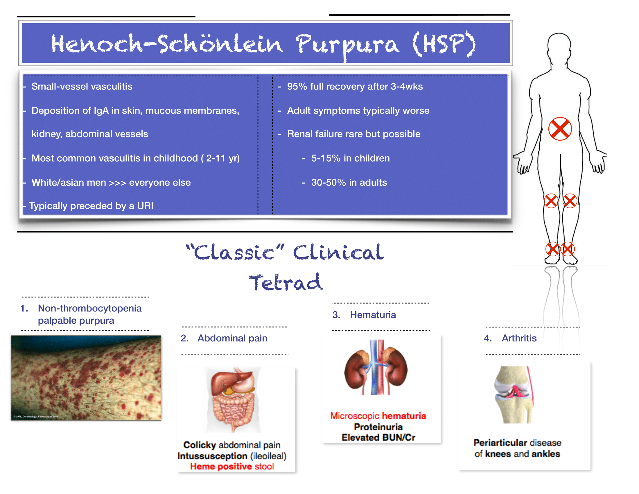
Thank you for providing this succinct review of HSP. This is not a diagnosis you will encounter frequently, but it’s an important one not to miss because, as you point out, there are several key implications for outpatient monitoring and follow-up. Here are a few additional tips when it comes to the diagnosis and initial management of HSP:

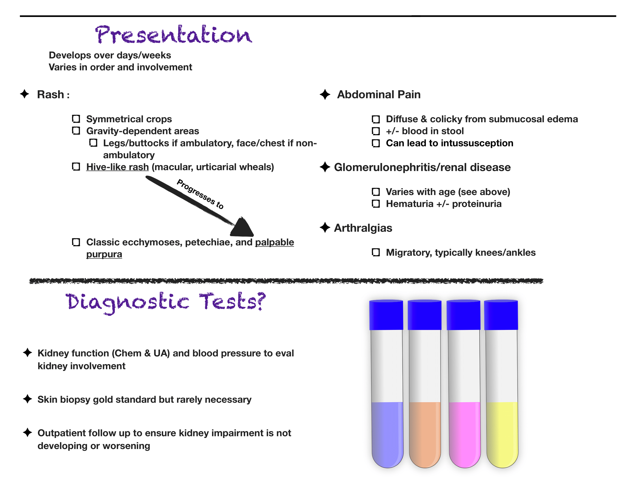
The diagnosis is truly a clinical one. If you have bilateral lower extremity petechiae/purpura plus belly pain, arthritis/arthralgia, or renal involvement, then you’ve made your diagnosis. Outside a urinalysis to evaluate for hematuria/proteinuria and checking a blood pressure to look for hypertension, there is limited utility for other diagnostic tests. Don’t forget to consult your favorite reference for normal pediatric blood pressure values to ensure you aren’t missing hypertension.

Renal disease is less common in kids compared to adults with HSP, but does happen. If you have hematuria/proteinuria or hypertension, go ahead and at least check a chemistry to look at BUN/Cr. Then talk with your favorite local pediatric nephrologist (if available) or PEM doc at your pediatric referral center to determine need for transfer versus close outpatient follow-up.

You appropriately point out that steroids are rarely indicated, and that hydration and NSAIDs will be your primary management. If you feel compelled to start steroids for severe abdominal pain (once you are sure it is not due to intussusception), you may need a longer (e.g. 4-8 week) taper, given the risk of rebound pain if tapered too quickly.

As always, set clear expectations with families and make sure they have good follow-up. I find that this can be a tough diagnosis to explain to parents, who are often quite scared about the rash. Spending some extra time talking with families once you’ve made the diagnosis can really go a long way. Warn parents that the rash is likely to persist for weeks and that the development of some additional petechiae/purpura is okay- you will prevent some unnecessary ED return visits this way. Strict return precautions for severe belly pain are key as HSP-associated intussuception is a very real complication. Every discharged patient should be seen by their PCP within about 1 week for a repeat UA and BP check- even if patients do not have evidence of renal disease at the time of diagnosis, it may develop later on, and close outpatient monitoring is critical.

Kristen_Loftus.png
 

Kirsten V. Loftus, MD, MEd

Attending Physician Emergency Medicine

Ann & Robert H. Lurie Children’s Hospital of Chicago

Instructor of Pediatrics (Emergency Medicine)

Northwestern University Feinberg School of Medicine


How to Cite This Post

[Peer-Reviewed, Web Publication] Kiesel, B, Kaltman, D. (2020, May 18). Henoch-Schonlein Purpura. [NUEM Blog. Expert Commentary by Loftus, K]. Retrieved from https://www.nuemblog.com/blog/hsp


Assisted Reproductive Technology

Written by: Jesus Trevino, MD (NUEM ‘19) Edited by: Keith Hemmert, MD (NUEM ‘18) Expert Commentary by: Lia Bernardi, MD

Written by: Jesus Trevino, MD (NUEM ‘19) Edited by: Keith Hemmert, MD (NUEM ‘18) Expert Commentary by: Lia Bernardi, MD

In 2015, assisted reproductive technology (ART) resulted in 72,913 live births, comprising approximately 1.6% of all infants born in the US (CDC 2017).  As ART is becoming increasingly common, it is important that emergency medicine providers are familiar with this treatment modality to appropriately diagnose and manage maternal complications.  This article will review the components and complications of ART that may present in the Emergency Department with a focus on the ovarian hyperstimulation syndrome (OHSS).

    ART encompasses therapies that address all causes of infertility, which span both the male factor (e.g., sperm motility) and female factor (e.g., mechanical, ovulatory).  In vitro fertilization is a common treatment strategy that overcomes reproductive barriers and involves: 1) controlled ovarian hyperstimulation, 2) oocyte retrieval, 3) oocyte fertilization and 4) oocyte implantation.  Below is an outline of these in vitro fertilization steps along with associated complications.

Controlled ovarian hyperstimulation

There are numerous protocols available to recruit ovarian follicles and they differ in the type and intensity of exogenous stimulation (Speroff).  Protocols are graded in order of increasing intensity (and success rate):

  • Natural cycle (i.e., no exogenous stimulation)

  • Minimal (i.e., clomiphene citrate)

  • Mild (i.e., clomiphene citrate + low-dose exogenous gonadotropins)

  • Aggressive (i.e., high-dose gonadotropins +/- gonadotropin-releasing hormone agonist or antagonist).

The most life-threatening complication of these strategies is ovarian hyperstimulation is OHSS (Adams):

  • Incidence - 0.5-5% of ART cycles; 0.1-2% involve severe presentations (Weinerman).

  • Pathophysiology - increased capillary permeability leads to edema, ascites, pleural and pericardial effusions; this inflammatory state may result in renal failure, respiratory failure and/or thromboembolism.

  • Timing - typically occurs within a week of exogenous HCG administration or in the peri-implantation period due to increases in endogenous HCG.

  • Risk factors - < 35 years, low BMI, gonadotropin-releasing hormone and/or analogues, hyperstimulation of ovarian follicles (detected via pelvic US), elevated estradiol levels

  • Presenting symptoms & signs - abdominal distention, rapid weight gain, peripheral edema, dyspnea, pulmonary edema/effusions, oliguria; minimize pelvic exams to avert ovarian cyst rupture and hemorrhage

  • Labs - +/- hyponatremia, AKI, estradiol > 3000 pg/mL (typically not practical in ED evaluation)

  • Management -

    • Criteria for outpatient management: normal VS, renal and hepatic labs, ovaries < 5 cm

    • Criteria for inpatient management: ovaries > 5 cm, ascites; admission is required for serial exams and pain control

    • Criteria for ICU: palpable ovaries, pleural effusions, ARDS, oliguria, hypotension, AKI, hepatic dysfunction; these patients require fluid resuscitation, +/- therapeutic thora/paracentesis, likely termination of ART cycle

    In addition, controlled ovarian hyperstimulation has an increased incidence of ovarian torsion - 0.08% without OHSS and 3% with OHSS (Weinerman).

Oocyte retrieval

This step is usually performed under conscious sedation with ultrasound-guided, transvaginal needle aspiration.  Complications may include (incidence %):

  • Vaginal puncture site bleeding (8%, Speroff)

  • Intraperitoneal bleeding (0.04-0.07%, Speroff)

  • Bowel perforation (0.04%, Weinerman)

  • Infection (0.3-0.6%, Speroff). Half of infections may present as TOA within 1-6 weeks after retrieval. 

Oocyte fertilization

The in vitro part - sperm meets egg.  There are rarely major maternal complications associated with this treatment step that present in the ED.

Oocyte implantation

Implantation occurs via a transcervical catheter under transabdominal US-guidance.  As the success rate for single fertilized embryos is 10-25%, multiple embryos are implanted to increase implantation yield (Adams).  Complications may include:

  • Multigestational pregnancy - 31-41% of IVF infants develop from multigestational pregnancies (Adams)

  • Ectopic pregnancy (0.7-4%, Speroff, Adams)

  • Heterotopic pregnancy (0.2-1%, Adams, Weinerman)

Lastly, patients with ART-facilitated pregnancies are at risk of thromboembolism (0.04-0.2%), especially in the presence of OHSS (4%, Weinerman).


Expert Commentary

This is an important review of complications that may arise in patients undergoing in vitro fertilization (IVF). Although patients who are planning for IVF can be assured that it is a generally safe process, there are medical issues that can occur throughout: during the ovarian stimulation phase, as a result of the oocyte retrieval or embryo transfer, or after a pregnancy is confirmed. 

During the ovarian stimulation phase of the process, few medical complications typically arise. The most common reason that a patient would seek emergency care would be for ovarian torsion. Given that ovarian size increases significantly, any patient who presents with severe pain during stimulation should be assessed for this.

The most likely time a patient undergoing IVF would present to the ED would be following the oocyte retrieval. Complications can take place after the conclusion of stimulation or due to issues from the retrieval itself.  One of the main issues that patients present for is ovarian hyperstimulation (OHSS). The most common time for this to happen is shortly after the oocyte retrieval, but patients may present in early pregnancy as well given that a rising hCG level worsens the syndrome. Evaluation and management of OHSS is reviewed succinctly above. Complications may also arise from the oocyte retrieval. Transvaginal ultrasound guided aspiration of ovarian follicles is performed using a needle that passes through the vagina. Possible complications include bleeding, infection, and/or or injury to other organs. Given that the needle is entering the vagina and the ovaries, bleeding can occur from the vagina or within the abdomen. Vaginal bleeding is typically quickly recognized and corrected prior to completing the procedure. Intraabdominal bleeding can be more difficult to identify and can potentially worsen after the patient is discharged. Patients with intraabdominal bleeding may present to the ED with symptoms of pain or hypotension. Imaging is generally helpful as part of the evaluation, but it is important to know that some bleeding generally occurs post-procedure even in an uncomplicated oocyte retrieval. Therefore imaging may reveal free fluid in a patient who does not have clinically significant ongoing bleeding.  It is also important to be aware that if a patient has intraabdominal bleeding, surgical intervention is not always required. Given the complexities of surgical exploration in these patients, the goal is to expectantly manage those with intraabdominal bleeding unless surgery is absolutely necessary. In addition to bleeding, infection, ovarian torsion and cyst rupture can also occur following oocyte retrieval. Finally, some patients will have extreme constipation due to the IVF process and may present with abdominal pain after oocyte retrieval as a result.  

Embryo transfers are a generally safe and low risk procedure. The procedure involves insertion of a sterile, soft catheter into the uterine cavity under ultrasound guidance with subsequent embryo release. Given the low risk nature of the procedure, complications after embryo transfer are very rare. Infection is theoretically possible, but unlikely. 

The final IVF related complications to consider are those that occur in pregnancy. As ectopic pregnancies are possible after IVF, any pregnant woman who presents with symptoms concerning for an extrauterine pregnancy should be evaluated accordingly. Heterotopic pregnancies can also occur and should remain on the differential diagnosis if a woman has concerning pain after an intrauterine pregnancy is confirmed. Given that a woman’s ovaries remain enlarged after ovarian stimulation if a pregnancy is achieved, ovarian torsion should also be considered in patients with abdominal pain. Another complication that can develop at any point during the IVF treatment process, including during pregnancy, are VTEs. Given supraphysiolgic estrogen levels that occur due to ovarian stimulation, providers must bear in mind that this complication can arise.  

Efficient recognition and treatment of the complications that can result from the IVF process are more likely when providers are well educated. Hopefully this review will improve the ability for patients undergoing IVF to be evaluated and treated most effectively when they present to the ED.

image-asset.png
 

Lia Bernardi, MD

Assistant Professor

Department of Obstetrics and Gynecology

Feinberg School of Medicine


How to Cite This Post

[Peer-Reviewed, Web Publication] Trevino, J, Hemmert, K. (2020, May 11). Assisted Reproductive Technology. [NUEM Blog. Expert Commentary by Bernardi, L]. Retrieved from https://www.nuemblog.com/blog/assisted-reproductive-tech


References

Yang-Kauh C. Complications of gynecologic procedures, abortion, and assisted reproductive technology. Chapter 125.  Emergency Medicine, Ed 2, 2013.

Speroff L, Fritz MA. Assisted Reproductive Technologies. Chapter 32. Clinical Gynecologic Endocrinology and Infertility, Ed 8, 2011.

Weinerman R, Grifo J. Consequences of superovulation and ART procedures. Semin Reprod Med. 2012 Apr;30(2):77-83.

ART Success Rates [Internet]. Centers for Disease Control and Prevention. Centers for Disease Control and Prevention; 2017 [cited 2017May2]. Available from: https://www.cdc.gov/art/artdata/index.html

Posted on May 11, 2020 and filed under Obstetrics & Gynecology.

Eyelid Lacerations

eyelid lac image.png

Written by: Maurice Hajjar, MD (PGY-2)  Edited by: Jessica Bode, MD (NUEM ‘19)  Expert Commentary by: Rehan Hussain

Eyelid Lacerations.png

Expert Commentary

Thank you for this excellent diagram, which demonstrates a thorough and systematic approach to eyelid lacerations encountered in the Emergency Room. I have a few extra pearls that may aid ER physicians in management of eyelid lacerations.

Be suspicious in glancing blunt trauma to the cheek or zygoma. This type of blow puts a great deal of stress on the medial canthal anatomy and may result in avulsion of the medial canthus with coexistent canaliculus laceration. This type of laceration may be missed because of the blunt mechanism and because the medial canthal tissues often reappose into a reasonable position, masking the extent of the injury. Look for displacement, excessive rounding, or abnormal laxity of the medial canthus.

Dog bites are notorious for causing canalicular lacerations. Canalicular probing should be performed in all such cases, even with lacerations that appear to be superficial. In some cases, debridement of necrotic tissue is warranted. With uncooperative children, conscious sedation or examination under anesthesia is often necessary to thoroughly examine the eyelids and globes. Administer systemic antibiotics if contamination or foreign body is suspected. For animal bites, consider rabies prophylaxis if warranted. Note that canalicular lacerations are not an ophthalmological emergency and repair can be delayed for 3-7 days without long term negative effects.

Visible orbital fat in an eyelid laceration indicates penetration of the orbital septum, and all such patients require CT imaging and documentation of levator and extraocular muscle function. Exploration of deeper tissue planes may be needed and ophthalmology consultation is warranted. Do not remove any foreign body prior to surgery if there is a possibility of globe penetration or extension into the orbit – this is best performed in a controlled OR environment. A multi-disciplinary approach may be necessary with ophthalmology and possibly ENT or neurosurgery depending on the extent of the injury.

When repairing lacerations, try to not overdo the subcutaneous lidocaine, because it can cause tissue distortion and make the repair more challenging, though you must find a balance to keep the patient comfortable during the repair. It is advisable to place a drop of proparacaine and a protective shell over the eye to prevent any inadvertent globe trauma. I prefer to use absorbable vicryl or gut sutures in children or if the patient seems unlikely to follow up. I enjoy your clever OPTIC mnemonic, and agree all of those listed scenarios should result in an ophthalmology consult.

rehan hussain.png
 

Rehan M. Hussain, MD

Ophthalmology

Retina Associates, Ltd


How to Cite this Post

[Peer-Reviewed, Web Publication] Hajjar, M, Bode, J. (2020, May 4). Eyelid Lacerations [NUEM Blog. Expert Commentary by Hussain, R]. Retrieved from http://www.nuemblog.com/blog/eyelid-lac


D-Dimer How To

d-dimer image.png

Written by: Pete Serina, MD, MPH (PGY-2)  Edited by: Laurie Aluce, MD (PGY-3)  Expert Commentary by: Timothy Loftus, MD, MBA


D-Dimer How To.png

Expert Commentary

Kudos to Drs. Aluce and Serina on a well-written, visually appealing infographic on the use and application of d-dimer testing in the ED. I would like to add a couple points of emphasis and elaboration, albeit in a less visually appealing and therefore more cumbersome format…

1. The most important step in any diagnostic algorithm for PE is the first question -- do you really think this patient could have a PE? It seems that PE is considered on the differential for nearly every patient in the ED. There’s plenty of data out there to suggest that even seasoned clinicians drastically overestimate the probability of PE.

2. Risk stratification - whether using an experienced physician’s clinical gestalt, Wells, or Revised Geneva Score - is the first step prior to the potential (mis)application of PERC. This can be a common pitfall in the diagnostic evaluation of PE, as PERC is only recommended in the low-risk patient population. There is no evidence to convincingly support its use in non-low-risk populations. Take for example a young cancer patient with dyspnea and pleuritic chest pain - a mistake would be to apply PERC to this patient prior to appropriate risk-stratification.

3. PERC is not perfect - however the evidence is pretty robust. Use caution in settings with a relatively high prevalence of PE. Additionally, PERC is a rule-out criteria, not a risk stratification tool.

4. While the authors did not mention specifically the use of high sensitivity d-dimer testing in pregnant patients, this is a topic of much discussion as of late. The first study to prospectively evaluate the utility of d-dimer testing in pregnancy was published in 2018 by Righini and co-authors (of Revised Geneva Score fame). Interestingly, the use of d dimer testing in pregnancy is a practice currently recommended against by the American Thoracic Society 2011 guidelines. In the 2018 study, the authors found a clinically meaningful (11%) proportion of patients in whom d-dimer testing could be safely used to exclude PE. As you might imagine, most of this utility was identified in those patients in the first trimester, as d-dimer levels rise during pregnancy (Kline even recommends trimester based cutoffs of 750/1000/1250 although this has yet to be prospectively studied). Further, PE has been cited as the #1 cause of obstetric mortality, which is no laughing matter in the United States where we have many opportunities for improvement with respect to maternal mortality. Muddying the waters further, the YEARS algorithm was also adapted for use during pregnancy. Ultimately, many of us await the next iteration of guidelines to support or optimize our diagnostic decision making for VTE in pregnancy, although the data seem very promising for using d-dimer testing in low to moderate risk patients.

5. I would echo the authors for those in the back - age-adjusting the d-dimer threshold is guideline recommended. Unfortunately, significant variability remains given local practice pattern variation, malpractice environment differences, and differences in assay use.

6. The recent PEGeD study (2019) has furthered the discussion on raising d-dimer thresholds for those with low clinical pretest probability (PTP). Importantly, the authors excluded pregnant patients and those who received “major surgery” within the past 3 weeks from this study. Essentially, this was a study that looked at the application of a higher d-dimer threshold in low PTP patients, also known as a risk-adjusted d-dimer approach. This has the potential to reduce CT imaging by 33% with 0 cases of VTE diagnosed at 3 month follow up.

7. Speaking of reducing CTPA imaging, Dr’s Kline, Courtney, and co-authors have recently published that 2.3% of ED patients undergo CTPA scanning, d-dimer was used in <50% of those patients, and increased d-dimer usage was associated with higher PE yield rate. This finding certainly supports local quality improvement efforts aimed at optimizing the utilization of CTPA within the ED….

Unfortunately, at the end of the day, up to 50% of PEs are diagnosed in patients with no apparent risk factors. That makes everything crystal clear, right?

Great job again by Dr’s Aluce and Serina on a concise, visually appealing, excellent overview of d-dimer testing in for PE in the ED.

References:

Kline J. et al. D-dimer concentrations in normal pregnancy: new diagnostic thresholds are needed. Clin Chem. 2005 May;51(5):825-9. PMID: 15764641

Leung AN et al. An Official American Thoracic Society/Society of Thoracic Radiology Clinical Practice Guideline: Evaluation of Suspected Pulmonary Embolism in Pregnancy. Am J Respir Crit Care Med 2011. Nov 15;184(10):1200-8 PMID: 22086989

Righini, M., et al. Diagnosis of Pulmonary Embolism During Pregnancy. A Multicenter Prospective Management Outcome Study. Ann Intern Med. 2018 Dec 4;169(11):766-773 PMID: 30357273

van der Pol, L. M., et al. Pregnancy-Adapted YEARS Algorithm for Diagnosis of Suspected Pulmonary Embolism. N Engl J Med. 2019 Mar 21;380(12):1139-1149 PMID: 30893534

White RH. The epidemiology of venous thromboembolism. Circulation. 2003;107(23 Suppl 1):I4-8.

Loftus.png

Timothy Loftus, MD, MBA

Assistant Professor

Department of Emergency Medicine

Northwestern University


How to Cite This Post

[Peer-Reviewed, Web Publication] Serina P, Aluce, L. (2020, April 27). D-Dimer How To. [NUEM Blog. Expert Commentary by Stelter, J]. Retrieved from http://www.nuemblog.com/blog/dimer


Other Posts You Might Enjoy

Posted on April 27, 2020 and filed under Pulmonary.

Marathon: The Collapsed Athlete

marathon image.png

Written by: Zach Schmitz, MD (PGY-3)  Edited by: Andrew Berg, MD (NUEM ‘19)  Expert Commentary by: Jake Stelter, MD


Marathon: The Collapsed Athlete

You’ve been enjoying a beautiful, 71 degree day wrapping ankles and rehydrating runners at the last marathon medical tent, just one mile from the finish. Suddenly, you get a different, more concerning type of call - there’s a runner down about a block south.

You fight against the flow of runners and finally see your patient on the left side of the course. He’s laying on his back and a bystander has placed an ice bag on his head. He tells you his name is Tony, but can’t tell you where he is or what he was doing. A friend says he was slowing down and looking unsteady before sitting on the curb. He’s sweating, working a little hard to breathe, and he has a 2+ radial pulse. What is your approach?

What to rule out first:

Just as with other ED patients, the first thing to do is rule out or intervene on life-threatening causes of runner’s collapse. Collapse during exercise is particularly concerning. There are five main causes of downed runners in that category: Sudden Cardiac Arrest, Exertional Heat Stroke, Anaphylaxis, Hypoglycemia, and Hyponatremia [1]. Below is an approach aimed toward addressing these concerns.

Marathon Algorithm.png

1. Sudden Cardiac Arrest

  • Suggested by an absent pulse and/or abnormal respirations.

  • Do not delay treatment. Start ACLS/BLS as your training and equipment allows and transport to a nearby ED.

  • Extremely rare[2]

2. Anaphylaxis

  • Suggested by any combination wheezes/stridor, shortness of breath, swelling, skin changes, nausea/vomiting, and altered mental status.

  • Treatment will likely be limited to IM epinephrine, as antihistamines, H2 blockers, and steroids are not routinely stocked in medical tents.

3. Exertional Heat Stroke

  • Suggested by altered mental status and a rectal temperature of > 104 degrees F.

  • These patients should be placed in an ice bucket immediately – any delay will risk permanent neurologic dysfunction.

  • Ice bucket immersion has been shown to reduce core body temperature 3x faster than ice towels and 15x faster than ice packs over major arteries[3].

  • Rapid on-site cooling is associated with better outcomes than immediate transfer to an emergency department for cooling. Those on site cooling end-points are controversial, but getting below 102 degrees F consistently leads to a safe transfer.[4]

  • You have to use rectal temperatures, as other temperature measurements have proven unreliable in a marathon setting.[4}

4. Hypoglycemia

  • Suggested by a spectrum from tremor, anxiety, diaphoresis, and altered mental status, up to seizure and coma.

  • Patients should be treated with glucose and transferred to a nearby medical facility.

5. Hyponatremia

  • Suggested by paresthesias, nausea/vomiting, and altered mental status, up to seizure and coma.

  • In one study of the Boston Marathon, 13% of runners had sodium values < 130, and 0.6% had critical values < 120. Those with longer race times, weight gain during the race, and those at the extreme ends of the BMI scale were more likely to have problems.[5]

  • Normal saline should be started for patients with initial Na of 130 or below, and 3% NS may be considered if Na < 125.


Thankfully, the above conditions comprise the minority of visits to medical tents at the marathon (including for downed runners). So what do you do with someone who is down and lightheaded but with a temperature of 99.3, sodium of 138, glucose of 98, and no signs of anaphylaxis? Collapse during exercise is still concerning, even after ruling out the causes above. You’ll want to confirm the patient can tolerate oral rehydration, place in a Trendelenberg position, and likely refer for further testing.

Collapse after exercise is more common, and, fortunately, often benign. Exercise associated collapse is likely to be the most frequent condition you encounter if you are in the final medical tent.


Exercise Associated Collapse (EAC)

Although considered to be more a chief complaint than diagnosis, EAC is defined as “a collapse in conscious athletes who are unable to stand or walk unaided as a result of light headedness, faintness and dizziness or syncope causing a collapse that occurs after completion of an exertional event.”[4] In one study, it accounted for 59% of patient presentations at the final medical tent.[1]

While running, increased oxygen demand by muscle leads to increased cardiac output and decreased peripheral vascular resistance. Skeletal muscle works as “second heart” for the race, overcoming this decrease in PVR to increase venous return. This mechanism is lost when running stops, and blood pools in the lower extremities. Cardiac output cannot be maintained, and perfusion is decreased. Further, the baroreceptor reflex controlling this mechanism is often compromised during long exercise. [6]

It is a fairly simple mechanism to reverse, and therefore a simple condition to treat. Placing the patient in a Trendelenberg position with the legs above the heart will usually achieve a fluid equilibrium in 10-30. Holtzhausen showed that these patients have no different electrolyte concentrations and are no more volume depleted than runners who finished the race without complication, so IV fluids are unnecessary[7]. However, keep in mind these people just ran a marathon, so they could probably use a little oral rehydration.

These patients should prove capable of sitting, then standing, then walking and eating before being discharged from the tent. If they show signs of altered mental status, vital sign abnormalities, or electrolyte imbalances, they should be treated appropriate and then transferred to an emergency department.


The vast majority of runners visiting a marathon medical tent are fully capable of finishing the race and just need help working out a cramp, covering up a blister, or grabbing some gel to cool a sore muscle. However, serious conditions do happen, and it is important to keep them in mind the next time you volunteer at your local marathon.


Take away points:

  • Life-threatening pathology is certainly possible in this relatively healthy cohort

  • Sudden cardiac arrest, exertional heat stroke, anaphylaxis, hypoglycemia, and hyponatremia should be considered for every down runner with altered mental status

  • Approach to the down runner: make sure you don’t need ACLS on scene > transfer to tent for rectal temp > Na, Glucose

  • If rectal temp is > 104, go directly to ice bath. Fully cool before transferring patient from tent

  • Although syncope post race can be scary, EAC is likely to resolve with 10-30 minutes of raised legs and oral rehydration


Expert Commentary

This is a great review of managing marathon runners who are acutely ill. It is important to keep in mind the diagnoses pointed out when dealing with a collapsed athlete.

1. Sudden Cardiac Arrest: This should be treated promptly following BLS/ACLS protocols. In this situation, the goal is to get to early defibrillation if possible as the most common cause is going to be a shockable arrhythmia, either ventricular fibrillation or ventricular tachycardia. The resources immediately available to you will vary depending on where on the course the patient goes down. Early activation of EMS is critical as they will bring with them both the means of transportation as well as ACLS supplies to aid in resuscitation.

2. Altered Mental Status: In a marathon athlete, the most important and life-threatening cause of altered mental status that needs to be ruled out is exertional heat stroke. As correctly pointed out, a core rectal temperature should be obtained on any athlete that is altered. Once identified as having a core temp over 103F, the athlete should be immediately cooled in an ice water tub until their temperature is 102F. At this point, the athlete should be removed from the water. Cooling below 102F can cause rebound hypothermia as cool peripheral blood shunts to the core. Avoid starting IV’s in runners prior to cooling, as getting blood into the tubs will contaminate them. If the athlete is normothermic and altered, check for hypoglycemia and treat accordingly.

3. Hyponatremia: Exercise-Induced Hyponatremia (EIN) is a relatively rare but very serious complication of endurance events. It is generally caused by excess sodium loss (sweating) that is often accompanied by excess free water intake. In a patient that is having signs and symptoms of neurologic dysfunction that is normothermic and not hypoglycemic, consider EIN. Common signs and symptoms include paresthesias, confusion, muscle weakness, cramping and seizures. If you have the ability to check a rapid sodium, then you can treat accordingly. If the patient’s sodium level is below 130 WITHOUT neurological symptoms, restrict free water intake and consider oral rehydration with electrolyte solutions. Sparingly administer isotonic IV fluids, no more than 250-500mL at a time and recheck the sodium level after each small bolus. If a patient falls into the category of hypervolemic hyponatremia, they may actually have an excess of ADH hormone and giving fluid may precipitate an even further drop in sodium. If a hyponatremic patient is having any neurological manifestation, especially seizures, the treatment is administration of 3% sodium chloride solution in 50-100mL boluses.

Also, as pointed out, be sure to consider other potential causes of your patient’s symptoms, including but not limited to cardiac pathology, trauma, stroke, and exercise associated collapse. Patients that are undifferentiated will often need to be transported to the nearest Emergency Department, as you are unlikely to have the resources to complete a diagnostic work-up in your course medical tent.

Jake Stelter.png

Jacob Stelter, MD

Instructor of Clinical Emergency Medicine

Primary Care Sports Medicine Fellow

University of Cincinnati

Medical Committee - Lead ICU Tent Coordinator

Bank of America Chicago Marathon


How to Cite This Post

[Peer-Reviewed, Web Publication] Schmitz Z, Berg, A. (2020, April 20). Marathon: The Collapsed Athlete. [NUEM Blog. Expert Commentary by Stelter, J]. Retrieved from http://www.nuemblog.com/blog/marathon


Other Posts You Might Enjoy:


References

[1] Roberts W, O’connor F, Grayzel J. Preparation and management of mass participation endurance sporting events. UpToDate. May 23 2017. https://www.uptodate.com/contents/preparation-and-management-of-mass-participation-endurance-sporting-events

[2] Roberts W.O., and Maron B.J.: Evidence for decreasing occurrence of sudden cardiac death associated with the marathon. J Am Coll Cardiol 2005; 46: pp. 1373-1374

[3] Casa D et al. Exertional Heat Stroke: New Concepts Regarding Cause and Care. Curr Sports Med Rep. 2012 May-Jun;11(3):115-23.

[4] Childress MA, O'Connor FG, Levine BD. Exertional collapse in the runner: evaluation and management in fieldside and office-based settings. Clin Sports Med. 2010 Jul;29(3):459-76. doi: 10.1016/j.csm.2010.03.007.

[5] Almond et al. Hyponatremia among runners in the Boston Marathon. N Engl J Med. 2005 Apr 14;352(15):1550-6.

[6] Asplund CA, O'Connor FG, Noakes TD Exercise-associated collapse: an evidence-based review and primer for clinicians Br J Sports Med 2011;45:1157-1162.

[7] Holtzhausen LM1, Noakes TD, Kroning B, de Klerk M, Roberts M, Emsley R. Clinical and biochemical characteristics of collapsed ultra-marathon runners. Med Sci Sports Exerc. 1994 Sep;26(9):1095-101.

[8] Madan S., Chung E., The Syncopal Athlete. American College of Cardiology. http://www.acc.org/latest-in-cardiology/articles/2016/04/29/19/06/the-syncopal-athlete. Apr 29 2016. Accessed 10 1 2017.

Posted on April 20, 2020 and filed under Environmental.

Is Fracture Healing Impaired by NSAIDs?

clincal question image.png

Written by: Andra Farcas, MD (PGY-3)  Edited by: Jessica Bode, MD (NUEM ‘19)  Expert Commentary by: Matthew Levine, MD


Clinical Question:

Are we impeding our patients’ fracture healing by giving them NSAIDs?

Why is this important?

Broken bones hurt. A lot. And we want to do something about that to make our patients feel better. In the context of the current opioid crisis and the controversy of prescribing opioids, we need good non-opioid alternatives. Enter NSAIDs. Multiple studies have investigated whether nonsteroidal anti-inflammatory drugs (NSAIDs) are efficient pain relievers in multiple scenarios, including fractures, and the consensus seems to lean towards yes. However, this is not a discussion about their effectiveness but rather an attempt to finally answer the question that emergency docs seem to have different answers to: will giving patients NSAIDs for their fracture-related pain actually lead to worse outcomes in terms of fracture healing?

Mechanism of action

NSAIDs work by inhibiting the COX enzyme that catalyzes the conversion of arachidonic acid into prostaglandins. In turn, prostaglandins work as inflammatory response mediators. When a bone is fractured, the healing process involves an inflammatory response. Giving NSAIDs alters that inflammatory response by decreasing prostaglandin production. This is why it’s been proposed that giving NSAIDs to patients with bone fractures will affect their healing.

Mechanism.png

Additionally, prostaglandins also modify the expression of bone morphogenic proteins (BMPs), which are involved in the bone healing process. This is another mechanism by which NSAIDs may affect fracture healing.

What the Research Shows

There have been a multitude of animal and human studies investigating the effect of NSAIDs on bone fracture healing, mostly in the orthopedic surgery community. The chart below has short summaries of some of the ones that are most relevant to emergency medicine.

What the Research Shows.png

Studies with Animals

Animal studies in general tend to use mice or rats with induced long bone fractures. The animals are exposed to NSAIDs or placebo and various bone characteristics are measured at various time frames. Capello, 2013 and Utvag, 2010 showed that Ketorolac, Parecoxib, and Diclofenac had no effect on strength, stiffness, or bone mineralization in rats with tibia fractures. On the other hand, Lu, 2012 and Murnaghan, 2006 showed that Indomethacin was associated with decreased bone and cartilage formation and that Rofecoxib was associated with slower and poorer healing in mice with tibia and femoral fractures, respectively.


Studies with Humans

While studies with animals have a lot of advantages, we’re often more interested in clinical outcomes than physiological nuances. DePeter 2016 in a retrospective chart review showed no association between ibuprofen exposure in kids with various fractures and healing complications like nonunion, delayed union, or re-displacement. However, there was no specification of the timeframe of treatment, and some patients received ibuprofen in the ED while others were sent home with it without a clear defined use period. Adolphson, 1993 showed there was no significant difference in bone mineral decrease in postmenopausal women with displaced Colles’ fractures that used Piroxicam for 8 weeks after the fracture compared to women who used a placebo. Giannoudis, 2000 found greater proportion of NSAID use in patients with nonunion of a femur shaft fracture compared to those who had union and found that in patients who had union, those who used NSAIDs took longer to achieve it. Bhattacharyya 2005 found that NSAID use at 61-90 days post fracture was associated with nonunion. However, an important thing to point out is that association does not equal causation. Is it that the NSAID use caused the poor outcome? Or is it that the poor outcome was more painful and thus those patients used pain medication for longer. To that point, Bhattacharyya, 2005 also found that there was an association between opioid use at 61-90 days and nonunion.


The Conclusion

The evidence isn’t slam-dunk in either direction on whether using NSAIDs impedes the fracture healing process. There aren’t many randomized control trials to explore causation (versus association) of NSAID use with fracture healing outcome. The one RCT I could find (Adolphson, 1993) leans towards no difference in outcome between NSAID users and placebo users. My takeaway: if my patients have no other contraindications to using NSAIDs and if their pain is well-controlled with said medication, then I’m going to advise they can use it for a short term and advise them to seek medical attention if they’re still needing to use NSADs regularly a few weeks out.


Expert Commentary

As is frequently the case in medicine, confounding factors make seemingly simple questions have not-so-simple answers.

Some animal studies suggested that NSAIDS can do harm to healing fractures. Others did not. The benefit of the animal study approach is that more variables can be controlled - the site of fracture, NSAID type, dose, frequency, duration of therapy. These animals were also surely more compliant with medications and follow up than our patients. However, animal studies do not necessarily translate to human outcomes. And these animal studies were less than definitive anyway.

The human studies were also less than definitive. Human studies are difficult for many reasons. Do NSAIDS affect pediatric, adult, elderly bone healing outcomes the same? Does it matter which NSAID, how often it is taken, the dose, the duration of use, which bone is fractured? When taking all these factors into consideration, it becomes more clear just how unclear the answer to this question is.

So at the end of the day, we do what we do time and time again in medical decision making – a risk-benefit analysis:

Option 1: Give NSAIDS. Risk causing an uncertain NSAID-related complication such as poor bone healing or a known NSAID complication such as a cardiovascular/GI/renal issue. Avoid narcotics.

Option 2: Give acetaminophen. Little downside as long as the patient can follow the directions you give them or on the bottle to avoid overdosing. Avoid NSAIDS and narcotics.

Option 3: Give narcotics. Avoid complications of NSAIDS. Expose to complications of narcotics. I don’t need to list these.

Option 4: Some combination of options 1, 2, and 3 because your gestalt is telling you that acetaminophen or NSAIDS alone won’t cut it for some cases.

I have different patients that end up falling into each of those options. This also raises another question - how do NSAIDS, acetaminophen, and opiates compare to each other for control of fracture pain? These scenarios and questions again demonstrate that medicine is often not a robotic one-size-fits-all, one-answer-to-every-question field, or else Dr. Google would replace us. Until more definitive RCTs come along, you will be required to use your judgment, or as I like to call it, expertise.


Matthew Levine, MD

Associate Professor

Department of Emergency Medicine

Northwestern University


How to Cite This Post

[Peer-Reviewed, Web Publication] Farcas A, Bode, J. (2020, April 6). Clinical Question: are we impeding our patients’ fracture healing by giving them NSAIDs? [NUEM Blog. Expert Commentary by Levine, M]. Retrieved from http://www.nuemblog.com/blog/fx-nsaids


Other Posts You Might Enjoy…


References

1. Adolphson, P., Abbaszadegan, H., Jonsson, U., Dalen, N., Sjoberg, H.E., Kalen, S. No effects of piroxicam on osteopenia and recovery after Colles’ fracture: A randomized, double-blind, placebo-controlled prospective trial. Archives of Orthopaedic and Trauma Surgery, 1993; 112: 127-130.

2. Bhattacharyya, T., Levin, R., Vrahas, M.S., Solomon, D.H. Nonsteroidal Antiinflammatory Drugs and Nonunion of Humeral Shaft Fractures. Arthritis & Rheumatism (Arthritis Care & Research), 2005; 53(3): 364-367.

3. Cappello, T., Nuelle, J.A.V., Katsantonis, N., Nauer, R.K., Lauing, K.L., Jagodzinski, J.E., Callaci, J.J. Ketorolac Administration Does Not Delay Early Fracture Healing in a Juvenile Rat Model: A Pilot Study. Journal of Pediatric Orthopaedics, 2013; 33(4): 415-421.

4. DePeter, K.C., Blumberg, S.M., Becker, S.D., Meltzer, J.A. Does the use of ibuprofen in children with extremity fractures increase their risk for bone healing complications? The Journal of Emergency Medicine, 2017; 52(4): 426-432.

5. Dodwell, E.R., Latorre, J.G., Parsini, E., Zwettler, E., Chandra, D., Mulpuri, K., Snyder, B. NSAID Exposure and Risk of Nonunion: A Meta-Analysis of Case-Control and Cohort studies. Calcific Tissue International, 2010; 87: 193-202.

6. Giannoudis, P.V., MacDonald, D.A., Matthews, S.J., Smith, R.M., Furlong, A.J., De Boer, P. Nonunion of the femoral diaphysis: the influence of reaming and non-steroidal anti-inflammatory drugs. The Journal of Bone & Joint Surgery, 2000; 82-B(5): 655-658.

7. Lu, C., Xing, Z., Wang, X., Mao, J., Marcucio, R.S., Miclau, T. Anti-inflammatory treatment increases angiogenesis during early fracture healing. Artchives of Orthopaedic and Trauma Surgery, 2012; 132: 1205-1213.

8. Murnaghan, M., Li, G., Marsh, D.R. Nonsteroidal Anti-Inflammatory Drug-Induced Fracture Nonunion: An Inhibition of Angiogenesis? The Journal of Bone and Joint Surgery, 2006; 88-A(3): 140-147.

9. Utvag, S.E., Fuskevag, O.M., Shegarfi, H., Reikeras, O. Short-Term Treatment with COX-2 Inhibitors Does Not Impair Fracture Healing. Journal of Investigative Surgery, 2010; 23: 257-261.

10. Yates, J.E., Shah, S.H., Blackwell, J.C. Do NSAIDs impede fracture healing? The Journal of Family Practice, 2011; 60(1):41-42.


Posted on April 5, 2020 and filed under Orthopedics.

ED Boarding

boarding.jpg

Written by: Julian Richardson, MD (PGY-3) Edited by: Luke Neill, MD (PGY-4) Expert commentary by: Tim Loftus, MD, MBA


Emergency Department Boarding

Emergency Department Boarding.png

Emergency Department boarding is the process of holding patients in the Emergency Department after the decision is made to admit the patient due to a lack of inpatient beds. 

Although boarding is often viewed as a problem specific to the Emergency Department, in actuality it represents a hospital wide problem which requires a concerted institutional effort to solve. 

 A True Medical Emergency

Obstructive Shock

ED boarding1.png

An institution encountering boarding must ensure to treat it as the true medical emergency that it is. Using this line of thinking, boarding can be thought of as analogous to obstructive shock. When the hospital is in a shock state, the Emergency Department is unable to effectively “circulate” patients through the hospital. As the demand for hospital beds outstrips supply, boarding becomes even worse, similar to the lactate elevation when tissue demand for oxygen cannot be met. As boarding worsens further, the Emergency Department soon becomes a heart in full cardiac tamponade. Emergent intervention is required to prevent this from occurring.

What Contributes to Boarding?

Some Emergency Department level variables that have been found to be associated with longer boarding times are hospitals located in an urban location, hospitals in the northeast, and the proportion of non-Hispanic blacks, though this may be a confounder with urban location. Emergency Departments with longer boarding times have a disproportionate number of patient visits, higher proportions of urgent visits, longer wait times to be seen, higher average hospital occupancies, greater hospital admission rates, and longer lengths of stays in the hospital. Specific patient characteristics that contribute to boarding are older age, arrival by EMS, and need for advanced imaging. Specific patient characteristics not associated with boarding are sex, race, payer type, triage category, ICU admissions, and whether a patient was seen by a resident or intern.

Why Do We Care About Emergency Department Boarding?

Emergency Department boarding has serious consequences including an increase in patient mortality. A study by Sun et al found that patients had a 5% greater risk odds of inpatient death. This study also showed that these conditions led to longer lengths of stay, a 1% increased cost per admission. Over a 1 year period that analyzed approximately 1 million patient visits, there were 300 inpatient deaths, 6,200 hospital days, and $17 million in costs that could be attributed to Emergency Department boarding.

How Can We Solve This Issue?

To intervene, an organization must recognize where to place its resources. Eliyahu Goldratt introduced the theory of constraints in “The Goal”, which is a methodology used to identify the most important limiting factor when encountering a problem. When analyzing Emergency Department boarding, it is clear that it is an output problem. When patients are unable to move out of the Emergency Department, a bottleneck is soon created.

ACEP has investigated high impact solutions to ED boarding as shown below which provides a great foundation upon which to build solutions: 

High Impact Solutions 

- Moving patients who had been admitted but boarding to inpatient spaces

- Coordinate discharges before noon

- Coordinate schedules of elective and surgical patients

Additional Solutions

- Bedside registration

- Fast track units: moving patients with non-urgent medical solutions to a separate area of the emergency department

- Observation units

- Physician triage

Ineffective Solutions

  • Expanding the emergency department to increase capacity

    • Observation units have been found to be more effective

  • Ambulance diversion

    • Harmful to patients and ineffective

Although there is no one perfect solution, with a concerted effort by an entire healthcare organization, we hope to see the permanent resolution of Emergency Department boarding in healthcare.


Expert Commentary

Thank you to Doctors Richardson and Neill on this excellent, succinct summary of the challenges we face in the ED with inpatient boarding.  I would like to highlight a few key themes and summarize some thoughts.

Words Matter

Firstly, I would offer that words in this context matter, and the way we frame this message impacts our ability to create our “burning platform” (1), foster buy-in, and ignite change. As many emergency physicians and hospital leaders can attest, often the most difficult step in improving ED crowding and inpatient boarding is to create a unified vision with shared goals.  In that respect, this is not “ED boarding” but rather hospital or inpatient boarding leading to ED crowding. The verbiage of “ED boarding” creates the connotation that it is an ED problem and only up to us to solve. Rather, and more accurately, it is inpatient boarding in the ED, leading to ED crowding. Illustratively, a 2009 Government Accountability Office (GAO) report confirmed that the most important cause of ED crowding is the lack of access to inpatient beds (2). How we message this to leaders, create this burning platform, and speak to this concept with colleagues, learners, and patients is a purposeful choice to create this unified vision. 

What’s the Current State?

The most recent data from the Emergency Department Benchmarking Alliance (EDBA) (3) demonstrates the following aggregate numbers for ED’s similar to NMH (80-100k visits):

  1. Median length of stay  for all ED patients 246 minutes 

  2. 4.4% left without being seen (LWBS) – about ~3500 patients annually for 80k annual volume ED) (NB:  LWBS predictably increases in a linear fashion as ED waiting room time increases )

  3. We are seeing more patients that are older, higher acuity, and subsequently receive more ED testing. 

  4. 65% of hospital admissions come through the ED (compared to 10% direct admissions, 8% transfers, and 15% L&D)

  5. Median boarding time 169 minutes (41% of ED LOS for admitted patients is boarding time)

As was mentioned, increased duration and incidence of boarding is associated with urban high-volume EDs as well as in those patients who arrive by EMS, during office hours, are older, and receive advanced imaging (2). Longer boarding time is associated with higher volumes, acuity, and admission rates; longer hospital lengths of stay, and being seen by a resident or intern (2). 

A critical framework to consider in this current state  is the possible return on investment for various solutions.  One adage is that “the cheapest hospital bed is the one in the ED hallway,” which gets at the concept that boarding in the ED is so prevalent because hospitals maximize revenue by prioritizing non-ED admissions at the expense of caring for inpatients in ED hallways.  One study (4) looked at this, using a high volume urban academic hospital with a typical revenue of non-ED admissions double that of ED admissions, and the authors found that by reducing boarding time by 1 hr it would result in $9k-$13k additional daily revenue from capturing LWBS and diverted patients.  To meet this additional ED demand, dynamic bed management policies were simulated, and the optimal strategy that reduces ED boarding time, LWBS, and diverted patients, increasing ED arrivals, and optimizing non-ED admissions would generate an additional $2.7 to $3.6M annual revenue

Boarding Effects on Patients

Why such a fuss?  Other than lack of control over the clinical environment being a leading driver of burnout among physicians, (5) EM near the top with respect to prevalence of burnout, (6) and burnout contributing to detrimental patient-centered outcomes, there are additional patient-centered outcomes that are directly impacted – negatively – by inpatient boarding in the ED.  

If you take nothing else away from this topic, here is the punchline:

Boarding inpatients in the ED causes care delays, adverse outcomes across a variety of conditions,7 increased medication errors, (8) increasing rates of delirium, (9) worsens door to doctor time, increases ED LOS for all ED patients, increases inpatient LOS, and worsens hospital mortality.10  Put another way – boarding inpatients in the ED causes death. 

Solutions

Alas, it is not all doom and gloom.  Solutions are many but variable in use and impact.  As many would offer, change starts with us. ED presence and leadership in these discussions and initiatives is a necessity. Leading with persistence, effective communication, advocacy, empathy, and the ability to tell a story that is patient-oriented to drive change and create our burning platform is a must. 

One study (11) found that no single strategy was consistently effective at alleviating hospital boarding and ED crowding. Rather, four broad organizational characteristics were associated with better ED performance – a direct surrogate for hospital performance – senior executive involvement, hospital-wide strategies, data-driven management, and performance accountability.  In high performing hospitals, executives identified crowding as a top priority, clearly articulated performance goals, provided resources, and had leadership on the floor to monitor performance. 

Further, researchers at one community ED in the Kaiser system found that a hospital leadership-based program aimed at reducing admit wait times was associated with a significant decrease in boarding time, ED LOS, LWBS rate, and ambulance diversion as well as an increase in patient experience (12). 

More strategically speaking, surgical schedule smoothing has been shown to significantly impact boarding times and ED crowding, among a bevy of other financial and operational metrics (13-14). 

Full capacity protocols (FCPs), such as Beds in Progress (BIP) has demonstrated safety, success, and satisfaction (15). Patients prefer to board in the inpatient hallway rather than the ED, and yet only 20% of hospitals have successfully implemented this strategy (16-17).

ED Admissions, Hospital Discharges, and Flow

Strategies to optimize a variety of constraints in this process are numerous and often innovative, including interventions such as discharges by 10 or 11, post acute care preferred provider networks to facilitate disposition in those who require advanced rehab or nursing services, multidisciplinary outpatient pathways (low risk chest pain or TIA, AFib, VTE, pneumonia, sickle cell), community paramedicine, health system capacity alignment utilizing command centers and throughput committees, and optimizing demand-capacity alignment in the inpatient setting (timely and effective consults, procedures, tests, etc).  All have shown varying degrees of impact, safety, and success in improving hospital boarding and ED crowding (10,18).

Fixing Our Shop First

In the end, successful physician leaders have demonstrated excellent clinical acumen as well as a track record of leadership within their own environment.  Effective change management, engagement, and creation of a “burning platform” will fall on deaf ears unless we demonstrate an endless desire, effort, and dedication towards optimizing ED operations.  Successful strategies, endorsed by national organizations such as IHI or ACEP and grounded in LEAN thinking, include those as you have mentioned above including: direct bedding with bedside triage and registration, separating flows such as fast tracks, super tracks, vertical 3’s, or split flow models, provider in triage (PIT), CDU creation and optimization, and aligning demand-capacity relationships can all effect powerful change, improve ED LOS, decreased LWBS, decrease wait times, and improve patient and staff experience. 

Strategies to Avoid

Framing this issue as ED Boarding, and thus an ED problem that is up to the ED to solve, is often a short-sighted and limited perspective.  Placing the blame entirely on the ED, or even the patients who choose to utilize the ED, can create winds of change around initiatives with little or no impact such as diverting low acuity patients, financially disincentivizing care ambulance diversion or increasing ED bed capacity.19

Thank you again to Doctors Richardson and Neill for summarizing a topic of perhaps the most importance to emergency physicians, as we attempt to drive change around the concepts of flow, boarding, and crowding for the safety and satisfaction of ourselves and our patients. 

Literature Cited:

  1. Guarisco J. Cracking the code: fixing the crowded emergency department, part 1 –building the burning platform. CommonSense 2013;September/October. 18-20. https://www.aaem.org/resources/publications/common-sense/right-column-items/cracking-the-code/archive

  2. Pitts SR, Vaughns FL, Gautreau MA, Cogdell MW, & Maisel Z. A cross-sectional study of emergency department boarding practices in the United States. Acad Emerg Med. 2014;21:497-503. 

  3. ED Benchmarking Alliance. 2018 Emergency Department Performance Measures. https://www.edbenchmarking.org/ 

  4. Pines JM, Batt RJ, Hilton JA, & Terwiesch C. The financial consequences of lost demand and reducing boarding in hospital emergency departments. Ann Emerg Med. 2011;58:331-340. 

  5. West CP, Dyrbye LN, Shanafelt TD. Phyisican burnout: contributors, consequences and solutions. J Intern Med. 2018;283(6):516-529.

  6. Medscape National Physician Burnout, Depression, and Suicide Report 2019. 

  7. Pines JM, Pollack CV, Diercks DB, Chang AM, Shofer FS, & Hollander JE. Association between emergency department crowding and adverse cardiovascular outcomes in patients with chest pain. Acad Emerg Med. 2009;16:617-625.

  8. Kulstad EB, Sikka R, Sweis RT, Kelley KM, & Rzechula KH. ED overcrowding is associated with an increased frequency of medication errors. AJEM.2010;28:304-309. 

  9. Singla A, Sinvani L, Kubiak J, Calandrella C, Brave M, Li T, et al. Emergency department hallway bed time is associated with increased hospital delirium. Ann Emerg Med. 2019;74(4S):S33. 

  10. Morley C, Unwin M, Peterson GM, Stankovich J, & Kinsman L. Emergency department crowding: a systematic review of causes, consequences, and solutions. PLoS ONE. 2018;13(8):e0203316. 

  11. Chang AM, Cohen DJ, Lin A, Augustine J, Handel DA, Howell E, et al. Hospital strategies for reducing emergency department crowding: a mixed-methods study. Ann Emerg Med. 2018;71:497-505. 

  12. Patel PB, Combs MA, & Vinson DR. Reduction of admit wait times: the effect of  a leadership-based program. Acad Emerg Med. 2014;21:266-273. 

  13. Litvak E. Innovations: surgical smoothing. Urgent Matters. https://smhs.gwu.edu/urgentmatters/news/innovations-surgical-smoothing

  14. Ryckman FC, Adler E, Anneken AM, Bedinghaus CA, Clayton PJ, Hays KR, et al. Cincinnati Children’s Hospital Medical Center: redesigning perioperative flow using operations management tools to improve access and safety. In Managing Patient Flow in Hospitals: Strategies and Solutions. 2nd ed. 97-111. 

  15. Vicellio A, Santora C, Singer AJ, Thode HC, Henry MC. The association between transfer of emergency department boarders to inpatient hallways and mortality: a 4-year experience. Ann Emerg Med. 2009;54:487-491. 

  16. Garson C, Hollander JE, Rhodes KV, Shofer FS, Baxt WG, & Pines JM. Emergency department patient preferences for boarding locations when hospitals are at full capacity. Ann Emerg Med. 2008;51:9-12. 

  17. Vicellio P, Zito JA, Savage V, et al. Patients overwhelmingly prefer inpatient boarding to emergency department boarding. J Emerg Med. 2013;45(6):942-946. 

  18. ACEP EM Practice Committee. 2016. Emergency department crowding: high impact solutions. 

  19. Han JH, Zhou C, France DJ, Zhong S, Jones I, Storrow AB, et al. The effect of emergency department expansion on emergency department overcrowding. Acad Emerg Med. 2007;14:338-343.

Untitled.png
 

Timothy Loftus

Assistant Professor

Department of Emergency Medicine

Feinberg School of Medicine


References

1.     Ramlakhan, S., Qayyum, H., Burke, D., & Brown, R. (2015). The safety of emergency medicine. Emerg Med J. doi: 10.1136/emermed-2014-204564

2.     Asplin, B. R., Magid, D. J., Rhodes, K. V., Solberg, L. I., Lurie, N., & Camargo, C. A., Jr. (2003). A conceptual model of emergency department crowding. Ann Emerg Med, 42(2), 173-180. doi: 10.1067/mem.2003.302

3.     ACEP. (2008). Emergency department crowding: high-impact solutions.

4.     Pitts, S. R., Vaughns, F. L., Gautreau, M. A., Cogdell, M. S., & Meisel, Z. (2014). A cross-sectional study of emergency department boarding practices in the Unites States Academic emergency medicine, 21(5), 6.

5.     Sun, B. C., Y., H. R., Weiss, R. E., Zingmond, D., & Han, W. (2013). Effect of emergency department crowding on outcomes of admitted patients. Ann Emerg Med, 61(6), 6.

6.     Vieth, T. L., & Rhodes, K. V. (2006). The effect of crowding on access and quality in an academic ED. Am J Emerg Med, 24(7), 787-794. doi: 10.1016/j.ajem.2006.03.026

7.     Carter, E. J., Pouch, S. M., & Larson, E. L. (2013). The relationship between emergency department crowding and patient outcomes: a systematic review. Journal of Nursing Scholarship, 46(2), 9.

8.     Blom, M. C., Jonsson, F., Landin-Olsson, M., & Ivarsson, K. (2014). The probability of patients being admitted from the emergency department is negatively correlated to in-hospital bed occupancy - a registry study. International Journal of Emergency Medicine, 7(8), 7.

9. Schull, M. J., Lazier, K., Vermeulen, M., Mawhinney, S., & Morrison, L. J. (2003). Emergency department contributors to ambulance diversion: a quantitative analysis. Ann Emerg Med, 41(4), 467-476. doi: 10.1067/mem.2003.23

10. Falvo, T., Grove, L., Stachura, R., & Zirkin, W. (2007). The financial impact of ambulance diversions and patient elopements. Acad Emerg Med, 14(1), 58-62. doi: 10.1197/j.aem.2006.06.056

11. Han, J. H., Zhou, C., France, D. J., Zhong, S., Jones, I., Storrow, A. B., & Aronsky, D. (2007). The effect of emergency department expansion on emergency department overcrowding Academic emergency medicine, 14(4), 6.

12. Grouse, A. I., Bishop, R. O., Gerlach, L., de Villecourt, T. L., & Mallows, J. L. (2014). A stream for complex, ambulant patients reduces crowding in an emergency department. Emerg Med Australas, 26(2), 164-169.


How to Cite this Post

[Peer-Reviewed, Web Publication] Richardson, J, Neill, L. (2020, Mar 30). ED Boarding. [NUEM Blog. Expert Commentary by Loftus, T]. Retrieved at https://www.nuemblog.com/blog/ed-boarding


Posted on March 30, 2020 and filed under Administration.

Blood Cultures in Suspected Simple Cystitis vs Pyelonephritis

simple cystitis.png

Written by: Em Wessling, MD (PGY-2) Edited by: Will Ford, MD (NUEM ‘19) Expert commentary by: Justin Morgenstern, MD


While the Joint Commission historically focused their urinary interests on CAUTI protection, there is room for improvement in how we care for those with community acquired urinary tract infections. In many emergency departments, patients with suspected pyelonephritis are having blood cultures drawn to screen for bacteremia. However, these cultures may be unnecessary and costly for most patients.

The Choosing Wisely campaign from the ABIM Foundation started in 2012 with the goal to reduce “the overuse [of medical testing] that does not add value for patients” (1). In 2015, Choosing Wisely, in collaboration with the Society for Healthcare Epidemiology of America, recommended that blood cultures should not be performed unless there are appropriate symptoms due to false positives leading to over treatment (2).  When looking at emergency department data, the lack of utility of blood cultures in general holds true. A study from Glasgow found that only 1.4% of all blood cultures drawn in the emergency department were true positives. Of these, less than 15% (or less than 0.2% of all cultures drawn) were used to guide clinical treatment, regardless of the suspected source of infection (3). 

Similarly, Choosing Wisely Australia recommends avoiding blood cultures if patients are not systemically septic and have a “direct specimen for culture,” including urine (4). However, researchers in Australia continue to debate if this Choosing Wisely recommendation is based on enough evidence to apply broadly, or if blood cultures would still be useful for specific, more complicated populations. 

In patients with pyelonephritis, we can see that blood cultures rarely add clinical value. In 2017, it was shown that less than 10% of patients who were hospitalized for community acquired acute pyelonephritis had positive blood cultures (5). In the same study, only 2.3% of the cases had differing blood cultures when compared to urine cultures that resulted in a change of care (5).   This was also demonstrated in a review article from 2005 that looked at the utility of blood cultures in immunocompetent, non-pregnant, adult patients and concluded that there was no use for blood cultures in this population (6). Blood cultures also have limited utility in predicting prognosis in patients with pyelonephritis. A recent study from Spain looked at all-cause mortality in pyelonephritis and urinary sepsis patients with bacteremia vs those without and found that here was no change (7). The same prospective study found no significant difference in length of stay and ICU transfers (7). 

Blood cultures do occasionally have a role to play in the treatment of pyelonephritis. While the average person with an uncomplicated UTI or pyelonephritis may not have an indication for blood cultures, there are select populations for whom blood cultures show a distinct benefit. Initially, it was postulated that those groups would include those with instrumentation of the Genito-urinary tract and those who are immunocompromised (6).  Recent studies suggest that blood cultures may also be helpful in patients recently treated with antibiotics, as they are at a higher risk for sterile urine culture but may still have a positive blood culture. Additionally, chronically ill patients may have polymicrobial urine cultures, for whom a single clinically relevant organism may be able to be isolated from a blood culture (8). 

While there is a plethora of research to demonstrate that in pyelonephritis for which a urine cultures is available, blood cultures are often not clinically significant, researchers are still trying to parse out which select groups would benefit from them.


Expert Commentary

This is an excellent post that clearly comes to the right conclusion: blood cultures are not necessary for most patients with pyelonephritis. (In fact, I think it’s likely that even urine cultures are overused.)

Whenever we order a test, we should consider: how will the results change my management? 

Blood cultures are occasionally used diagnostically (for endocarditis), but pyelonephritis is a clinical diagnosis. The results of the blood culture is not going to change our final diagnosis. Therefore, the only management change we could possible make based on the blood cultures is a change in antibiotics. 

Our initial antibiotics cannot be guided by cultures, but luckily our empiric antibiotics are incredibly effective. There are only a handful of bacterial species that routinely cause urinary tract infections, and we have a handful of commonly used antibiotics, so we choose correctly most of the time. Even when the chosen antibiotic is reported as resistant on the culture, you will frequently find that the patient is better clinically. (In vitro antibiotic resistance is not the same as in vivo resistance.)

Only a small number of patients will have a positive blood cultures. Only a smaller number will have a positive culture demonstrating resistance to the original antibiotic. And an even smaller number will still be sick at the time that the culture is reported. For this small minority of patients, the culture will guide our new antibiotic choice, but considering the limited menu of antibiotics we use for UTIs, we probably could have made the same decision empirically, and we would be right most of the time. (Even in the era of multidrug resistance and ESBL, you should have a general sense of what antibiotics work in your community.)

However, that entire line of logic is unnecessary if you already took a urine culture. (The same line of reasoning can demonstrate why urine cultures are probably also overused, but I will admit that although I never send cultures in simple UTIs, I still send them in pyelonephritis.) Considering that it is the actual source of the infection, the urine culture is far more likely to grow the causative organism. So if you already have a test that will guide your antibiotic change in the case of resistance, the blood culture is completely redundant. It cannot help. So we should stop sending them. 

Untitled.png
 

Justin Morgenstern, MD

Emergency Medicine

Toronto, Canada


References

  1. Levinson, Wendy, et al. "‘Choosing Wisely’: a growing international campaign." BMJ Qual Saf 24.2 (2015): 167-174.

  2. Society for Healthcare Epidemiology of America. “Don’t Perform Urinalysis, Urine Culture, Blood Culture or C. Difficile Testing Unless Patients Have Signs or Symptoms of Infection. Tests Can Be Falsely Positive Leading to over Diagnosis and Overtreatment.” Choosing Wisely - An Initiative of the ABIM Foundation, ABIM Foundation, 1 Oct. 2015, www.choosingwisely.org/clinician-lists/shea-urinalysis-urine-culture-blood-culture-or-c-difficile-testing/.

  3. Howie, Neil, Jan F. Gerstenmaier, and Philip T. Munro. "Do peripheral blood cultures taken in the emergency department influence clinical management?." Emergency Medicine Journal 24.3 (2007): 213-214.

  4. Denny, Kerina J., and Gerben Keijzers. "Culturing conversation: How clinical audits can improve our ability to choose wisely." Emergency Medicine Australasia 30.4 (2018): 448-449.

  5. Kim Y, Seo MR, Kim SJ, Kim J, Wie SH, Cho YK, Lim SK,
Lee JS, Kwon KT, Lee H, Cheong HJ, Park DW, Ryu SY,
Chung MH, Pai H. Usefulness of blood cultures and radiologic imaging studies in the management of patients with community-acquired acute pyelonephritis. Infect Chemother 2017;49:22-30.

  6. Mills, Angela M., and Suzanna Barros. "Are blood cultures necessary in adults with pyelonephritis?." Annals of emergency medicine 46.3 (2005): 285-287.

  7. Artero, Arturo, et al. "The clinical impact of bacteremia on outcomes in elderly patients with pyelonephritis or urinary sepsis: A prospective multicenter study." PloS one 13.1 (2018): e0191066.

  8. Karakonstantis, Stamatis, and Dimitra Kalemaki. "Blood culture useful only in selected patients with urinary tract infections–a literature review." Infectious Diseases 50.8 (2018): 584-592.


How to Cite This Post

[Peer-Reviewed, Web Publication] Wessling, E, Ford, W. (2020, Mar 26). Blood Cultures in Suspected Simple Cystitis vs Pyelonephritis. [NUEM Blog. Expert Commentary by Morgenstern, J]. Retrieved from https://www.nuemblog.com/blog/bcx-cystitis


Posted on March 26, 2020 and filed under Infectious Disease.

Cold Injury Management in the ED

Written by:&nbsp;Sean Watts, MD (PGY-2)&nbsp; Edited by:&nbsp;David Kaltman, MD (PGY-4)&nbsp; Expert commentary by: Pinaki Mukherji, MD

Written by: Sean Watts, MD (PGY-2)  Edited by: David Kaltman, MD (PGY-4)  Expert commentary by: Pinaki Mukherji, MD

Learning Objectives:

Recognize the signs and symptoms of freezing injury 

Understand the pathophysiology of  freezing cold injury and how it manifests in the delineated signs and symptoms

Discuss the treatment practices for managing freezing injury in the emergency department 

• Discuss barriers to effective treatments for these conditions, and current research to improve outcomes

 Introduction 

As interests in outdoor activities and the rates of homelessness have increased over the past twenty years, so has the level of patients presenting with cold injuries. Due to this increasing prevalence, it is important for emergency physicians to understand the signs, symptoms, and management of these injuries. This is especially true for emergency physicians working in areas where mountaineering, skiing, ice-climbing and other outdoor activities are popular, or in areas with significant cold exposure and large populations of undomiciled patients.

Cold injuries get divided into two categories: non-freezing injuries and freezing injuries. Non-freezing injuries include trench foot, pernio, panniculitis, and cold urticaria and are generally due to prolonged exposure to damp, non- freezing conditions.  Freezing injuries include frostbite and its associated severity classifications. Of these categories frostbite remains the most severe and, and can present in a range of symptoms from clear blisters and cyanosis, to hard non-deforming necrotic skin.

Treatment of both non-freezing cold injuries and frostbite are similar, and co-presentation often occurs. However, if a patient presents with concomitant hypothermia, this should be treated first. The course of treatment for hypothermia should be guided by a patient's level of consciousness, shivering intensity, and cardiovascular stability in the field since accurate temperature readings cannot always be obtained. A rectal temperature should be obtained in the emergency department and active rewarming measures such as heat pads, or heated humidified oxygen should be utilized, and application of these rewarming devices should be applied to the areas of the body with the potential for greatest convective heat loss—back, axilla. While rewarming takes place, the practitioner should monitor vitals and provide CPR, AED, and intubation, as necessary.

Freezing Injury

The  mechanism that accounts for frostbite is due to direct cell death due cold exposure and further cell death due to ischemia. As water freezes in tissues it expands—poking holes in the cellular membrane—leading to hyperosmotic cell death. This cell death is further exacerbated by what is known as the hunting reaction—an alternating freeze/ thaw cycle due to local alternating vasoconstriction and vasodilation. Emboli form in the vasculature due to endothelial damage, resulting in ischemia, leading to destruction of the microvasculature and localized cell death.

Frostbite can occur anywhere on the body but generally occurs on the distal extremities, face, nose, and ears. The injured area often appears pale and feels stiff and cold, and patients endorse stinging and numbness. 

Now, frostbite gets divided into three zones: the zone of coagulation, the zone of hyperemia, and the zone of stasis. The zone of coagulation is distal and where the cellular damage is most severe. The zone of hyperemia is superficial, proximal, and has the least cellular damage. The zone of stasis is between the two and has the most potential for intervention to salvage tissue.

Frostbite also gets characterized into four different classifications  schemes based on severity of injury and prognosis of recovery. First-degree frostbite, also referred to as frostnip, is characterized by partial skin freezing with erythema, edema, and has excellent outcomes. Second-degree injury is defined by full-thickness skin freezing, formation of substantial edema, erythema, and the formation of clear blisters. Second degree injury has the most potential for intervention with modest outcomes. Third-degree injury is defined by damage that extends into the subdermis with associated hemorrhagic blisters  and necrosis of skin and necrosis of skin, appearing as a blue-gray discoloration. Fourth-degree injury is characterized by further extension into adipose tissue, muscle, and bone, with little edema that forms a dry black eschar. Fourth degree frostbite tends to have the “mummified” appearance of dry gangrene.

First Degree Frostbite

First Degree Frostbite

Second degree frostbite--note the clear filled blisters characteristic of this degree of frostbite

Second degree frostbite--note the clear filled blisters characteristic of this degree of frostbite

Third degree frostbite--note the areas of hemorrhagic blisters, characteristic of third degree frostbite

Third degree frostbite--note the areas of hemorrhagic blisters, characteristic of third degree frostbite

Fourth Degree Frostbite

Fourth Degree Frostbite

In the field and  the emergency department, treatment  should be focused on preventing refreezing injury.  It is imperative that active thawing measures not be initiated unless the thawed state can be maintained (remember the hunting reaction!). Preliminary measures to help thawing include hydration, administration of low molecular weight dextran (it has been shown to reduce blood viscosity and decrease thrombi), and NSAIDS that will reduce prostaglandin and thromboxane release. If it is possible to maintain a thawed state, active thawing can take place by submersion in a water bath maintained between 37 C and 39 C. The emergency physician will know the rewarming process is complete when the affected area becomes red or purple and is soft and pliable. If the affected area is an extremity, it should be elevated in order to prevent dependent edema from forming. When rewarming occurs patients will often note severe pain, and patients should be treated with parenteral opioid therapy. Other post-thaw therapy includes antithrombotic drugs—tPa has been used widely in addition to heparin, as well as vasodilating agents. When patients present within 24 hours, with multiple digits affected, or evidence of multiple limbs affected, intra- arterial tPA can be utilized along with intra arterial heparin. Iloprost has also been suggested for grade 2-4 frostbite when patients present <48 hours after injury.  If blisters form, they should be treated with topical aloe vera cream every 6 hours, and tetanus immunization status should be assessed and given if needed. 

One question that commonly arises in the emergency management of frostbite is does this injury need surgery? In general, early surgical intervention is not indicated for the management of frostbite. Studies have demonstrated that early surgery contributes to unnecessary tissue loss and poor cosmetic results. This stems from the inability to assess the depth of frostbite at its early stages and that tissue below blackened necrotic tissue is regenerating. Technetium (Tc)-99m scintigraphy often gets used after maximum rewarming therapy to predict long-term viability of affected tissue. Escharotomy is the only early surgical intervention indicated if the patient has range of motion or circulation abnormalities. Most patients with frostbite can be discharged from the emergency department with good follow up--barring a situation where the individual will simply be re exposed to cold temperatures or they require admission for pain management. 

Future Directions

While non-freezing and freezing injuries continue to become common occurrences in austere environments and amongst undomiciled patient populations, developmental therapies to improve  outcomes continue to be researched. For example, iloprost, as been gaining popularity, as it was recently used in Sweden with success. It is a prostacyclin analogue that mimic the effects of a sympathectomy and helps to prevent emboli from forming. In several studies it has proven more effective than tPA administration; however, it is not a currently approved FDA drug. More large scale clinical trials and cohort studies are needed, as many of these trials have low sample sizes.

Summary and Pearls

  • Suspect non-freezing or freezing injury in undomiciled patients or in patients with prolonged exposure in cold environments

  • Perform a thorough neurovascular exam of the afflicted digit/extremity, and attempt to grade if consistent with frostbite

  • Only begin rewarming if the warm state can be maintained

  • Frostbite, even when severe is not a surgical emergency

  • Consider iloprost or tPA for appropriate candidates


Expert Commentary

This is a nice overview of the spectrum of presentation of freezing injury. I would reinforce a few key points to give practical context in the treatment of these patients.

  • Systemic signs always take priority when resuscitating a cold injured patient. As such, the rewarming measures described will utilize dry heat, and target mental status and cardiovascular status. In contrast, in the rewarming of freezing injury, moist heat is always preferred.

  • As mentioned here, non-freezing and freezing injury coexist, and are not always easy to tell apart. While non-freezing injury may be outside of the scope of this blog post, severe cases of trench foot can appear similar to 2nd or 3rd degree frostbite injuries. Blister formation in non-freezing injury is rare, but can occur, and the sloughing of skin that occurs can be mistaken for ruptured blisters.

  • While the tissue damage of freezing injury can be severe, the deep necrosis that results is typically dry gangrene, whose natural course is auto-amputation. While a non-freezing injury like trench foot typically has better outcomes, severe cases can leave patients with circulatory compromise and non-intact skin, leading to wet gangrene and potentially sepsis.

  • Rapid rewarming of freezing injury (15 to 60 min.) is supported by animal models, with immersion being the preferred modality. The author above writes that rewarming should commence “if it is possible to maintain a thawed state.” For patients who have treatment initiated in the field, or en route to comprehensive care, this is a judgment not to be made lightly, as refreezing leads to much worse tissue destruction.

  • NSAIDs are indicated in the treatment of freezing injury, with an excellent safety to benefit ratio. The use of other agents aimed at improving consequences of thrombosis are less certain and should be reserved for more severe cases. Both tpa and iloprost have been associated with lower amputation rates in small studies and case series, along with PGE1 and isosorbide dinitrate as potential agents showing promise.

  • Low molecular weight dextran is recommended by Wilderness Medicine Society guidelines, with a minimal bleeding risk, but is often avoided if the patient is being considered for antithrombotic treatments such as tpa.

References

Sachs, C., Lehnhardt, M., Daigeler, A., Goertz, O. (2015). The Triaging and Treatment of Cold-Induced Injuries. Dtsch Arztebl Int., 112(44), 741-747. Doi:10.3238/arztebl.2015.0741

Petrone, P., Asensio, J., Marini, C. (2014). Management of accidental hypothermia and cold injury. Oct, 51(10):417-31. Doi: 10.1067/j.cpsurg.2014.07.004. 


Imray, C., Grieve, A., Dhillon, S. (2009). Cold damage to the extremities: frostbite and non-freezing cold injuries. Postgrad Med J., Sep; 85(1007):481-8. Doi: 10.1136/pgmj.2008.068635

Untitled.png
 

Pinaki Mukherji, MD

Assistant Professor

Department of Emergency Medicine

Department of Internal Medicine

Donald and Barbara Zucker School of Medicine at Hofstra/Northwell


References

Management of accidental hypothermia and cold injury. Petrone P, Asensio JA, Marini CP. Curr Probl Surg. 2014 Oct;51(10):417-31. doi: 10.1067/j.cpsurg.2014.07.004. Epub 2014 Jul 29.

Wilderness Medical Society practice guidelines for the out-of-hospital evaluation and treatment of accidental hypothermia: 2014 update.Zafren K1, Giesbrecht GG2, Danzl DF3, Brugger H4, Sagalyn EB5, Walpoth B6, Weiss EA7, Auerbach PS7, McIntosh SE8, Némethy M8,McDevitt M8, Dow J9, Schoene RB10, Rodway GW11, Hackett PH12, Bennett BL13, Grissom CK14.

Wilderness Environ Med. 2014 Dec;25(4 Suppl):S66-85. doi: 10.1016/j.wem.2014.10.010

Wilderness Medical Society Practice Guidelines for the acute treatment of Frostbite:2014 update. McIntosh SE1, Opacic M2, Freer L3, Grissom CK4, Auerbach PS5, Rodway GW6, Cochran A7, Giesbrecht GG8, McDevitt M9, Imray CH10, Johnson EL11, Dow J12, Hackett PH13. Wilderness Environ Med. 2014 Dec;25(4 Suppl):S43-54. doi: 10.1016/j.wem.2014.09.001.

Frostbite: a practical approach to hospital management. Handford C1, Buxton P2, Russell K3, Imray CE4, McIntosh SE5, Freer L6, Cochran A7, Imray CH8.Extrem Physiol Med. 2014 Apr 22;3:7. doi: 10.1186/2046-7648-3-7. eCollection 2014Gross, E., & Moore, J. (2012).

Using thrombolytics in frostbite injury. Journal of Emergencies, Trauma, and Shock, 5(3), 267. doi:10.4103/0974-2700.99709 

Tintinalli, J. E., & Stapczynski, J. S. (2016). Tintinalli's emergency medicine: A comprehensive study guide. New York: McGraw-Hill. 

Wrenn, K. (1991). Immersion foot. A problem of the homeless in the 1990s. Archives of Internal Medicine, 151(4), 785-788. doi:10.1001/archinte.151.4.785 

Petrone, P., Kuncir, E., & Asensio, J. A. (2003). Surgical management and strategies in the treatment of hypothermia and cold injury. Emergency Medicine Clinics of North America, 21(4), 1165-1178. doi:10.1016/s0733-8627(03)00074-9


Gonzaga, T., Jenabzadeh, K., Anderson, C. P., Mohr, W. J., Endorf, F. W., & Ahrenholz, D. H. (2016). Use of Intra-arterial Thrombolytic Therapy for Acute Treatment of Frostbite in 62 Patients with Review of Thrombolytic Therapy in Frostbite. Journal of Burn Care & Research, 37(4). doi:10.1097/bcr.0000000000000245


How to Cite this Post

[Peer-Reviewed, Web Publication] Watts, S, Kaltman, D. (2020, Mar 16). Cold Management in the ED. [NUEM Blog. Expert Commentary by Mukherji, P]. Retrieved from https://www.nuemblog.com/blog/cold-injury


Posted on March 16, 2020 and filed under Environmental.

AV Fistulas and Grafts

AV fistulas.png

Written by: Adesuwa Akhetuamhen, MD (PGY-3)  Edited by: Jordan Maivelett, MD (PGY-4)  Expert commentary by: Joel Topf, MD, FACP


AV fistulas and AV grafts are permanent vascular access for patient’s who need hemodialysis. More than 1 million North American patients initiated dialysis of the past decade. As such EM doctors should be aware of common AV fistula/graft complications and how to manage them.

Bleeding

  • Most commonly caused by uremic platelet dysfunction. If the patient is uremic and hemostasis is difficult to achieve, consider a dose of Vasopressin/Desmopressin (DDAVP).

  • Minor oozing can be managed with application of topical hemostatic agents

  • Larger bleeds can be sutured (e.g.,  purse string or figure-of-8). If unsuccessful, vascular surgery should be consulted for surgical repair.

Extremity swelling and thoracic central vein occlusion

  • Mild to moderate swelling of the upper extremity and chest wall is common after AV access creation and usually subsides within 2 weeks

  • If swelling is persistent beyond 2 weeks, an anatomic problem such as thoracic central vein occlusion (i.e., stenosis or thrombosis of a central vein) is present in 25% of cases

  • Massive edema of the extremity with AV access is pathognomonic for thoracic central vein occlusion

  • Vascular duplex ultrasound can evaluate for localized clotting but is unable to visualize the central thoracic veins

  • The gold standard for diagnosing thoracic central vein occlusion is venography, with CT venography being a potential option as well

  • Treatment involves endovascular or surgical intervention

AV fistula or graft thrombosis

  • Presents as an AV fistula/graft with limited or nondetectable flow at the dialysis center, decreased or absent palpable thrill, and decreased or absent bruit

  • Vascular duplex ultrasound can evaluate clot burden

  • Thrombosis is more common in AV grafts than in AV fistulas (the thrombosis rate in fistulas is 1/6th the rate seen in grafts)

  • Can lead to AV fistula/graft failure

  • Treatment is endovascular or surgical

AV fistula/graft failure

  • Most common complication of AV fistulas and grafts

  • Definitions vary, with emphasis placed on time of creation and history of use

    • Primary failure (a.k.a., early failure or premature failure): failure before use or within 3 months of use

    • Mature failure: failure after 3 months of use

  • Often due to an anatomic problem, including:

    • Pre-graft arterial stenosis

    • Post-graft venous stenosis

    • In-graft stenosis or thrombosis

  • Presents as an AV fistula/graft with limited or nondetectable flow at the dialysis center, decreased or absent palpable thrill, and decreased or absent bruit

  • Work-up includes vascular duplex ultrasound and angiography

  • Treatment is endovascular or surgical with the goal of salvaging the graft if possible

Infection

  • Bacteremia related to AV fistula cannulation is uncommon, with a rate of about 2% per 100 access days

  • Most commonly due to Staph aureus and Staph epidermidis

  • Bacteremia is thought to be related to concomitant seeding of heart valves and endocarditis. As such, the recommended treatment duration is 6 weeks of antibiotics.

  • Surgical removal of the AV fistula is recommended if septic emboli are present

  • Localized infection of an AV fistula itself is rare, involves pus/abscess formation, and requires surgical drainage

  • AV grafts carry a higher infection risk than AV fistulas. In addition, secondary infection of a clot within the graft is more common than in AV fistulas.

Aneurysm

  • Defined as a focal dilation >1.5x the normal diameter of the vessel

  • Main complications include rupture, infection, and erosion of the overlying skin

  • Risks of rupture include nonhealing ulcer, spontaneous bleeding, and rapid expansion of the size

  • Requires surgical revision prior to rupture

  • Localized infection of an AV graft is also surgical 

Vascular steal

  • Most commonly affects the distal extremity due to shunting of blood flow through the AV fistula and away from said extremity

  • Often worse during dialysis sessions due to increased shunting

  • Symptoms are related to ischemic changes (e.g., pain, paresthesias, numbness, weakness) 

  • On physical exam, above symptoms may improve with manual compression of the AV fistula

  • Digital waveforms and pressures with and without AV fistula compression can also be used as a screening test to rule out the diagnosis of vascular steal

  • Vascular duplex ultrasound is indicated to evaluate for distal arterial stenosis or flow reversal that could be contributing to vascular steal

  • Treatment options vary and depend on the severity of symptoms. If mild and intermittent, close observation over weeks to allow for collateral vessels to form is possible. If ischemic symptoms are severe or persistent, vascular surgery should be consulted to evaluate for revascularization options.

  • Special case: if a patient has an internal mammary coronary artery bypass graft on the same side as their AV fistula, shunting from the fistula can result in coronary steal with subsequent myocardial ischemia.

Heart failure

  • AV fistula placement results in decreased systemic vascular resistance, which lowers blood pressure and results in a compensatory increase in sympathetic tone. The fistula also results in increased venous return, which can lead to right ventricular (RV) overload.

  • The above effects can result in high output heart failure with associated RV dysfunction and dilation

  • Initial treatment is medical and targeted towards volume control with diuretics and dialysis, blood pressure control, and correction of anemia

  • Surgical options include closing unused fistula sites, decreasing flow through an active high flow fistula, or when all else fails closing a fistula

Neuropathy

  • Caused by local amyloid deposition

  • Usually causes median nerve dysfunction (i.e., carpal tunnel)


Untitled.png
 

Joel M Topf, MD, FACP


References:

1) UpToDate Arteriovenous fistula creation for hemodialysis and its complications. https://www.uptodate.com/contents/arteriovenous-fistula-creation-for-hemodialysis-and-its-complications?search=fistula%20dialysis&source=search_result&selectedTitle=2~150&usage_type=default&display_rank=2#H3359368070

2) UpToDate Arteriovenous graft creation for hemodialysis and its complications. https://www.uptodate.com/contents/arteriovenous-graft-creation-for-hemodialysis-and-its-complications?search=av fistula&topicRef=115151&source=see_link - H1354144314

3) UpToDate Thoracic central vein occlusion associated with hemodialysis access. https://www.uptodate.com/contents/thoracic-central-vein-occlusion-associated-with-hemodialysis-access?search=fistula%20dialysis&topicRef=1917&source=see_link#H204766401


4) UpToDate Primary failure of the hemodialysis arteriovenous fistula. https://www.uptodate.com/contents/primary-failure-of-the-hemodialysis-arteriovenous-fistula?search=av fistula&topicRef=1917&source=see_link - H60151140

5) UpToDate Failure of the mature hemodialysis arteriovenous fistula. https://www.uptodate.com/contents/failure-of-the-mature-hemodialysis-arteriovenous-fistula?search=av fistula&topicRef=1917&source=see_link - H24

6) Lok CE, Foley R. Vascular access morbidity and mortality: trends of the last decade. Clin J Am Soc Nephrol 2013; 8: 1213–19

7) UpToDate Evaluation and management of heart failure caused by hemodialysis arteriovenous access. https://www-uptodate-com/contents/evaluation-and-management-of-heart-failure-caused-by-hemodialysis-arteriovenous-access?search=av fistula&topicRef=1917&source=see_link - H4285282968


How to Cite this Post

[Peer-Reviewed, Web Publication] Akhetuamhen, AT; Maivelett, J. (2020, Mar 9). AV Fistulas and Grafts. [NUEM Blog. Expert Commentary by Topf, J]. Retrieved from https://www.nuemblog.com/blog/av-fistulas


Posted on March 9, 2020 .

Temporomandibular Joint Reductions

TMJ.png

Written by: Trish O’Connell, MD (PGY-2)  Edited by: Jacob Stelter, MD (PGY-3)  Expert commentary by: Matt Levine, MD


tmj_33754972.png

Expert Commentary

That was a high yield visual guide to TMJ reductions.  Earlier in my career I was often frustrated by these cases.  I was taught the traditional reduction method. I found myself having to sedate the patient and stand on the bed to generate enough downward force.  This was awkward and I was still usually unsuccessful. The wrist pivot method really changed my practice. I found it required less sedation, easier to generate force without awkward positioning, required less physical strength, and led to higher success rates for me.  My current practice is to have a patient roll a 10mL syringe in their mouth while I am setting up, which usually doesn’t work, and to proceed with the wrist pivot technique if still dislocated.  

While procedural sedation has evolved away from versed and more towards agents such as ketamine, etomidate, and propofol, versed remains an ideal agent for TMJ reduction.  It provides good anxiolysis and is a better muscle relaxant than etomidate and ketamine.  The deep sedation of propofol is unnecessary.

When the sedated patient awakens, beware of “the yawn”!  I’ve had patients dislocate again that way, to the chagrin of the patient (and the benefit of the procedure-seeking resident).  Wrapping Kerlex gauze under the chin and around the top of the head until the patient is alert enough to avoid full yawning will prevent “the yawn.”

The main role of imaging is to rule out associated fracture.  Plain films are generally inadequate to confirm or rule out TMJ dislocation.  If you really need imaging, CT is the best test. If the patient had no trauma but their mouth is stuck open, you usually won’t need imaging.

wrist fractures.png
 

Matthew R. Levine, MD

Assistant Professor

Department of Emergency Medicine

Northwestern University


How to Cite this Post

[Peer-Reviewed, Web Publication] O’Connell, T, Stelter, J. (2020, Mar 2). Temporomandibular Joint Reductions. [NUEM Blog. Expert Commentary by Levine M]. Retrieved from https://www.nuemblog.com/blog/tmj-reduction


Other posts you may enjoy

Posted on March 2, 2020 and filed under Procedures.

Malingering in the Emergency Department

Malingering in the ED.png

Written by: Aaron Wibberley, MD (PGY-2)  Edited by: Kaitlin Ray, MD (PGY-4)  Expert commentary by: Chris Lipp, MD


Malingering.png

Expert Commentary

Malingering is a patient behaviour with a profound hazard: the misdiagnosis of a “deceptive” patient who in reality has a serious medical illness. Chief complaints associated with malingering may coincide with a vast differential of possibilities: neck pain, symptoms after head trauma, and abdominal pain. Just like musculoskeletal back pain is a diagnosis of exclusion for a patient presenting with acute discomfort, malingering can be considered when a patient has been thoroughly assessed based on their history and physical examination (with appropriate diagnostic testing). Tools exist to help psychiatrists, neurologists and occupational physicians in diagnosing malingering, but these are largely out of the skill set of most emergency physicians. To determine if malingering should be suspected there are several questions to consider: are there any rewards the patient may be seeking after? What incentive may the patient have to seek after hospitalization, time off work, or addictive medication prescriptions? In most cases a team-based approach involving interdisciplinary professionals and sufficient collateral information are required to (1) make the diagnosis of malingering substantiated and (2) free from the excessive medicolegal risks of misdiagnosis. Emergency department clinicians must vigilantly consider malingering, factitious disorders, and other psychiatric illness as diagnoses of exclusion.

Chris Lipp.png
 

Chris Lipp, MD

Attending Physician

Calgary Emergency Medicine

Author at CanadiEM

Co-Founder of CRACKCast


How To Cite This Post

[Peer-Reviewed, Web Publication] Wibberley, A. Ray, K. (2020, Feb 24). Malingering in the ED. [NUEM Blog. Expert Commentary by Lipp, C]. Retrieved from http://www.nuemblog.com/blog/malingering


Other Posts You Might Enjoy…

Posted on February 24, 2020 and filed under Psychiatry.

In-Flight Medical Emergencies

Environmental_inflight.png

Written by: Andra Farcas, MD (PGY-3)  Edited by: Phil Jackson, MD (PGY-4)  Expert commentary by: William Brady, MD


In-Flight Medical Emergencies

You’re at 35,000 feet reclining in your tiny seat sipping your free ginger ale when suddenly: “Is there a medical professional on board?”


How often can you expect to encounter this?

The incidence of in-flight medical emergencies (IMEs) is difficult to accurately assess, as there is no mandated reporting and most studies gather data from ground medical support, which is not always contacted in flight. Studies estimate 1 in 604 flights will have an IME. Others report anywhere from 24-130 per 1 million passengers; since roughly 4 billion passengers fly annually, this means anywhere from 260-1420 emergencies daily worldwide. Thus, depending on how often you fly, there’s a good chance you’ll be called upon to volunteer.


What will you most commonly see?

The most common reason for an IME is syncope/pre-syncope, which make up about 33-37% of all IMEs. Other common complaints include GI symptoms (15%), respiratory symptoms (10-12%), cardiovascular complaints (7-8%), seizure or post-ictal state (6%), psychiatric (4%), allergic reaction (2%), suspected stroke (2%), diabetes complications (2%), obstetric emergencies (0.7%), and cardiac arrest (0.2-0.3%). About 4% of IMEs require a diversion of the flight.


How is the environment different?

Commercial air travel cabins are pressurized at 5000-8000ft elevation. At this pressure, gas expands by about 30%. Anatomical spaces containing gas, such as sinuses and middle ear, may be affected, as well as non-physiologic gas collections like a pneumothorax or post-operative air collections.

The partial pressure of oxygen is also low at this elevation so even a healthy individual will be mildly hypoxic to 90-93%.

The air is re-circulated, so passengers may be exposed to allergens even if they are many rows away. The re-circulated air is de-humidified so dehydration is also more likely.


Do you have to volunteer? Are you protected if you do?

Ethical obligation aside, do you have a legal obligation to volunteer? In the United States (as well as in Canada, England, and Singapore), physicians have no legal obligation to assist in an IME. In Australia, some European countries, and the Quebec province of Canada, however, they do.

If you do decide to volunteer, you are usually not required to have proof of your credentials in US flights but that may differ based on airline.

In the US, medical professionals who volunteer in IMEs are protected from liability by the Good Samaritan provision of the Aviation Medical Assistance Act except in cases of gross negligence or willful misconduct (such as intoxication, willfully harmful behavior, etc.). Note that if you seek compensation for helping, such as miles, seat upgrade, monetary, etc., you may jeopardize your standing under these immunity laws.

While airlines can get sued for conduct during IMEs, there has only been one reported case in the US of a medical professional being sued for volunteering to assist, and the case was dismissed without hearing.


What equipment and medications do you have?

In the US, all airlines must have the minimum required equipment, which includes basic supplies for assessment, airway/breathing, and intravenous access. Depending on the airline, you may find additional supplies as well.

Internationally, the International Civil Aviation Organization (ICAO), a United Nations agency, regulates international flight safety and recommends that “adequate” medical supplies be on board. While they have recommendations for what those supplies should be, these are non-mandatory and therefore the supplies will vary from country to country and airline to airline.

There are also minimum requirements for on board medications, and additional meds will vary based on airline and internationally.

In-Flight Equipment and Meds.png

What support do you have?

Flight attendants in the US are trained in CPR and operating the AED every 2 years. They are familiar with the supplies on board and can be a valuable resource.

If the IME is significant, you can also get assistance from medical ground support. These are medical professionals employed by companies who are contracted by airlines to help with IMEs. They are familiar with the equipment available on the flight, as well as medical resources at nearby airports. The flight crew will tell the pilot, who will contact the med support center and the airline operations center. The crew will then be your communication to ground med support. Since 45% of IMEs are responded to by flight crew alone (with the rest split between doctors and nurses/paramedics), the crew will likely consult ground med support before any interventions (meds, procedures) are taken.


What do you need to know about some specific scenarios?


Cardiac Arrest

Cardiac arrests are rarely the cause of IMEs (0.2-0.3%) but account for 86% of in-flight deaths. The crew should be trained in CPR and operating the AED so your time will be best spent on tasks like IV access and medication administration. Compression-only CPR is a reasonable option, although there are CPR masks on board if there are enough assistants. Terminating a resuscitation effort is tricky in an IME, but some authors suggest it is reasonable after 20-30 minutes of resuscitative effort without return of spontaneous circulation. Only a physician can pronounce death in flight.

Respiratory Compromise

Aircraft are not required to have oximeters. While there are oxygen tanks on US planes, they can only be set to low (2L/min) or high (4L/min) flow and the supply is limited.

COPD exacerbations are not uncommon and there should be bronchodilators on board.

It may be possible to request a descent to a lower altitude to improve oxygenation, although this is complicated given that lower altitude flight uses more fuel.

Pneumothoraces have been reported. If significant, there should be needles on board to perform needle thoracostomy. Descending to a lower altitude may also help in this instance with oxygenation and gas expansion.


Acute Coronary Syndrome

If ACS is suspected, there is aspirin 325mg on board which may be administered if no active bleeding or true allergy. While there is sublingual nitrogen on board, it should be used with caution, as there is no way to rule out an RV infarct.

Altered mental status

Glucometer availability may vary depending on the airline. A solution could be to ask other passengers, though this could compromise sterility and threaten patient privacy.

The seizure threshold may be lowered by in-flight hypoxemia and disturbance in circadian rhythms, and thus a high index of suspicion is advised. Suspected stroke is also one of the common reasons for diversion.

When is diversion reasonable?

The decision to divert a flight for a medical emergency is ultimately made by the captain of the aircraft with recommendations from ground med control and medical volunteers. There are many factors that go into consideration, including cost (anywhere from $20,000 to $725,000 has been reported), fuel amount (planes often take off with more fuel than it would be safe to land with), ability to land at the closest airport, and medical resources available at that airport. One study reported that even when there is a diversion for IME, only 1/3 of patients are actually taken to the hospital by EMS and only 1/3 of those transported are admitted. Most common causes for diversion include cardiac arrest, OB emergencies, cardiac symptoms, suspected stroke or other neuro symptoms, and respiratory symptoms.


Summary

IMEs can be daunting medical encounters to undertake given that you will be faced with limited diagnostic capabilities and a finite arsenal of medications and supplies to treat a wide variety of potentially life-threatening conditions. Regardless, we as emergency physicians are perhaps the most well-equipped medical professionals to handle this strenuous circumstance. If you provide reasonable care that meets your usual standards, then it is extraordinarily unlikely that you will ever be faced with liability. Always, be very cautious when accepting compensation as this may threaten your immunity. Remember that you are never alone and the decision to divert an aircraft will never rest on your shoulders without the support of the ground medical team.


Expert Commentary

Inflight medical emergencies (IME), an unanticipated medical event occurring on a commercial aircraft while in flight, represent a challenge to the volunteer healthcare provider.[1] The challenges are numerous, including the austere environment of the aircraft cabin, the almost complete absence of diagnostic studies, a very limited cache of medical supplies, and the distance from / time to definitive medical care. The aircraft cabin is cramped with little privacy and limited ability to place the patient in a comfortable, supine position; in addition, the ambient noise level is prohibitive to certain physical examination maneuvers, such as auscultation and blood pressure determination.

Diagnostic devices are largely non-existent; the most useful tool is your brain. Medications, basic supplies, and an automatic external defibrillator (AED) are present on all US commercial aircraft and many non-US airlines. While the AED is useful in the happily rare cardiac arrest, other supplies and medications are not particularly useful. In fact, the volunteer healthcare provider should not anticipate the presence of adequate supplies and medications…the aircraft is not an air ambulance or other medical facility. The Table lists required and additional medications and equipment. Please realize that the additional supplies are NOT present on US airlines for a range of reasons, most of which are appropriate and understandable. For example, if diazepam is included in the medical kit, significant risk for the airline is present regarding appropriate storage and use of this potent medication.

The volunteer healthcare provider, at times, may recommend an emergent landing. The volunteer healthcare provider should state his/her case with recommendations in basic, lay terminology. The decision to land the aircraft, however, is made by the captain of the vessel; should the captain disagree with the recommendation to urgently land the aircraft, do not argue. And also realize that during certain flights, there may not be an opportunity to divert and land…for instance, if the flight path is trans-Atlantic.

Lastly, as noted, volunteer healthcare provider on a US aircraft is protected from civil liability, assuming that he/she is not acting negligently in the approach to patient care.

Oddly, as I finished writing this commentary, on a flight to Munich, the announcement “Is there a doctor on board” interrupted my morning coffee. An adult female with a history of epilepsy was having a generalized tonic-clonic seizure. By the time that we (a paramedic and I) responded, she had stopped convulsing; she was post-ictal and gradually awakened over about 15 minutes. Our intervention was placing her in a safety position and maitianing an open airway during her initial posit-ictal period. By the time that we landed, she was alert without complaint. An ambulance met us at the gate; we provided a report on her condition and turned care over to the German medics. Afterwards, I documented the event in my own record and declined the airline’s offer of bonus frequent flyer miles.

References

Nable, J.V., Tupe, C.L., Gehle, B.D., Brady, W.J. In-Flight Medical Emergencies during Commercial Travel. NEJM. 2015;373(10):939-945

Brady.png
 

William J. Brady, MD

Professor of Emergency Medicine, Medicine, & Nursing

Department of Emergency Medicine

University of Virginia


How To Cite This Post

[Peer-Reviewed, Web Publication] Farcas, A. Jackson, P. (2020, Feb 17). In-Flight Medical Emergencies. [NUEM Blog. Expert Commentary by Brady, W]. Retrieved from http://www.nuemblog.com/blog/in-flight


Other Posts You Might Enjoy


References

  1. Aerospace Medical Association Air Transport Medicine Committee. Medical Emergencies: Managing In-flight Medical Events (Guidance materials for health professionals). Aerospace Medical Association. 2016. http://www.asma.org/publications/medical-publications-for-airline-travel/managing-in-flight-medical-events. 

  2. Kodama, D., Yanagawa, B., Chung, J., Fryatt, K., Ackery, A.D. “Is there a doctor on board?”: Practical recommendations for managing in-flight medical emergencies. Canadian Medical Association Journal. 2018;26(190):E217-222

  3. Martin-Gill, C., Doyle, T.J., Yealy, D.M. In-Flight Medical Emergencies: A Review. JAMA. 2018;320(24):2580-2590

  4. Nable, J.V., Tupe, C.L., Gehle, B.D., Brady, W.J. In-Flight Medical Emergencies during Commercial Travel. NEJM. 2015;373(10):939-945

A Practical Approach to Abdominal Imaging

a practical approach to abdominal imaging.png

Written by: Zach Schmitz MD (PGY-3) Edited by: David Kaltman, MD (PGY-4) Expert commentary by: Samir Abboud, MD


I often find myself in a gray zone when it comes to imaging abdominal pain. Any third year medical student worth their salt can tell you to get the RUQ ultrasound for the fat, fertile, forty year-old female with RUQ abdominal pain, fever, positive Murphy’s sign, and leukocytosis. However, my patients don’t usually fit the textbook, and I’m often thinking about what I might miss or see with test X vs test Y. Below, I’ll touch on a few common dilemmas where the optimal choice of imaging modality isn’t immediately clear by focusing on what you actually gain or lose by ordering one imaging test over another.


Scenario 1: Stone or Appendicitis?

Case: 62 year old female with HTN and HLD presents with RLQ pain. The pain woke her this morning and has been intermittent all day, occurring exclusively when she urinates. It is sharp, non-radiating, and increasing in intensity. She never had a pain like this and can now barely sit still. She has thrown up a few times over the past few hours. Vitals are stable and she is afebrile. She appears uncomfortable with RLQ tenderness but no rebound or guarding. Labs show slight leukocytosis, and urine has no blood.

If I suspect stone over appendicitis, will a CT without contrast miss appendicitis?

  • CT, MR, and US are well studied in their ability to detect and accurately diagnose appendicitis.[1] 

    • CT with IV contrast is 96-100% sensitive and 91-100% specific. Per the American College of Radiology’s (ACR) appropriateness system, this is the most appropriate initial test for suspected appendicitis in adults.[2]

    • MR is 96% sensitive and 96% specific.[3]

    • Ultrasound has a wide range of data, with sensitivity ranging from 21-95.7% and specificity of 71-97%.[2]

  • CT without oral or IV contrast is nearly as useful for diagnosing appendicitis

    • A meta-analysis by Xiong et al included seven original studies investigating a total of 845 patients.[4]

      • Pooled sensitivity - 0.90 (95% CI: 0.86-0.92)

      • Pooled specificity - 0.94 (95% CI: 0.92-0.97)

      • Pooled positive likelihood ratio - 12.90 (95% CI: 4.80-34.67)

      • Pooled negative likelihood ratio - 0.09 (95% CI: 0.04-0.20)

Will a contrast enhanced CT for appendicitis ruin my chance to catch a kidney stone?

  • Non-contrast CT is the emergency standard in diagnosing nephrolithiasis with good reason - it is 97% sensitive and 95% specific.[5]

  • Will contrast ruin the ability to detect a stone?

    • This makes theoretical sense as stones and contrast are both hyper-intense on CT.

    • Sensitivity is decreased for small stones with contrast enhanced studies.

    • However, for stones > 3mm, sensitivity remains 95%.[5]

    • Only about 5% of stones that small ultimately require intervention.

Takeaways: You sacrifice a bit with a non-contrast study looking for appendicitis and a contrast enhanced study looking for stone, but both still work well. The American Urology Association recommends consultation for stones > 10mm.[6] Urology would also need to be involved with signs of sepsis, abscess, deterioration in renal function, intractable symptoms, or a transplant/solitary kidney. It seems I am very likely to see a stone requiring something other than watchful waiting on a CT with contrast. It is worse to miss an appendicitis than a 2mm stone, so contrast might make more sense if it’s close. 


Scenario 2: RUQ Ultrasound after Negative CT San

Case: 84 year old male with a history of prostate cancer and hypertension presents from a nursing home with 4 days of diffuse abdominal pain. He has had no vomiting or bowel movements over this time. No urinary symptoms. He is hemodynamically stable, and his abdomen is diffusely tender (maybe worse in the RUQ) and distended but overall not terribly impressive. You order a CT for possible obstruction and it just shows a large stool burden. The gallbladder was visualized and looked normal. 

If a CT is negative, should I get a RUQ US to look for cholecystitis?

  • RUQ Ultrasound

    • Per ACR, this is the most appropriate initial study for RUQ pain and suspected biliary disease.[7]

    • A 2012 meta analysis showed a sensitivity of 81% (95% CI 75-87%) and specificity of 88% for acute cholecystitis.[8]

    • It has the advantage of being dynamic, with a sonographic Murphy sign independently showing an 86% sensitivity and 35% specificity, positive predictive value of 43%, and negative predictive value of 82%.[9] 

  • Computed Tomography (CT) 

    • The same 2012 meta analysis only had one study with CT, but noted a sensitivity of 94% with fairly broad confidence intervals (95% CI 73-99) and a specificity of only 59%.[8]

    • ACR notes CT’s NPV for acute cholecystitis approaches 90%.[7]

    • A 2015 study looked at 101 patients who went to the OR and got both a CT and US. For acute cholecystitis, the sensitivities for CT and US were 92% and 79% respectively. For cholilithiasis, sensitivities for CT and US were 60% and 89% respectively.[10,11]

    • ACR states it is “usually appropriate” to proceed with CT for RUQ pain and suspected biliary disease with a negative or equivocal ultrasound.[7]

    • Although it lacks a sonographic murphy’s sign equivalent, its advantage is to help in operative planning and seeing complications, such as perforation or gangrene.

  • MRI has a sensitivity of 85% and a specificity of 81%. It is also considered “usually appropriate” by ACR if ultrasound is negative or equivocal[7]

  • Cholescintigraphy is the best imaging, showing 97% sensitivity and 90% specificity for acute cholecystitis. It is also the most appropriate study if you suspect acalculous cholecystitis.[7]

Takeaways: There are a few interesting points from this set of data. First, CT seems to have at least as good of ability to pick up cholecystitis compared to ultrasound. However, it is much worse in detecting gallstones themselves, which may be very relevant to a patient with abdominal pain. Second, the sensitivity of both RUQUS or CT isn’t really that great and we are probably missing a few episodes of cholecystitis. If there is a very high index of suspicion but negative imaging, it may be worthwhile to pursue additional workup. Overall, if the CT shows a normal gallbladder, and you are not worried about intractable biliary colic, the ultrasound probably won’t add much. 


Scenario 3: Female Pelvic Pain

Case: 33 year old female with a history of chlamydia infection presenting with right sided abdominal pain. The pain has gradually been getting worse for 1 day. She has had a few episodes of vomiting. There is some white vaginal discharge she always has. On exam, she is tachycardic, normotensive, and febrile to 101.5. She has RLQ tenderness with voluntary guarding. On pelvic exam, there is some white vaginal discharge, CMT, R adnexal tenderness that seems less intense than her RLQ tenderness, and no masses noted.

If this patient had a normal appendix and ovaries after a contrast enhanced CT for appendicitis, how useful is an additional transvaginal ultrasound to rule out gynecologic pathologies?

For ovarian torsion:

  • A retrospective study of 834 patients showed the NPV of a contrast enhanced CT of the pelvis for ovarian torsion is 100%.[12]

  • A prospective study of 199 patients showed doppler ultrasound has a sensitivity and specificity for torsion of 100 and 97%.[13]

For Tubo-Ovarian Abscess (TOA):

  • CT is thought to be between 78 and 100% sensitive.[14]

  • 2011 literature review gives a broad range of sensitivity and specificity for US in TOA with a sensitivity of 56-93% and specificity from 89-98%.[15]

Takeaways: ACR appropriates rates ultrasound as the most appropriate test for female pelvic pain.[14] However, it also rates CT with contrast as more appropriate for suspected appendicitis.[2] This patient raises concerns for both, and a CT was done first. CT is good for finding intra abdominal and pelvic abscess. It is more difficult to assess how useful ultrasound is for TOA, as many studies in the literature review were either before year 2000 or used a transabdominal approach. Overall, if someone has a CT scan for appendicitis that shows normal ovaries, the transvaginal ultrasound seems to add little for either torsion or TOA.


One potential dangerous conclusion from this set of data is that we should just CT everyone up front. While CT shows good sensitivities for many of the pathologies in question, simply ordering a CT first ignores the many good reasons - such as cost, radiation dose, speed, improved specificity and comparable sensitivity, resource utilization, sonographic murphy sign - RUQUS and pelvic ultrasound are the most appropriate initial tests for suspect biliary and pelvic pathology. That said, it a patient has an entirely normal CT that was already performed for other indicated reasons, the use of additional imaging may be unnecessary and should be considered carefully. Overall, the question of exactly what imaging test to order when ruling out common, emergent, abdominal pathologies is often a difficult one with shades of gray. By having a better understanding of exactly what type of information we are getting and missing from each test we order, emergency physicians can more quickly, safely, and accurately diagnose and treat our patients.


Expert Commentary

This is a thoughtful, well-reasoned approach to optimizing the imaging strategy in challenging, atypical clinical scenarios. To add a few nuances to some of the points raised:

When considering a contrast-enhanced versus non-contrast CT (both IV and PO) in the clinically ambiguous scenario, it is important to consider your patient’s body habitus. Figure 1 includes representative images from a non-contrast enhanced CT of a patient with a BMI above 25. You can clearly see the inflammatory stranding in the right lower quadrant mesenteric fat (Figure 1a) and portions of an appendicolith (Figure 1 b), in this patient who ultimately proved to have appendicitis. The natural contrast provided by the patient’s mesenteric fat in this scenario helps us work around the absence of IV contrast.

Figure 1a

Figure 1a

Figure 1b

Figure 1b

Figure 2 includes representative images from a contrast enhanced CT of a very thin patient, with a relative paucity of intra-abdominal fat. In this patient, the relative absence of natural contrast would greatly reduce our chances to diagnose appendicitis (or even identify the appendix) in the absence of IV contrast. PO contrast is additionally likely to be most helpful in very thin patients [Alabousi 2015].

Figure 2

Figure 2


The author asks (and answers) a very insightful question with regards to identifying kidney stones on contrast enhanced CT. A few points to add:

Assuming the contrast enhanced study is obtained prior to the excretory phase of imaging (and most routine studies are) ureteral stones should still be largely visible - the stones that will generally be more difficult to identify will be the non-obstructing stones still within the collecting system. Additionally, while there is indeed a small sacrifice in sensitivity for small stones with contrast enhanced studies, the identification of secondary complications is much improved.

Consider Figure 3, which demonstrates a 2 mm stone in the proximal left ureter identified on a contrast enhanced study. Notice the slightly delayed nephrogram on the left relative to the right, which could indicate a component of obstructive uropathy. Similarly, identification of such complications as pyonephrosis, pyelonephritis, and perinephric abscess is much improved with contrast enhanced images. For this reason, I would suggest that in the clinically ambiguous scenario, erring on the side of the contrast enhanced study would be wise.

Figure 3

Figure 3


It is important to note that the CT scanner installed in our emergency department is a dual-energy machine. Many of our other departmental scanners are dual-energy as well. With these scanners, we are able to apply algorithms to deconstruct the elemental composition of stones and provide more information than simply size and location - i.e. uric acid or non-uric acid stone - if requested. We can additionally generate virtual non-contrast images from the contrast-enhanced images, without exposing our patients to additional radiation. While it is tempting to think that we could recapture some of the sensitivity for renal stones using these virtual non-contrast images, this has unfortunately not been borne out in the literature at this time [Vrtiska 2010], though remains an area of continued investigation as imaging technology is further improved.

The advantages of dual-energy imaging are not only limited to the kidneys. With regards to the evaluation of biliary colic, virtual monochromatic images can be generated with resulting increased conspicuity of gallstones, even those that appear isodense to bile on the conventional images [Ratanaprasatporn 2018].

In general, if you find yourself with a high degree of suspicion for any disease process and discordant imaging findings, I would encourage you to call your radiologist. The additional clinical information exchanged during such a call may direct what additional data sets should be generated and what additional imaging studies may be of most benefit. Last, but certainly not least, that “second look” armed with additional clinical information can pick up on subtle findings that are, in isolation, entirely non-specific, but in a certain clinical scenario could clinch the diagnosis you are seeking.

References:

Alabousi A et al. Is Oral Contrast Necessary for Multidetector Computed Tomography Imaging of Patients With Acute Abdominal Pain? Canadian Association of Radiologists Journal. 2015;66(4): 318 - 322

Ratanaprasatporn L et al. Multimodality Imaging, including Dual-Energy CT, in the Evaluation of Gallbladder Disease. Radiographics 2018;38(1): 75-89

Vrtiska TJ et al. Genitourinary Applications of Dual-Energy CT. American Journal of Roentgenology. 2010;194: 1434-1442.

Abboud.png

Samir Abboud, MD

Assistant Professor of Radiology

Northwestern University


How To Cite This Post:

[Peer-Reviewed, Web Publication] Schmitz, Z. Kaltman, D. (2020, Feb 10). An Approach to Abdominal Imaging. [NUEM Blog. Expert Commentary by Abboud, S]. Retrieved from http://www.nuemblog.com/blog/abdominal-imaging.


Other Posts You Might Enjoy…


References

  1. Dahabreh IJ, Adam GP, Halladay CW, Steele DW, Daiello LA, Weiland LS, Zgodic A, Smith BT, Herliczek TW, Shah N, Trikalinos TA. Diagnosis of Right Lower Quadrant Pain and Suspected Acute Appendicitis. Comparative Effectiveness Review No. 157. (Prepared by the Brown Evidence-based Practice Center under Contract No. 290-2012-00012-I.) AHRQ Publication No. 15(16)-EHC025-EF. Rockville, MD: Agency for Healthcare Research and Quality; December 2015. www.effectivehealthcare.ahrq.gov/reports/final.cfm.

  2. American College of Radiology. ACR Appropriateness Criteria®: RLQ pain. Available at https://acsearch.acr.org/docs/69357/Narrative/ Accessed 5/10/19.

  3. Duke E, Kalb B, Arif-Tiwari H, et al. A Systematic Review and Meta-Analysis of Diagnostic Performance of MRI for Evaluation of Acute Appendicitis. AJR Am J Roentgenol 2016;206:508-17.

  4. Xiong B, Zhong B, Li Z, Zhou F, Hu R, Feng Z, Xu S, Chen F. Diagnostic Accuracy of Noncontrast CT in Detecting Acute Appendicitis: A Meta-analysis of Prospective Studies. Am Surg. 2015 Jun;81(6):626-9.

  5. Curhan G, Aronson M, Preminger G. Diagnosis and acute management of suspected nephrolithiasis in adults. UpToDate.com. April 30 2019. 

  6. Assimos D, Krambek A, Miller N et al. Surgical Management of Stones: AUA/Endourology Society Guideline (2016). https://www.auanet.org/guidelines/kidney-stones-surgical-management-guideline. Accessed 5/10/19.

  7. American College of Radiology. ACR Appropriateness Criteria®: RUQ pain. Available at https://acsearch.acr.org/docs/69474/Narrative/ .

  8. Kiewiet J.J., Leeuwenburgh M.M., Bipat S., et al: A systematic review and meta-analysis of diagnostic performance of imaging in acute cholecystitis. Radiology 2012; 264: pp. 708-720.

  9. Bree, Robert L. Further observations on the usefulness of the sonographic Murphy sign in the evaluation of suspected acute cholecystitis. Journal of Clinical Ultrasound. March/April 1995.

  10. Wertz JR1,2, Lopez JM3, Olson D4, Thompson WM1,2. Comparing the Diagnostic Accuracy of Ultrasound and CT in Evaluating Acute Cholecystitis. AJR Am J Roentgenol. 2018 Aug;211(2):W92-W97. doi: 10.2214/AJR.17.18884. Epub 2018 Apr 27.

  11. Fagenholz, P et al. Computed Tomography Is More Sensitive than Ultrasound for the Diagnosis of Acute Cholecystitis. Surg Infect (Larchmt). 2015 Oct;16(5):509-12. doi: 10.1089/sur.2015.102. Epub 2015 Sep 16. 

  12. Lam A1, Nayyar M2, Helmy M2, Houshyar R2, Marfori W2, Lall C2.Assessing the clinical utility of color Doppler ultrasound for ovarian torsion in the setting of a negative contrast-enhanced CT scan of the abdomen and pelvis. Abdom Imaging. 2015 Oct;40(8):3206-13. Doi: 10.1007/s00261-015-0535-4.

  13. Laufer, M. Ovarian and fallopian tube torsion. UpToDate. April 30 2019. https://www.uptodate.com/contents/ovarian-and-fallopian-tube-torsion?search=ovarian%20torsion&source=search_result&selectedTitle=1~70&usage_type=default&display_rank=1 .

  14. Beigi, R. Epidemiology, clinical manifestations, and diagnosis of tubo-ovarian abscess. UpToDate. April 30 2019. https://www.uptodate.com/contents/epidemiology-clinical-manifestations-and-diagnosis-of-tubo-ovarian-abscess?search=tuboovarian%20abscess&source=search_result&selectedTitle=2~24&usage_type=default&display_rank=2 .

  15. Lee DC1, Swaminathan AK. Sensitivity of ultrasound for the diagnosis of tubo-ovarian abscess: a case report and literature review. J Emerg Med. 2011 Feb;40(2):170-5. doi: 10.1016/j.jemermed.2010.02.033. Epub 2010 May 13.

  16. American College of Radiology. ACR Appropriateness Criteria®: Female Pelvic Pain. Available at https://acsearch.acr.org/docs/69503/Narrative/ .

Little Lungs, Little Differences: Initiating Emergency Department Mechanical Ventilation in the Pediatric Patient

peds vent (2).png

Written by: Matt McCauley MD (PGY-3) Edited by: Jacob Stelter, MD (NUEM ‘19) Expert commentary by: Katie Wolfe, MD


Airway management of pediatric patients is a reasonable source of anxiety for the emergency physician. Children are intubated three to six times less often than adult emergency department patients [1]. Hence, it stands to reason that EP experience with mechanically ventilated children can be scarce [2] . Additionally, evidence driven practice in pediatric mechanical ventilation is limited and practice patterns vary between institutions and providers. These unknowns can make the prospect of managing these patients even more intimidating [3].  However, pediatric ventilator management is largely driven by data extrapolated from adults, which should come as a relief to the EP [4]. By keeping in mind small differences in pediatric physiology and keeping the consulting intensivist (and Broselow Tape) close at hand, an EP can effectively initiate mechanical ventilation in the smallest and most anxiety-provoking patients. 


Getting Help

Like the Fat Man said in House of God: “[Disposition] comes first.” The intubated child is bound for a pediatric ICU and hopefully the accepting pediatric intensivist is already aware of any intubated patient and can be a great deal of help and support as you work together to make your patient safe for transfer upstairs or across town. Although the use of a Broselow tape and other height based methods of estimating body weight for drug dosing is fraught with error 5, the Broselow’s color coding allows for quick estimation of ideal body weight (IBW) that is required to calculate ideal tidal volumes.


Choosing a Mode  

As mentioned, practice patterns related to pediatric ventilator management vary greatly [3]. The most commonly used modes for emergency pediatric ventilation include pressure assist control ventilation (PCV), volume control ventilation (VCV), and pressure regulated volume control ventilation (PRVC) [2]. PCV is typically favored in neonates and infants while volume modes are preferred in larger children [2]. When utilizing PCV, the provider sets the, inspiratory rate, inspiratory time, and inspiratory pressure meaning that the delivered tidal volume is dependent on the lung compliance of the patient [6]. This means that worsening compliance results in low tidal volumes. (Table 1) 

Vt = Compliance x Delta Pressure


In contrast, VCV ventilation requires that the physician set the  inspiratory rate, inspiratory flow rate, tidal volume, and PEEP. The ventilator delivers a fixed flow of air until the desired tidal volume is reached. This means that worsening compliance results in higher airway pressures (Table 1) [1]. The final commonly used mode for ventilating pediatric lungs is PRVC which, rather than requiring a set inspiratory flow rate like most volume controlled modes, utilizes a set inspiratory time, a targeted tidal volume, and a range of allowed pressures. With each breath the ventilator delivers a decelerating breath over the set time at an inspiratory pressure within the allowed range. If the resulting tidal volume is too high, the next breath is delivered with less pressure, if the volume falls short of the targeted tidal volume, the next breath is delivered with more pressure (Table 1) [6].


Finally, synchronized intermittent mandatory ventilation (SIMV) is often added to the above modes in pediatric ventilation. In SIMV, any time the patient initiates breaths within the set  respiratory rate, a pressure supported breath (usually at 5-10 mmHg) is given rather than the full volume or pressure controlled breath. Pediatric patients are more likely than their adult counterparts to over-breathe the set respiratory rate, putting them at risk of breath stacking from large volume breaths. SIMV can help to mitigate this risk [2]. Despite all this complexity, there is a paucity of good evidence for or against any particular mode for ventilation the critically ill child [4]. This should reassure the EP to choose their most familiar ventilator mode in conjunction with their intensivist. 
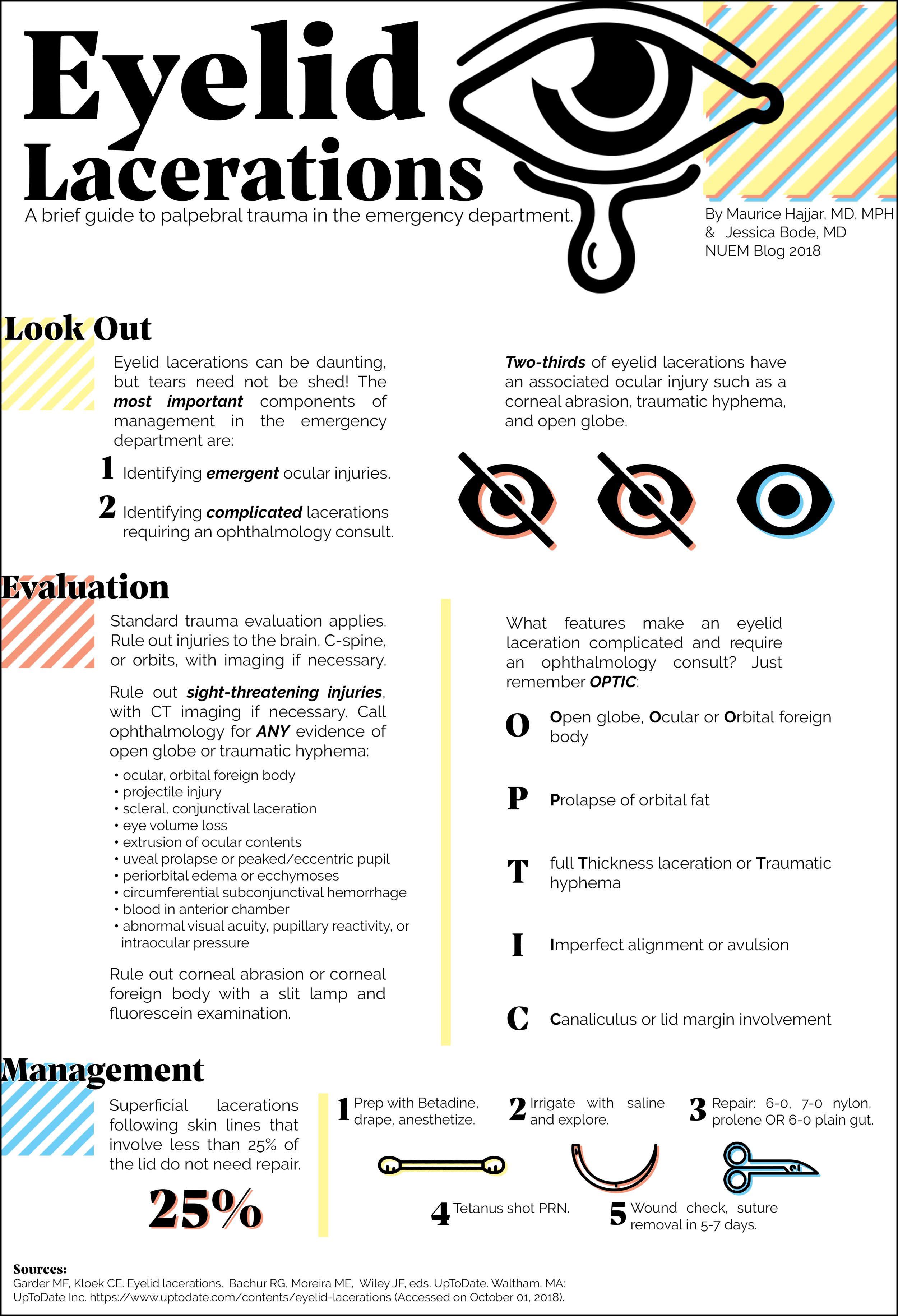
Screen Shot 2020-02-03 at 10.30.04 AM.png


Choosing Age-Appropriate settings

Since pediatric respiratory rates vary wildly from adults, one should take the patient’s age into account when initiating mechanical ventilation (table 2) . With the exception of children with obstructive pathophysiology, the physician should attempt to match the patient’s pre-intubation minute ventilation [2]. Tidal volume goals for pediatric patients do not vary much from adults with most data being extrapolated from adult studies [4] and large trials have been unable to establish as safe threshold for tidal volume [8].  The  Pediatric Acute Lung Injury Consensus Conference (PALICC) recommends targeting 5-8cc/kg of ideal body weight (IBW) for most children either by setting a tidal volume in VCV or altering driving pressures for pressure controlled modes to target tidal volumes in this range for most patients [9]. Just as in adult patients, the intubating physician can set initial FiO2 at 100% to overcome hypoxia caused by peri-intubation apnea and then quickly down titrate targeting a SpO2 of 92-97% [9] .

Screen Shot 2020-02-03 at 10.30.24 AM.png

If ventilating pediatric patients with pressure controlled ventilation, initially inspiratory times can be found on the Broselow or in a PALS manual 10 (table 3). If a volume controlled mode of ventilation is desired, inspiratory flow can be titrated to achieve a inspiratory to expiratory time ratio of 1:2 [2].  After initial setup, arterial blood gas analysis, continuous end-tidal CO2 measurement, and a chest X ray to evaluate tube positioning are just as critical here as they are in the adult patient. 

Screen Shot 2020-02-03 at 10.30.44 AM.png


Ventilating the Child with Refractory Hypoxemia 

While most pediatric patients will be relatively straightforward to ventilate, the patient intubated for infectious pathologies like pneumonia or bronchiolitis is at risk for ARDS and should be approached with lung-protective ventilation strategies in mind.  Victims of drowning are similarly at risk and fall under the category of patients requiring lung-protective settings [11]. There is no pediatric equivalent for the ARDSnet trial [12] so adult data has been extrapolated to be applied to pediatric patients [3] making this familiar territory for the adult emergency physician. 

While most children will tolerate levels of PEEP between 3-5cmH204, PALICC recommends that children at risk of ARDS receive moderately elevated levels of PEEP  between 10-15cmH2O with an SPO2 goal between 88-92% for kids requiring PEEP more than 10mmH2O [9]. In order to assess the extent of lung injury, an inspiratory hold maneuver can be used to determine lung compliance which is typically 1.5-3.0/cmH2O/kg for an infant [2]. If the EP notes decreased compliance, tidal volumes closer to 3-6cc/kg should be targeted [9]. If these measures fail to improve oxygenation, inspiratory time can be increased in order to target an inspiratory to expiratory ratio closer to 1:2. With the exception of children with elevated intracranial pressure (ICP), congenital heart disease, or  pulmonary hypertension, permissive hypercapnea is acceptable as long as the pH remains > 7.2 [4].  


Ventilating the Child with Obstructive Physiology 

Endotracheal intubation of the asthmatic child is thankfully a rare event but one that portends to a high mortality [13]. At baseline, children exhibit higher airway resistance than their adult counterparts [2] and the even higher airway resistance in asthmatic patients creates high levels of intrinsic PEEP while increases the risk of breath stacking and pneumothorax [14]. If the patient’s respiratory rate is too high, lungs will remain progressively inflated at end expiration. This increases intra-thoracic pressure thereby decreasing preload and precipitating cardiovascular collapse. The level of this intrinsic PEEP can be assessed with an expiratory hold maneuver (Figure 1). To do this, the ventilator occludes the expiratory port at the end of exhalation allowing the alveolar and airway pressures to equilibrate. The total pressure at this moment minus the set PEEP on the ventilator represents the intrinsic PEEP [7].  More simply, a flow/time curve that fails to return to baseline prior to the onset of inspiration may signal to the EP that there may be high levels of intrinsic PEEP 2. (Figure 2) 

Screen Shot 2020-02-03 at 10.32.23 AM.png
Screen Shot 2020-02-03 at 10.32.37 AM.png

To counteract this, asthmatics and other patients with obstructive physiology will need respiratory rates far below median age values. In one series of asthmatic children aged as young as nine months, rates as low as 8-12 breaths per minute were used [15]. In order to further facilitate full expiration, the I:E ratio should be increased to target values as low as 1:4-5 [15]. High levels of PEEP are typically not required in these patients and use of low to zero PEEP has been documented [14]. Hypercapnea should be expected and is allowable in these patients [14]. 

Prolonged mechanical ventilation of the pediatric patient exhibits far more complexities than this blog post covers and is beyond the scope of most emergency medicine practice. However, by relying on evidence driven practice for adult intubated patients with close guidance from a pediatric intensivist and pediatric resuscitation reference, the initial steps and safe monitoring of the intubated child are well within the abilities of the emergency physician. 

Screen Shot 2020-02-03 at 10.31.15 AM.png

Expert Commentary

Thank you for this concise summary of mechanical ventilation in children. As noted, while this is an infrequent occurrence, the initial management of a ventilated child is incredibly important. 

In choosing initial ventilator settings, the key is decision and reassessment. Most modes of ventilation will work in most children. However, careful attention to what support you’re providing your patient with and what the results of that support are, is vital. Personally, I like using PRVC mode because it adjusts support in children with changing lung compliance without a lot of manipulation required by the physician. But, in any mode of ventilation you can make adjustments as you note changes in compliance. In pressure mode, watch your tidal volumes and in volume control or PRVC, monitor your peak pressures (along with your saturations and end tidal) to see if you’re achieving your goals. Use of SIMV versus AC modes of ventilation are important in the weaning phase of ventilation but less important as you’re initiating mechanical ventilation as the patient is typically neuromuscularly blocked. I also want to emphasize the importance of weaning supplemental oxygen as soon as possible in order to understand the adequacy of your support from an oxygenation and ventilation standpoint. Hypoxemia is bad but so is hyperoxia and masking hypoventilation.

While the research in pediatric ARDS is not as robust as in adults, there is a growing body of literature describing epidemiology and current practice.[1] Current management strategies continue to be extrapolated from adult data- including lung protective strategies of permissive hypoxemia and hypercarbia (tidal volumes 3-6 cc/kg, saturations >92% in mild pARDS and >88% in severe pARDS, pH > 7.2 with exceptions for specific populations including those with pulmonary hypertension).[2] Restrictive fluid strategies (after initial resuscitation) and adequate sedation are recommended. There is ongoing research regarding the use of HFOV and prone positioning in pARDS but this is outside the scope of emergency department care.

The intubated asthmatic remains a source of anxiety among many pediatric intensivists. Key takeaways are low respiratory rate to allow for full exhalation and prevent air trapping and matching intrinsic PEEP. Permissive hypercapnia is appropriate in these patients and their CO2 should be measured by blood gas; recognizing that there is a significant amount of dead space and end tidal may be falsely reassuring/low. When intubating patient with obstructive physiology, it’s also important to ensure adequate preload and have a high suspicion for pneumothorax if they decompensate. Utilizing ketamine for sedation can be useful in these patients and has the advantages of bronchodilation and not significantly suppressing their respiratory drive, allowing them to participate in setting their inspiratory/expiratory times. 

Final thought: don’t hesitate to ask for help- from the pediatric intensivists in house or over the phone- we are happy to collaborate!

References:

  1. Khemani RG, Smith L, Lopez-Fernandez YM, et al. Paediatric acute respiratory distress syndrome incidence and epidemiology (PARDIE): an international, observational study. Lancet Respir Med. 2019 Feb;7(2):115-128. doi: 10.1016/S2213-2600(18)30344-8. Epub 2018 Oct 22.

  2. Orloff KE, Turner DA, Rehder KJ. The Current State of Pediatric Acute Respiratory Distress Syndrome. Pediatr Allergy Immunol Pulmonol. 2019 Jun 1; 32(2): 35–44. doi: 10.1089/ped.2019.0999. Epub 2019 Jun 17.

katie wolfe.png
 

Dr. Katie Wolfe, MD

Attending Physician

Pediatric Critical Care

Ann & Robert H. Lurie Children's Hospital of Chicago

Instructor of Pediatrics (Critical Care)

Northwestern University Feinberg School of Medicine


How To Cite This Post

[Peer-Reviewed, Web Publication] McCauley, M. Stelter, J. (2020, Feb 3). Initiating Emergency Department Mechanical Ventilation in the Pediatric Patient. [NUEM Blog. Expert Commentary by Wolfe, K]. Retrieved from http://www.nuemblog.com/blog/ped-mech-vent.


Other Posts You Might Enjoy…


References

  1. Losek J.D., Olson L.R., Dobson J.V., et al: Tracheal intubation practice and maintaining skill competency: survey of pediatric emergency department directors. Pediatr Emerg Care 2008; 24: pp. 294-299

  2. Pacheco, G. S., Mendelson, J., & Gaspers, M. (2018). Pediatric Ventilator Management in the Emergency Department.  Emergency Medicine Clinics of North America36(2), 401–413.  https://doi.org/10.1016/j.emc.2017.12.008

  3. Rimensberger, Peter C., Ira M. Cheifetz, and Martin C. J. Kneyber. “The Top Ten Unknowns in Paediatric Mechanical Ventilation.” Intensive Care Medicine 44, no. 3 (2018): 366–70. https://doi.org/10.1007/s00134-017-4847-4

  4. Kneyber, Martin C. J., Daniele de Luca, Edoardo Calderini, Pierre-Henri Jarreau, Etienne Javouhey, Jesus Lopez-Herce, Jürg Hammer, et al. “Recommendations for Mechanical Ventilation of Critically Ill Children from the Paediatric Mechanical Ventilation Consensus Conference (PEMVECC).” Intensive Care Medicine 43, no. 12 (December 2017): 1764–80. https://doi.org/10.1007/s00134-017-4920-z.

  5. Wells et al. The accuracy of the Broselow tape as a weight estimation tool and a drug-dosing guide – A systematic review and meta-analysis. Resuscitation. 2017 Dec;121:9-33.

  6. Singer, BD. Corbridge, TC. "Pressure modes of invasive mechanical ventilation" Southern Medical Journal"  104, no. 10 October 2011, pp 701-709 

  7. Singer, BD. Corbridge, TC. "Basic Mecahnical Ventilation" Southern Medical Journal"  102, no. 12 December 2009 , pp pp 1238-1245 

  8. de Jager P, Burgerhof JG, van Heerde M, et al: Tidal volume and mortality in mechanically ventilated children: A systematic review and meta-analysis of observational studies*. Crit Care Med 2014; 42:2461–2472

  9.  Rimensberger PC, Cheifetz IM. Ventilatory support in children with pediatric acute respiratory distress syndrome: proceedings from the pediatric acute lung injury consensus conference. Pediatr Crit Care Med.(2015) 16(5 Suppl. 1):S51–60. 10.1097

  10. Chameides L, Samson RA, Schexnayder SM, Hazinski MF (Eds).Pediatric Advanced Life Support Provider Manual, , American Heart Association, Dallas 2012.

  11. Semple-Hess, J., & Campwala, R. (2014). Pediatric submersion injuries: emergency care and resuscitationPediatric Emergency Medicine Practice, 11(6), 1–21

  12. Kneyber, Martin C. J. “Mechanical Ventilation for Pediatric Acute Respiratory Distress Syndrome: Few Known Knowns, Many Unknown Unknowns.” Pediatric Critical Care Medicine: A Journal of the Society of Critical Care Medicine and the World Federation of Pediatric Intensive and Critical Care Societies 17, no. 10 (2016): 1000–1001.

  13. Rampa S, Allareddy V, Asad R, et al. Outcomes of invasive mechanical ventilation in children and adolescents hospitalized due to status asthmaticus in United States: a population based study. J Asthma 2015; 52:423.

  14. Rubin, Bruce K., and Vladimir Pohanka. “Beyond the Guidelines: Fatal and near-Fatal Asthma.” Paediatric Respiratory Reviews 13, no. 2 (June 2012): 106–11. https://doi.org/10.1016/j.prrv.2011.05.003.

  15. Cox, R. G., G. A. Barker, and D. J. Bohn. “Efficacy, Results, and Complications of Mechanical Ventilation in Children with Status Asthmaticus.” Pediatric Pulmonology 11, no. 2 (1991): 120–26

Posted on February 3, 2020 and filed under Pediatrics.

Febrile Neutropenia in the ED

Written by: Nick Wleklinski, MD (PGY-2) Edited by: Steve Chukwuelebe, MD (NUEM ‘19) Expert commentary by: Sean Fox, MD, FACEP, FAAP

Written by: Nick Wleklinski, MD (PGY-2) Edited by: Steve Chukwuelebe, MD (NUEM ‘19) Expert commentary by: Sean Fox, MD, FACEP, FAAP

Febrile Neutropenia: Is Admission Always Necessary? 

Febrile Neutropenia: 

• Defined as an ANC <500 cells/μL with a temperature of >101F (38.8C) or >100.4F sustained for 1 hour 

• Most common in hematologic malignancies, however those with solid tumors are also at risk, especially after first round of chemotherapy (1) 

• Systemic dysfunction (i.e. hypotension, respiratory failure, renal insufficiency, etc.) seen in 25- 30% of cases and infection/severe sepsis carries a high mortality rate (2) 

Yet, not all “hot” neutropenics must be admitted and given IV antibiotics. Local practice patterns may vary, but there are guidelines defining those who are appropriate for outpatient management. 

How do you define a “low risk” Febrile Neutropenic? 

• Do they look sick, have signs of end organ dysfunction, and/or have a myriad of comorbidities? Then admission is the right call. 

• Do they have a knack for getting infected with resistant bugs? Are they already taking a fluoroquinolone to keep the bad bacteria at bay? Sorry, you’ll be coming into the hospital. 

• Do they live far away, alone, and/or have a history of not following up? ADMIT (1, 2) 

• For those who were able to answer “NO” to the above questions, then it’s time to use the MASCC (Multinational Association for Supportive Care in Cancer) and CISNE (Clinical Index of Stable Febrile Neutropenia) scores to further risk stratify. 

More reasons to Use MDCalc: 

MASCC Score: Utilizes subjective clinical data and non-laboratory objective data to help risk stratify patients. The study was validated in all cancer types (solid, hematologic, and bone marrow transplant). The higher the score, the better; a score >21 is considered low-risk. While this score has a decent sensitivity (60-95%) for identifying low risk patients, those classified as low risk still have ~10% risk of developing serious complications (i.e. hypotension, organ dysfunction) (1, 3). That is where the CISNE score comes in.

Untitled.png

CISNE score: Strictly uses objective data to help stratify patients. A CISNE of 0 indicates low risk; 1-2, intermediate; and ≥ 3, high risk. This score is applied to seemingly stable patients to better stratify their risk. In the validation study, the complication rate was significantly lower in the low risk group compared to the high risk, 1.1% vs. 32.5%, respectively, yielding a specificity of 96.6% when identifying low risk patients (4, 5).

Screen Shot 2020-01-25 at 6.36.41 PM.png

Using both: These scores can be used similarly to the Wells and PERC criteria, with MASSC ~ Wells and CISNE ~ PERC. This combination works well as the MASCC has a higher sensitivity but poor specificity when predicting poor outcomes for low risk patients while the CISNE is completely opposite (5, 6). The CISNE was not validated on patients with hematologic malignancies but can still help stratify the risk of discharging these patients (7).

Using the dynamic duo:

Screen Shot 2020-01-25 at 6.42.44 PM.png

Antibiotics:

Discharge: The goal- provide Pseudomonas coverage. It is still recommended these patients be observed for 4 hours prior to discharge (1).

Screen Shot 2020-01-25 at 6.43.20 PM.png

Admission: Start with monotherapy, unless the patient is severely ill or has focal infection (pneumonia, cellulitis, etc.); then provide additional coverage (i.e. Vancomycin). Make sure to consider previous infection history and local resistance patterns.

Screen Shot 2020-01-25 at 6.43.30 PM.png

Duration of antibiotics: Currently, it is recommended to continue antibiotics until the neutropenia has resolved. There has been some debate on earlier discontinuation prior to neutrophil recovery, but there is no solid evidence supporting this yet (8).

To Summarize:

• Some febrile neutropenics can be sent home, but require scrutiny and coordination with the patient’s oncologist for close follow up 

• MASCC and CISNE are a good tool to help risk stratify, but clinical judgement is still your best friend 

• Consider that discharging low-risk patients prevents exposure to nosocomial infections and the burden of a hospital stay, which tends to average about 4 days (9). 

Screen Shot 2020-01-26 at 5.00.40 PM.png

Expert Commentary

NUEM team,

Thank you for this concise review and reminder of the challenges inherent in managing patients who are neutropenic and febrile. Febrile neutropenia, deserves our vigilance as it is associated with substantial mortality! Unquestionably, these can be some of the most critically ill patients in your department but who may also be deceptively “well appearing.”

Similar to other conditions that have the high potential for severe M&M, febrile neutropenia nearly mandates an ultra-cautious approach: resuscitate, get lots of cultures, give antibiotics, and admit. That being noted, the management is continuing to evolve, and it is incumbent upon all of us to endeavor to stay abreast of the most current recommendations. Your post is a useful tool toward that end. 

Similar to other high-risk conditions, we are learning how to better define a low-risk population that does not benefit from aggressive management strategies.  I appreciate the summary flow diagram for the management suggestions for the febrile neutropenic patient. In my experience, physicians who often manage these patients would likely concur with it. The one key point that cannot be overstated is the importance of including the patient’s oncologist in the decision process early. The scoring systems include clinical estimations that are best made in concert with the oncologist. 

While we may be improving our ability to define the low-risk febrile neutropenic patient, I still assume the worst regardless of how well the patient appears. I actively search for any subtle sign of sickness. I anticipate the patient’s occult illness if they are well-appearing. If all of the stars align (and the moon and sun eclipse while a rainbow is overhead), then perhaps the oncologist and I will be able to define the patient as being low enough risk to go home after antibiotics.  

I appreciate this post as a means to help us all to continue to refine our knowledge base; as part of our career’s quest is to learn every day.

fox.png
 

Sean M. Fox, MD, FACEP, FAAP

Professor of Emergency Medicine & Professor of Pediatrics

Program Director, Emergency Medicine Residency Program

Department of Emergency Medicine

Carolinas Medical Center


References:

1. Taplitz, R. A., Kennedy, E. B., Bow, E. J., Crews, J., Gleason, C., Hawley, D. K., . . . Flowers, C. R. (2018). Outpatient Management of Fever and Neutropenia in Adults Treated for Malignancy: Amercian Society of Clinical Oncology and Infectious Diseases Society of America Clinical Practice Guideline Update. Journal of Clinical Oncology, 1443-1453. doi:10.1200/JCO.2017.77.6211 

2. Zimmer, A. J., & Freifeld, A. G. (2019). Optimal Management of Neutropenic Fever in Patients with Cancer. Journal of Oncology Practice, 15(1), 19-24. doi:10.1200/JOP.18.00269 

3. Klasktersky, J., Paesmans, M., Rubenstein, E., Boyer, M., Elting, L., Feld, R., . . . Talcott, J. (2000). The Multinational Association for Supportive Care in in Cancer risk index: A multinational scoring system for identifying low-risk febrile neutropenic cacner patients. Journal of Clinical Oncology, 18(16), 3038-3051. doi:10.1200/JCO.2000.18.16.3038 

4. Carmona-Bayonas, A., Jiménez-Fonseca, P., Echaburu, J. V., Antonio, M., Font, C., Biosca, M., Ayala de la Peña, F. (2015). Prediction of Serious Complications in Patients With Seemingly Stable Febrile Neutropenia: Validation of the Clinical Index of Stable Febrile Neutropenia in a Prospective Cohort of Patients From the FINITE Study. Journal of Clinical Oncology, 33(5), 465- 471. doi:10.1200/JCO.2014.57.2347 

5. Ahn, S., Rice, T., Yeung, S.-c., & Cooksley, T. (2018). Comparison of the MASCC and CISNE scores for identifying low-risk neutropenic fever patients: analysis of data from three emergency departments of cancer centers in three continents. Supportive Care in Cancer, 26(5), 1465-1470. doi:10.1007/s00520-017-3985-0 

6. Moon, H., Choi, Y. J., & Sim, S. H. (2018). Validation of the Clinical Index of Stable Febrile Neutropenia (CISNE) model in febrile neutropenia patients visiting the emergency department. Can it guid emergency physicians to a reasonable decision on outpatient vs. inpatient treatment? PLoS ONE. doi:https://doi.org/10.1371/journal.pone.0210019

7. Coyne, C., Le, V., Brennan, J., Castillo, E., Shatsky, R., Ferran, K., . . . Vilke, G. (2017). Application of the MASCC and CISNE Risk-Stratification Scores to Identify Low-Risk Febrile Neutropenic Patients in the Emergency Department. Annals of Emergency Medicine, 69(6), 755-764. doi:10.1016/j.annemergmed.2016.11.007 

8. Stern, A., Carrara, E., Bitterman, R., Yahav, D., Leibovici, L., & Paul, M. (2019). Early discontinuation of antibiotics for febrile neutropenia versus continuation until neutropenia resolution in people with cancer. Cochrane Database of Systematic Reviews. doi:10.1002/14651858.CD012184.pub 

9. Baugh, C. W., Wang, T. J., Caterino, J. M., Baker, O. N., Brooks, G. A., Reust, A. C., & Pallin, D. J. (2016). Emergency Department Management of Patients with Febrile Neutropenia: Guideline Concordant or Overly Aggressive? Academic Emergency Medicine, 24(1), 83-91. doi:10.1111/acem.13079


How to Cite This Post

[Peer-Reviewed, Web Publication] Wleklinski, N., Chukwuelebe, S. (2020, Jan 27). Febrile Neutropenia. [NUEM Blog. Expert Commentary by Fox, S]. Retrieved from http://www.nuemblog.com/blog/febrile-neutropenia


Posted on January 27, 2020 and filed under Infectious Disease.