Drug Interactions

Written by: Trish O’Connell, MD (NUEM ‘22) Edited by: Will Ford (NUEM ‘19) Expert Commentary by: Dion Tyler, PharmD and Bayan Al-Namnakani, PharmD

Written by: Trish O’Connell, MD (NUEM ‘22) Edited by: Will Ford (NUEM ‘19) Expert Commentary by: Dion Tyler, PharmD and Bayan Al-Namnakani, PharmD


Drug Interactions Final Draft.png

Expert Commentary

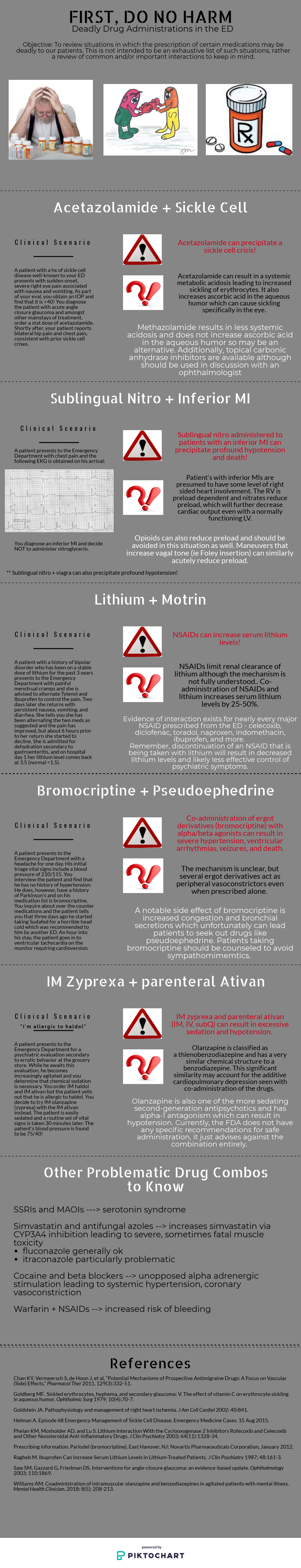
IM olanzapine + IM/IV benzodiazepines: The FDA-approved package insert for olanzapine currently recommends general avoidance of intramuscular olanzapine and parenteral benzodiazepines [1]. The European Medicines Agency (EMA) recommends waiting > 1 hour following IM olanzapine administration to administer benzodiazepines with careful monitoring for excessive sedation and cardiorespiratory depression, likely taking into account the 15-45 minute time to reach peak concentrations for IM olanzapine [2]. 

The warning against coadministration of intramuscular olanzapine and benzodiazepines (BZDs) arose from postmarketing data of adverse events in patients receiving intramuscular olanzapine for acute agitation [3]. This study reported that BZD use was associated with 51.7% (15/29) of fatal cases and 85.7% (24/28) of serious adverse drug reactions, defined as those that were life-threatening, extended the hospital stay, or resulted in a permanent disability. The authors thus recommended that the combination of IM olanzapine and benzodiazepines be avoided in the absence of further prospective data. However, it is important to note that many of these patients in this cohort had severe comorbidities, and BZD association included all instances (oral, IV, or IM) of BZD administration throughout their hospital stay. Some patients expired several days or weeks following their last olanzapine dose, making a causal association difficult to determine. Subsequent, smaller cohorts have found that oxygen desaturations are greatest in individuals who receive olanzapine and BZD therapy and have consumed ethanol, yet desaturation rates were similar with this combination in patients without ethanol intoxication when compared to olanzapine and haloperidol monotherapy as well as haloperidol and BZD combination therapy [4,5]. A retrospective, medication use evaluation (MUE) of IM olanzapine and lorazepam also demonstrated no incidences of hypotension or oxygen desaturation when the combination was administered within 1-hour or 24-hours of each other [6].  Lastly, a prospective cohort of individuals receiving IV olanzapine or IV droperidol followed immediately by IV midazolam compared with IV midazolam alone also demonstrated similar rates of oxygen desaturations and adverse events among all three groups [7]. However, IV olanzapine is not approved for use in acute agitation, and may display different pharmacokinetics compared to IM administration. 

While evidence supporting the safe use of this combination is growing, it may be prudent to use caution while coadministering IM olanzapine and BZDs in the absence of further controlled studies and in patients at greatest risk for adverse events, including the elderly and those who’ve consumed ethanol. 

References:

  1. Olanzapine [package insert]. Indianapolis, IN: Eli Lilly and Company, 2010.

  2. Zyprexa. European Medicines Agency. https://www.ema.europa.eu/en/medicines/human/EPAR/zyprexa. Accessed June 28, 2020.

  3. Marder SR, Sorsaburu S, Dunayevich E, et a.l. Case reports of postmarketing adverse event experiences with olanzapine intramuscular treatment in patients with agitation. J Clin Psychiatry. 2010;71(4):433-41.

  4. Wilson MP, MacDonald K, Vilke GM, et al. Potential complications of combining intramuscular olaznapine with benzodiazepines in emergency department patients. J Emerg Med. 2012;43(5):889-96.

  5. Wilson MP, MacDonald K, Vilke GM, et al. A comparison of the safety of olanzapine and haloperidol in combination with benzodiazepines in emergency department patients with acute agitation.  J Emerg Med. 2012;43(5):790-97.

  6. Williams AM. Coadministration of intramuscular olanzapine and benzodiazepines in agitated patients with mental illness. Ment Health Clin. 2018;8(5):208-13.

  7. Chan EW, Taylor DM, Knott JC, et al. Intravenous droperidol or olanzapine as an adjunct to midazolam for the acutely agitated patient: a multicenter, randomized, double-blind, placebo-controlled clinical trial. Ann Emerg Med. 2013;61:72-81. 


Myasthenia gravis and medications in the emergency department: Myasthenia gravis (MG) is an autoimmune disorder resulting in destruction of acetylcholine receptors at the neuromuscular junction (NMJ) and resultant muscular weakness. While not included in the original blog post, this is a drug-disease interaction where pharmacy services are frequently recruited for assistance in choosing medications that will not cause or exacerbate a myasthenic crisis. Although not all-inclusive, common agents that may be frequently encountered in the ED are listed below [1-3]:

  • Neuromuscular blocking agents (paralytics): Succinylcholine exerts its therapeutic effects through depolarization of the acetylcholine receptor at the NMJ causing sustained paralysis. In the setting of MG and reduced acetylcholine receptors, succinylcholine requirements may be increased, necessitating a higher dose of 1.5-2 mg/kg. Conversely, MG patients are more sensitive to nondepolarizing neuromuscular blockers,such as rocuronium and vecuronium, requiring a lower dose than normal. For rocuronium, a dose of 0.3-0.6 mg/kg may be considered for these patients.

  • Antibiotics: Several classes of antibiotics have been shown to prevent transmission of acetylcholine to the acetylcholine receptor at varying levels of risk listed below:

    • High risk: aminoglycosides, fluoroquinolones

    • Medium risk: Macrolides, polymixin B

    • Low risk: Penicillins, cephalosporins, carbapenems, nitrofurantoin, clindamycin, sulfonamides, doxycycline

  • Magnesium: Magnesium interferes with release of acetylcholine to the NMJ and may exacerbate a myasthenic crisis. A  higher threshold for repletion may be necessary in MG patients as well as avoidance of use for migraines, tachyarrhythmias, and as a component of laxatives.

  • Beta blockers: Beta blockers also appear to have an effect at the NMJ in preventing acetylcholine transmission, and have been found to exacerbate MG symptoms in patients with a variety of different agents in the class and routes of administration, such as ophthalmic timolol. 

  • Corticosteroids: While frequently used as treatment for a MG crisis, these agents may paradoxically worsen muscle strength through acetylcholine receptor blocking and effects on muscle contractility. 

References:

  1. Roper J, Fleming ME, Long B, et al. Myasthenia gravis and crisis: evaluation and management in the emergency department. J of Emerg Med. 2017;53:843-53.

  2. Ahmed A, Simmons Z. Drugs which may exacerbate of induce myasthenia gravis: a clinician’s guide. The Internet Journal of Neurology. 2008;10:e1-8.

  3. Singh P, Idowu O, Malik I, et al. Acute respiratory failure induced by magnesium replacement in a 62-year-old woman with myasthenia gravis. Tex Heart Inst J. 2015;42(5):495-97.


Lithium + ibuprofen (NSAIDs): Treatment with lithium could be quite challenging due to its extremely narrow-therapeutic index (0.5–1.2 mEq/L). Therefore, minor changes affect serum levels. The most common lithium poisoning occurs unintentionally (with chronic use) when the lithium intake exceeds its elimination such as in impaired kidney function or due to drug-drug interaction.1

Lithium is a water-soluble monovalent cation widely distributed in the body and it goes complete glomerulus filtration, 75 % of the ion is reabsorbed mainly in the proximal tubule. The exact mechanism is not fully understood. It appears that NSAIDs decrease the eGFR resulting in decreased lithium renal excretion. Some experts hypothesized that this is a result of the prostaglandin synthesis inhibition by NSAIDs which may lead to low renal blood flow and facilitate the reabsorption of sodium and lithium (theoretically). However, this hasn’t been proven [1].

This interaction is well known in clinical practice and most providers will be cautious when it comes to NSAIDs. Small prospective studies have shown large interindividual differences in lithium clearance associated with different NSAIDs. Those effects are highly variable and less predictable. It can occur with any NSAID and studies haven’t concluded a strong relationship with a specific agent. They have reported a reduction in lithium level by 10-25% in healthy volunteers [1-2], and up to 60% in another study [3]. A small retrospective study quantified the relative risk of lithium toxicity secondary to a medication new start in elderly patients who are on lithium. The relative risk was dramatically higher with ACEIs (RR=7.6, 95% CI=2.6–22.0) and loop diuretics (RR=5.5, 95% CI=1.9–16.1). Interestingly, NSAIDs and thiazides were not independently associated with increased risk of lithium toxicity [5].

Lithium levels and toxic effects should be monitored with concomitant NSAIDs initiation. Consider lithium dose reduction especially with NSAIDs new start or dose increase.

References: 

  1. Finley, P.R. Drug Interactions with Lithium: An Update. Clin Pharmacokinet 55, 925–941 (2016).

  2. Reimann IW, Diener U, Frölich JC. Indomethacin but not aspirin increases plasma lithium ion levels. Arch Gen Psychiatry. 1983;40(3):283–6.

  3. Ragheb MA. Aspirin does not significantly affect patients’ serum lithium levels. J Clin Psychiatry. 1987;48(10):425.

  4. Ragheb M. Ibuprofen can increase serum lithium level in lithium-treated patients. J Clin Psychiatry. 1987;48(4):161–3.

  5. Juurlink, D.N., Mamdani, M.M., Kopp, A., Rochon, P.A., Shulman, K.I. and Redelmeier, D.A. (2004), Drug‐Induced Lithium Toxicity in the Elderly: A Population‐Based Study. Journal of the American Geriatrics Society, 52: 794-798. doi:10.1111/j.1532-5415.2004.52221.x.

Nitroglycerin (NTG) and inferior MI: Great job to the authors on describing an interaction that comes up quite frequently in the emergency department. The ACC/AHA guidelines on acute STEMI recommend avoidance of NTG in patients with RV dysfunction, pre-existing hypotension, marked bradycardia or tachycardia, and use of phosphodiesterase-5 inhibitor (PDE5) use in the previous 24-48 hours [1]. Specifically, 24 hours should elapse following sildenafil use, and 48 hours following tadalafil use, due to the difference in pharmacokinetics between these PDE5s [2,3]. 

It appears that this recommendation has been challenged by a retrospective analysis conducted by Robichaud and colleagues assessing the incidence of hypotension in prehospital patients with inferior STEMI and acute chest pain receiving NTG compared with those who did not receive NTG [4]. The determination of STEMI was made by a computer-interpreted electrocardiogram (ECG) utilized by EMS while in the prehospital setting. The researchers found similar rates of hypotension between the two groups, but stated that a computer-interpreted ECG cannot be used as the sole predictor for patients who may be predisposed to hypotension following NTG administration. In the absence of controlled data, it may be necessary to exercise caution when considering NTG in STEMI patients with known RV involvement and avoid use in hypotensive patients. 

References:

  1. O'Gara PT, Kushner FG., Ascheim DD, et al. 2013 ACCF/AHA guideline for the management of ST-elevation myocardial infarction: A report of the American College of Cardiology Foundation/American Heart Association Task Force on practice guidelines. Circ. 2013;127:e362-e425.

  2. Sildenafil [package insert]. New York, NY: Pfizer Labs, 2014.

  3. Tadalafil [package insert]. Indianapolis, IN: Eli Lilly and Company, 2018.

  4. Robichaud L, Ross D, Proulx M, et al. Prehospital nitroglycerin safety in inferior ST elevation myocardial infarction, Prehosp Emerg Care. 2016;20(1):76-81.

Dion tyler final.PNG

Dion Tyler, PharmD

Emergency Medicine Pharmacy Specialist

Sinai Health System

Chicago, IL

Bayan Al-Namnakani, PharmD

PGY-2 Emergency Medicine Clinical Fellow

Northwestern Memorial Hospital Pharmacy


How To Cite This Post:

[Peer-Reviewed, Web Publication] O’Connell, T. Ford, W. (2020, Nov 29). Drug Interactions. [NUEM Blog. Expert Commentary by Tyler, D. Al-Namnakani, B]. Retrieved from http://www.nuemblog.com/blog/drug-interactions.


Other Posts You May Enjoy

Posted on November 30, 2020 and filed under Pharmacology.

Elderly Fallers

Written by: Nick Wleklinski, MD (NUEM ‘22) Edited by: Kumar Gandhi, MD, MPH (NUEM '20) Expert Commentary by: Scott Dresden, MD, MS

Written by: Nick Wleklinski, MD (NUEM ‘22) Edited by: Kumar Gandhi, MD, MPH (NUEM '20) Expert Commentary by: Scott Dresden, MD, MS


Oh How the Older Adults Fall

Introduction:

Older adults (>65yrs old) fall. In 2006, older adult patients who fell made up approximately 2.1 million of ED visits totaling $6.1 billion in health care dollars [1]. Falls are the most common cause of unintentional injury for older folks, accounting for 13% of all ED visits from 2008-2010 [2]. These numbers are only increasing as our population ages and it is predicted to double by 2030 [3]. The injuries incurred wildly vary, but these patients tend to fall into two buckets: Major injury/organic etiology à admit vs. simple mechanical fall à Discharge.

Common injuries requiring hospitalization:

Falls resulting in major injury carry significant morbidity and mortality. Hip fractures lead to deterioration in function and carry ~27% mortality at 1 year [4]. Head injuries account for a significant amount of fall-related deaths, making CT brain imagining imperative in most fall patients. Add a CT C-spine as these injuries are more common in the older adults, the Canadian and Nexus C-spine rules don’t work well for these patients [5]. Additionally, rib fractures are common and require significant analgesia to prevent splinting and subsequent complications. Be sure to consider blunt cardiac injury and pulmonary contusion! Given that falls are a frequent cause of trauma in older adult patients, it is important to keep the effects of aging in mind when running the ABC’s (Table 1) [6].

Table 1: Further considerations for ABC’s in older adult trauma patients.

Table 1: Further considerations for ABC’s in older adult trauma patients.

The tougher scenario: Those without any injuries:

Patients without any major injuries deserve more thought than simply ruling out organic etiologies (i.e. CVA, ACS, arrythmia, etc.) and major trauma. These patients are at high risk for subsequent falls and may even have underlying physiologic injuries. Using the term “mechanical fall” is risky as it can anchor providers into comfort. Therefore, having a more regimented approach can help better risk stratify these patients.

The fall itself:

  • Where did it happen?

    • Those in nursing homes/institutional setting fall more frequently than those in the community (60% vs ~33%, respectively) [7]

    • Falls at home should trigger need for home safety evaluation

  • Have you fallen before?

    • History tends to repeat itself, with nearly 50% of fallers falling again within 1 year [8]

  • Witnessed vs Unwitnessed?

    • Collateral information can provide key details if a patient is a fall risk and requires further evaluation by physical therapy

  • How long where you on the ground? [9, 10]

Figure 1: Increased time on the ground leads to worsening fall anxiety and increased risk of rhabdomyolysis and subsequent kidney injury

Figure 1: Increased time on the ground leads to worsening fall anxiety and increased risk of rhabdomyolysis and subsequent kidney injury

Evaluating the patient:

  • Outside of the obvious (CVA, ACS, etc.), it is important to also consider other common etiologies:

    • Hypotension

    • Arrythmias

    • Infection (PNA, UTI, pressure ulcers)

    • Vestibular dysfunction (i.e. BPPV)

    • Anemia

      • Ask about melena as this is a commonly not investigated [11]

    • Delirium

    • Malignancy

  •  Medications: Polypharmacy is a known issue in older adults, but there are certain medications to take note of. Antidepressants and antipsychotics are associated with the highest risk of falls while diuretics and narcotics didn’t have as much of a risk (Table 2) [12]. Additionally, who manages the meds and how are they organized at home?

Table 2: Common medications associated with falls

Table 2: Common medications associated with falls

  • What is their baseline? This is the meat and potatoes of the evaluation and where future risk factors can be identified and addressed.

    • How steady do they feel on their feet?

    • Decreased cognition (Dementia, Alzheimer’s, etc.) incurs increase fall risk [13]

    • Do they have arthritis/chronic pain?

      • Can result in unsteady gait from favoring certain part of body, increasing risk

    • Timed Up and Go Test:

      • A great way to evaluate lower extremity strength and balance (figure 2)

    • Visual and auditory impairment: Visual acuity should be addressed. Look at their eyewear as multifocal lenses increase fall risk [14].

    • Feet: check for neuropathy and ask about footwear.

    • Assist devices used for ambulation? Do they use these devices regularly and correctly?

    • Delirium screening

      • The Confusion Assessment Method is used in triage [15]

Figure 2: The Timed Up and Go Test.

Figure 2: The Timed Up and Go Test.

Things we can do:

Although continuity is not generally part of the EM specialty, we can help address future fall risk for these patients who we discharge after their fall evaluation. Recommending supplements such as vitamin D and calcium are helpful for reducing risk for fall-related injuries [7]. Balance training through outpatient physical therapy referrals can further help reduce fall risk. Follow up is imperative and these patients should see their PMD or a geriatrician soon after their discharge from the ER to continue their fall evaluation.  

Conclusion:

While major trauma from falls is exciting and straight forward, it is important to give more thought to those older adult patients deemed to have a “mechanical fall”. Gathering information about the fall and determining the patient’s baseline can help stratify future risk. The incidence of falls is only going to increase as our population ages, so having a regimented approach to these patients is imperative.


Expert Commentary

As was expertly described, falls in older adults lead to significant morbidity and mortality.  Unfortunately, in the ED they are often dismissed as “mechanical,” the injuries are treated, but the causes are never identified. The term mechanical fall is ambiguous and unhelpful and should not be used in the ED. Some mean that the fall was not a result of seizure or syncope, but it is not a clear term.  Additionally, it does not help with prognosis. There are no differences in adverse events at 6 months between “mechanical” and non-mechanical faller. For a great discussion of the Myth of the Mechanical Fall see Shan Liu’s presentation at IGNITE presentation at SAEM18 (https://saem-ondemand.echo360.org/media-player.aspx/5/13/431/1608).

Even if injuries are minor, patients often do poorly. Between 36% and 50% of patients have an adverse event such as a recurrent fall, emergency department revisit, or death within 1 year after a fall, including 25% who die within 1 year. As the CDC likes to remind us, every 20 minutes someone dies from a fall (https://www.cdc.gov/steadi/index.html).

So what do we do with this medical problem that has a 25% 1-year mortality? As with many problems in geriatrics, falls are a sentinel event, and deserve a sentinel response. It is our job to prevent the next fall. The Geriatric Emergency Department Guidelines provide a framework for a risk assessment after a fall. One might think that the cause of the fall is obvious (e.g. tripped over a crack in the sidewalk). However a thoughtful assessment begins by asking “if this patient was a health 20-year-old, would he or she have fallen? “ If the answer is no, then the assessment of the underlying cause of the fall should be more comprehensive and should include a thorough history of the fall and risk factors such as ability to perform Activities of Daily Living (ADLs), appropriate footwear, and medications. Physical exam should include orthostatic blood pressure, a head to toe exam even for patients with seemingly isolated injuries, a neurologic exam with special attention to neuropathy and proximal motor strength, and a safety assessment. Patients should be able to rise from the bed or chair, turn, and steadily ambulate in the ED before considering discharge (not while the nurse is handing the patient his or her discharge paperwork). For patients who are unable to safely ambulate, consideration of an assist device such as a cane or walker should be given, physical therapy (PT) and occupational therapy (OT) consultation, and possibly hospital admission.  All patients who are admitted after a fall should be admitted by PT and OT. Additionally, patients who fell should have home safety assessments which may be arranged through occupational therapy.

In addition to the GED guidelines, the CDC has developed the Stopping Elderly Accidents, Deaths & Injuries (STEADI) program. This program includes an algorithm for fall risk screening, assessment and intervention. For screening they recommend patients answer the Stay Independent screening (a 12 question tool), however if the patient is in the ED for a fall, this step can be omitted because the patient has already declared themselves as high risk for falls.  To evaluate gait, strength, and balance, the Timed Up & Go, 30-Second Chair Stand, or 4-Stage Balance Test are recommended. In addition, to the assessments mentioned previously (medications, orthostatics), asking about potential hazards such as throw rugs or slippery floors, and a visual acuity check are advised. Once risk factors are identified they should be addressed through physical therapy, exercise of fall prevention programs, medication optimization, home safety evaluation, discussion with outpatient clinicians regarding orthostatic hypotension, referral to a podiatrist for proper footwear, recommending a vitamin D supplement. Finally, ensuring close and enduring followup is important. Consider a referral to a geriatrician if the patient doesn’t already see one.

Scott_Dresden-29.png

Scott Dresden, MD, MS

Associated Professor of Emergency Medicine

Director of Geriatric Emergency Department Innovations (GEDI)

Northwestern Memorial Hospital


How To Cite This Post:

[Peer-Reviewed, Web Publication] Wleklinski, N. Gandhi, G. (2020, Nov 23). Elderly Fallers. [NUEM Blog. Expert Commentary by Dresden, D]. Retrieved from http://www.nuemblog.com/blog/elderly-falls.


Other Posts You May Enjoy

References

  1. Owens, P.L., et al., Emergency Department Visits for Injurious Falls among the Elderly, 2006: Statistical Brief #80, in Healthcare Cost and Utilization Project (HCUP) Statistical Briefs. 2006, Agency for Healthcare Research and Quality (US): Rockville (MD).

  2. Sapiro, A.L., et al., Rapid recombination mapping for high-throughput genetic screens in Drosophila. G3 (Bethesda), 2013. 3(12): p. 2313-9.

  3. Foundation, C.f.D.C.a.P.a.T.M.C. The State of Aging and Health in America 2007. 2007  [cited 2019.

  4. Cenzer, I.S., et al., One-Year Mortality After Hip Fracture: Development and Validation of a Prognostic Index. J Am Geriatr Soc, 2016. 64(9): p. 1863-8.

  5. Goode, T., et al., Evaluation of cervical spine fracture in the elderly: can we trust our physical examination? Am Surg, 2014. 80(2): p. 182-4.

  6. Carpenter, C.R., et al., Major trauma in the older patient: Evolving trauma care beyond management of bumps and bruises. Emerg Med Australas, 2017. 29(4): p. 450-455.

  7. Nagaraj, G., et al., Avoiding anchoring bias by moving beyond 'mechanical falls' in geriatric emergency medicine. Emerg Med Australas, 2018. 30(6): p. 843-850.

  8. Liu, S.W., et al., Frequency of ED revisits and death among older adults after a fall. Am J Emerg Med, 2015. 33(8): p. 1012-8.

  9. Austin, N., et al., Fear of falling in older women: a longitudinal study of incidence, persistence, and predictors. J Am Geriatr Soc, 2007. 55(10): p. 1598-603.

  10. Deshpande, N., et al., Activity restriction induced by fear of falling and objective and subjective measures of physical function: a prospective cohort study. J Am Geriatr Soc, 2008. 56(4): p. 615-20.

  11. Tirrell, G., et al., Evaluation of older adult patients with falls in the emergency department: discordance with national guidelines. Acad Emerg Med, 2015. 22(4): p. 461-7.

  12. Woolcott, J.C., et al., Meta-analysis of the impact of 9 medication classes on falls in elderly persons. Arch Intern Med, 2009. 169(21): p. 1952-60.

  13. Muir, S.W., K. Gopaul, and M.M. Montero Odasso, The role of cognitive impairment in fall risk among older adults: a systematic review and meta-analysis. Age Ageing, 2012. 41(3): p. 299-308.

  14. Lord, S.R., J. Dayhew, and A. Howland, Multifocal glasses impair edge-contrast sensitivity and depth perception and increase the risk of falls in older people. J Am Geriatr Soc, 2002. 50(11): p. 1760-6.

  15. Han, J.H., et al., Diagnosing delirium in older emergency department patients: validity and reliability of the delirium triage screen and the brief confusion assessment method. Ann Emerg Med, 2013. 62(5): p. 457-465.

Posted on November 23, 2020 and filed under geriatrics.

The BICAR-ICU Trial and Practical Use of Bicarb in Metabolic Acidosis

Written by: Philip Jackson, MD (NUEM ‘20) Edited by: Katie Colton, MD  (NUEM ‘19) Expert Commentary by: Benjamin Singer, MD

Written by: Philip Jackson, MD (NUEM ‘20) Edited by: Katie Colton, MD (NUEM ‘19) Expert Commentary by: Benjamin Singer, MD


Introduction

 Until recently, there has been a paucity of high-quality data to inform the use of intravenous sodium bicarbonate in severe metabolic acidosis.  This has resulted in a lack of universal practice guidelines to inform clinicians in emergency medicine and other specialties when caring for some of their sickest patients.

 Historically there have been two camps of thought when approaching the use of sodium bicarbonate in the sick, acidotic patient.  Severe acidemia results in protein dysfunction, potentially leading to arrhythmia, cardiovascular collapse, multi-organ failure and eventual death. [1-6] Thus, correcting acidemia with alkalotic bicarbonate solutions could prevent these compounding complications.  However, growing evidence suggests that the deleterious effects associated with profound acidemia may be more strongly associated with the underlying physiological insult than the acidosis itself.  Therefore, treating the acidemia without addressing the underlying pathology may expose the patient to side effects including hypernatremia, hypocalcemia, and exacerbation of CNS cellular acidosis (due to increased levels of carbon dioxide) without resulting in a net benefit. [1,2]


The BICAR-ICU Trial

 A recent randomized, prospective, multi-center trial by Jaber et al. evaluated the effects of bicarbonate administration to ICU patients with metabolic acidemia. The study randomized 389 ICU patients with severe metabolic acidemia (pH ≤7⋅20, PaCO ≤45 mm Hg, and sodium bicarbonate concentration ≤20 mmol/L), a total Sequential Organ Failure Assessment score of 4 or more or an arterial lactate concentration of 2 mmol/L or more into a treatment group receiving 4.2% sodium bicarbonate (<1L per day) or a control group receiving an equivalent volume of standard crystalloid solution.

 There was no significant difference between the two groups in the primary outcome, a composite of all-cause mortality at day 28 and the presence of organ failure at day 7.  Interestingly, bicarbonate administration decreased the need for renal replacement therapy (RRT) and in a sub-group of patients with acute kidney injury bicarbonate infusion improved mortality and decreased vasopressor requirements.

 It is important to note that the study excluded patients with significant urinary or digestive tract losses of bicarbonate (two important causes of non-anion gap metabolic acidosis) and patients that had already been treated with bicarbonate or RRT. Furthermore, a significant proportion of patients in the control group (24%) received bicarbonate at some point during the study and only 60% of the treatment group actually maintained the goal pH of >7.30. These factors skew the study towards a negative outcome, as they presumably blunt the effects of administration of bicarbonate. Thus, it is possible that more of a benefit may have been observed without these disruptions.

 The study did not differentiate between etiologies of metabolic acidemia, though ketoacidosis was also excluded, and thus it is difficult to draw conclusions on the value of bicarbonate in various pathologic conditions. There was no specific protocol regarding timing of administration or concentration, making the study difficult to replicate in the emergency setting. Nevertheless, it was essentially the first large, multi-center RCT evaluating this topic and so some practical conclusions can be drawn; these should be interpreted in the context of each individual patient.


Practice recommendations in special situations

  • Anion Gap Metabolic Acidosis: The human body maintains an essentially neutral net electrical charge through the retention and excretion of ions (notably H+) and anions (notably Cl- and HCO3-). Addition or retention of other anions will increase the anion gap and cause a net negative charge, causing retention of  H+ ions and leading to a metabolic acidosis. In general, administration of bicarbonate to this scenario will balance the pH but will not remove the additional anions that are the root cause of the pathologic acidosis and would presumably provide little benefit to patient outcomes.

  • Lactic Acidosis: Previous to the BICAR-ICU trial, most available data suggests no benefit of bicarbonate. Notably two small prospective physiological studies of 14 and 10 patients, respectively, demonstrated no hemodynamic response or difference in response to catecholamines. [7,8]  Additional retrospective and observational studies did not result in clear conclusions. [9,10] The BICAR-ICU trial did not specifically evaluate this population and thus current guidelines recommend against bicarbonate administration unless pH falls below 7.15 or bicarbonate falls below 5 mEq/L (at which point small changes in bicarbonate concentration can lead to potentially fatal changes in pH). [11]

    • In general, DON’T GIVE IT

  • Diabetic and Alcoholic Ketoacidosis: A prospective RCT of 21 patients in severe DKA showed no benefit of bicarbonate therapy. [12] Limited data in pediatric DKA and adult AKA populations show similar findings. [13,14] There is evidence that bicarb administration is associated with worse outcome in pediatric patients. It is feasible to give bicarbonate to patients in extremis (pH<6.9) in the adult population to theoretically prevent cardiovascular collapse.

    • In general, DON’T GIVE IT except when pH<6.9 and with extreme caution in the pediatric patient

  •  Toxic Ingestions (methanol, ethylene glycol, toluene, salicylates, etc.): In general, along with specific therapies, bicarbonate infusion is a mainstay of therapy as systemic and urinary alkalinization removes these anions through ion trapping of metabolites. [15]

    • GIVE IT, along with specific antidotes and possible dialysis

  •  Uremic Acidosis:  Uremic acidosis results from the inability of the injured kidney to excrete anions such as phosphates, sulfates, and nitrates and so removal of these substances is the mainstay of therapy. Administration of bicarbonate does not directly impact this end, and data supporting its use is limited. [16] However, it is the current practice of many nephrologists to treat uremic acidosis with bicarbonate infusion to prevent the need for RRT.  It is intuitive that bicarbonate can prevent RRT as bicarbonate therapy both corrects pH and also temporarily improves hyperkalemia (depending on the concentration of the solution). This was again demonstrated in the BICAR-ICU trial with a reduced need for RRT in the treatment group, as well as a mortality benefit in a subgroup with AKI. Though further investigation is needed, it is reasonable to give bicarbonate in this population in consultation with nephrology.

    • GIVE IT, judiciously in severe acidemia and in consultation with a nephrologist

  • Non-Anion Gap Metabolic Acidosis: In general, this results from loss of total body bicarbonate or retention of additional chloride. It is thus, theoretically reasonable to treat this population with bicarbonate because you are directly addressing the underlying pathophysiology. [1,2]

  •  Renal Losses, including Renal Tubular Acidosis (RTA): Several types exist, but the pathophysiology lies in the inability of the kidneys to re-absorb bicarbonate resulting in increased urinary losses. The mainstay of therapy is bicarbonate, both oral and IV if severe. [17]

    • GIVE IT

  •  Gastrointestinal Losses (pancreatic fistula, diarrhea, uretal diversion, etc.): Excessive loss of bicarbonate through the GI tract causes a systemic acidosis. Removing the offending pathology (repairing the fistula) is the mainstay of therapy with bicarbonate replacement as a temporizing measure. [18,19]

    • GIVE IT, in severe cases

  • Hyperchloremic Metabolic Acidosis: Usually, as the result of iatrogenic over-administration of chloride rich fluids (normal saline). Therapy involves stopping administration of high chloride content fluids and/or switching to a more pH neutral solution such as Lactated Ringer’s or sodium bicarbonate in dextrose. [20,21]

    • GIVE IT, in severe cases


Conclusions

•   Administration of sodium bicarbonate is recommended along with therapies targeting specific etiologies of acidemia in severe cases of non-anion gap metabolic acidosis and anion gap metabolic acidosis secondary to most toxic ingestions.

•   Bicarbonate administration is reasonable in severe metabolic acidemia secondary to uremic acidosis and in patients with both AKI and acidemia.  Further research is needed to elucidate protocols and to clearly demonstrate benefits.

•   Bicarbonate administration is rarely recommended in both ketoacidosis and lactic acidosis unless the patient is in extremis as it has shown no clear benefit and may cause harm.


Expert Commentary

In this trial there was no attempt to differentiate the cause of acidosis a priori, but the type of metabolic acidosis matters when considering bicarb administration. Why?

a) Metabolic acidosis without elevation in the anion gap is more likely to respond to bicarb administration than acidosis with an elevated anion gap. You can think of non-gap acidosis as bicarb deficiency; by administering bicarb, you are repleting bicarb.

b) The trial supports the use of bicarb for uremic acidosis, which tends to be a mix of non-gap- and gap-associated phenomena (renal tubular acidosis combined with an increase in unmeasured anions). Note that the number-needed-to-treat was six patients to prevent one of them from going on dialysis in the AKI subgroup.

c) Lactic acidosis is a misnomer in that the process that creates an elevation in blood lactate anions is physiologically separate from the process generating protons. [1,2] Lactate elevation occurs because of shunting of glycolysis-generated pyruvate away from oxidative metabolism and toward lactate production. This shunting can occur in states of hypoxia (oxidative metabolism shut down, usually Type A) or normoxia (so-called aerobic glycolysis, usually Type B). Lactate is a weak base, so why is there often an associated acidosis? The proton comes from hydrolysis of ATP, which cannot be rapidly replenished under conditions that also favor lactate production (e.g., hypoxia).

So, why does bicarb administration not work well for lactic acidosis? Because even if you titrate off those extra protons using huge amounts of bicarb, you will not rebalance hydrolysis and re-generation of ATP until you fix the underlying problem (ischemia, sepsis, etc.). The rationale for avoiding bicarb in ketoacidosis is similar. Hence, I agree with the recommendation to use bicarb in patients with severe non-uremic anion-gap-associated acidemia only as a temporizing measure while working to reverse the underlying cause.

References

1. Mizock BA. Controversies in lactic acidosis. Implications in critically ill patients. JAMA. 1987;258:497-501.

2. Andersen LW, Mackenhauer J, Roberts JC, Berg KM, Cocchi MN, Donnino MW. Etiology and therapeutic approach to elevated lactate levels. Mayo Clin Proc. 2013;88:1127-1140. PMCID: PMC3975915.

Benjamin Singer.PNG

Benjamin Singer, MD

Assistant Professor of Medicine

Pulmonary and Critical Care

Biochemistry and Molecular Genetics

Northwestern University


How To Cite This Post:

[Peer-Reviewed, Web Publication] Jackson, P. Colton, K. (2020, Nov 16). The BICAR-ICU Trial and Practical Use of Bicarb in Metabolic Acidosis. [NUEM Blog. Expert Commentary by Singer, B]. Retrieved from http://www.nuemblog.com/blog/BICAR-ICU-trial.


Other Posts You May Enjoy

References

 1. Adams, J, et al.  Emergency Medicine: Clinical Essentials.  2013; 2nd edition: 1363-1378.

2. Arbo, J, et al.  Decision Making in Emergency Critical Care: An Evidence-Based Handbook.  2015; 1: 496-499.

3. Jaber S, Paugam C, Futier E, et al. Sodium bicarbonate therapy for patients with severe metabolic acidemia in the intensive care unit (BICAR-ICU): a multi-center, open-label, randomized controlled, phase 3 trial. Lancet. June 2018.

4. Berend K, de Vries APJ. Physiological approach to assessment of acid–base disturbances. N Engl J Med 2014; 371: 1434–45.

5. Kraut JA, Madias NE. Lactic acidosis. N Engl J Med 2014; 371: 2309–19.

6. Jung B, Rimmele T, Le Goff C, et al. Severe metabolic or mixed acidemia on intensive care unit admission: incidence, prognosis and administration of buffer therapy: a prospective, multiple­center study. Crit Care 2011; 15: R238.

7. Cooper DJ, Walley KR, Wiggs BR, Russell JA. Bicarbonate does not improve hemodynamics in critically ill patients who have lactic acidosis. A prospective, controlled clinical study. Ann Intern Med 1990; 112: 492–98.

8. Mathieu D, Neviere R, Billard V, Fleyfel M, Wattel F. Effects of bicarbonate therapy on hemodynamics and tissue oxygenation in patients with lactic acidosis: a prospective, controlled clinical study. Crit Care Med 1991; 19: 1352–56.

9. El­Solh AA, Abou Jaoude P, Porhomayon J. Bicarbonate therapy in the treatment of septic shock: a second look. Intern Emerg Med 2010; 5: 341–47.

10. Kim HJ, Son YK, An WS. Effect of sodium bicarbonate administration on mortality in patients with lactic acidosis:  a retrospective analysis. PLoS One 2013; 8: e65283.

11. Gauthier P, Szerlip H. Metabolic acidosis in the intensive care unit. Crit Care Clin.  2002; 18: 289-308.

12. Morris LR, Murphy MB, Kitabchi AE: Bicarbonate therapy in severe diabetic ketoacidosis. Ann Intern Med. 1986; 105: 836-840.

13. Green SM, Rothrock SG, Ho JD, et al.: Failure of adjunctive bicarbonate to improve outcome in severe pediatric diabetic ketoacidosis. Ann Emerg Med. 1998; 31: 41-48 

14. Hojer J: Severe metabolic acidosis in the alcoholic: differential diagnosis and management. Hum Exp Toxicol. 1996; 15: 482-488.

15. O’Malley G. Emergency department management of the salicylate-poisoned patient. Emerg Med Clin North Am.  2007; 25(2): 333-346

16. Roderick PJ, Willis NS, Blakeley S, Jones C, Tomson C. Correction of chronic metabolic acidosis for chronic kidney disease patients. Cochrane Database of Systematic Reviews 2007, Issue 1. Art. No.: CD001890. DOI: 10.1002/14651858.CD001890.pub3

17. Morris C, Low J. Metabolic acidosis in the critically ill: Part 2.  Causes and treatment.  Anesthesia.  2008; 63: 396-411.

18. Callery M, et al. Prevention and management of pancratic fistula. J Gastrointest Surg.  2009; 13(1): 163-173

19. Davidson T, et al.  Long-term metabolic and nutritional effects of urinary diversion. Urology.  1995; 46: 804-809.

20. Kellum J. Saline induced hyperchloremic metabolic acidosis. Crit Care Med.  2002; 30: 259-261.

21. Prough D, Bidani A. Hyperchloremic metabolic acidosis is a predictable consequence of intraoperative infusion of 0.9% saline.  Anesthesiology.  1999; 90: 1247-1249.

Posted on November 16, 2020 and filed under Critical care.

Preterm Neonatal Resuscitation

Written by: Aaron Wibberley, MD (NUEM ‘22) Edited by: Vidya Eswaran, MD '20 Expert Commentary by: Spenser Lang, MD

Written by: Aaron Wibberley, MD (NUEM ‘22) Edited by: Vidya Eswaran, MD '20 Expert Commentary by: Spenser Lang, MD


Peterm Neonatal Resuscitation Blog_1.jpg

Expert Commentary

Thanks to Dr. Wibberley and Dr. Eswaran for providing this infographic on a tough topic – neonatal resuscitations.

Usually, deliveries in the emergency department cause a dichotomy of emotions – initial anxiety, then relief and happiness. Most of our deliveries tend to be quick, precipitous, with hopefully just enough warning for us to grab gloves and remember where the baby warmer is. Unfortunately, when babies decide to struggle with their first few minutes of life, this becomes a lot more stressful for everyone.

Fair warning – though I am an emergency medicine physician, and prepared to deal with emergent situations of any age, I think there are very few of us who feel as comfortable with neonatal resuscitations as we do with critically ill trauma or cardiac arrest patients. Especially if your department sees very little pediatrics, it is completely normal to feel anxiety when imagining resuscitating a neonate, and even more so a pre-term baby. This is OK! In fact, this should motivate you to get familiar with NRP, and provides a perfect opportunity for spaced repetition throughout your career to enhance recall.

Here are my broad strokes steps for a fresh neonate requiring resuscitation.

#1: Know your resources! The first step in managing a neonatal resuscitation occurs far before the patient shows up in your department. Where is your baby warmer? Where are your teeny-tiny BVM’s? What’s the smallest ETT and intubating blade you stock, and where?  I promise you, the hardest part of intubating this baby won’t be the actual mechanics of placing an ETT – it will be in the preparation and supply gathering. Don’t rely on your nurses to know everything when seconds count – know where this stuff is yourself.

#2: Call for help, early and often. Many emergency departments have some type of OB/imminent delivery response – hopefully this brings in a pediatrician well trained in neonatal resuscitation as well. Hopefully, this also brings a nurse who is used to placing IV’s in these itty bitty babies. If this doesn’t describe your hospital, call to start the transfer process, and move on to #3…

#3: Dry and stim. Nearly all babies respond to drying and stimulation. Please don’t start bagging a poor newborn before drying it off and giving it a good rub for 30-60 seconds (unless it’s extremely pre-term – try to avoid rubbing all the skin off of a 25-weeker, this is bad form.) At the same time, keep in mind that these babies will need some form of external thermoregulation so make sure the warmer is actually functioning.

#4: When in doubt, fix the breathing. As is obvious when scanning through NRP guidelines, 95% of managing a sick newborn lies in assisting the respirations. Poor tone? Fix the breathing. Initial HR below 100? Try to fix the breathing. Poor color? You get it. Don’t be afraid to escalate from blow by, to PEEP, to BVM. If the baby has little to no respiratory effort, a couple initial breaths via BVM can quickly improve the situation. But please, when you’re bagging a tiny neonate, use small breaths – this is not the typical 120 kg patient we are used to.

#5: In the short term, an IO is your friend. A UVC is golden, but not really possible in an active resuscitation. The good news is that most babies don’t need IV access in the short term – for my  reasoning, see #3 and #4. The literature suggests that placing the neonatal IO in the proximal tibia, distal tibia, or distal femur can be safe and effective.

#6: This is the time to debrief. Whether a happy or a tragic ending, this is a rare and emotional event in the emergency department. Debrief with your team. Talk to whoever you talk to about this stuff – spouse, friend, coworker. We are champions of compartmentalization in the emergency department out of necessity, but don’t bear the entirety of these encounters on yourself – lean on those around you.

Spenser Lang.PNG

Spenser Lang, MD

Assistant Professor

Department of Emergency Medicine

University of Cincinnati


How To Cite This Post:

[Peer-Reviewed, Web Publication] Wibberly, A. Eswaran, V. (2020, Nov 9). Preterm Neonatal Resuscitation. [NUEM Blog. Expert Commentary by Lang, S]. Retrieved from http://www.nuemblog.com/preterm-neonatal-resuscitation


Other Posts You May Enjoy

Posted on November 9, 2020 and filed under Pediatrics.

A “Pill-in-the-Pocket” Approach to Paroxysmal Atrial Fibrillation

Written by: David Feiger, MD (NUEM ‘22) Edited by: Jon Andereck, MD, MBA NUEM ‘19) Expert Commentary by: Kaustubha Patil, MD

Written by: David Feiger, MD (NUEM ‘22) Edited by: Jon Andereck, MD, MBA NUEM ‘19) Expert Commentary by: Kaustubha Patil, MD


The Case

A healthy 65-year-old male with paroxysmal atrial fibrillation presents to the emergency department in atrial fibrillation with rapid ventricular rate. His blood pressure is 135/83, heart rate 135, respirations 15 with an O2 saturation of 98% on room air. He states that he took his “pill-in-the-pocket” four hours prior to presentation and his symptoms did not resolve.

Atrial fibrillation

A study in the Western Journal of Emergency Medicine in 2013 observed the costs associated with emergency department (ED) treatment and discharge of patients presenting with atrial fibrillation (AF) or atrial flutter was $5,460 [10]. Those admitted to the hospital naturally incur far higher costs. For those eligible, a visit to the ED could be avoided with the “pill-in-the-pocket” approach.

What is the “pill-in-the-pocket” approach?

The “pill-in-the-pocket” approach is the administration of a prescribed class IC antiarrhythmic, either flecainide or propafenone, following recent onset of episodes of palpitations in patients with paroxysmal AF. It is generally initiated by the patient’s cardiologist after extensive cardiac evaluation to rule out structural disease and other conduction abnormalities. The idea is to terminate the suspected episode of AF without having to present to an ED or clinic. Several studies have investigated the safety of this approach and supported this method of outside-the-hospital termination of paroxysmal AF events [2, 14].

Who is eligible for the “pill-in-the-pocket” approach?

In a study in the New England Journal of Medicine supporting the feasibility and safety of this out-of-hospital treatment, only specific patients were selected to participate. Inclusion criteria included:

  • healthier patients between 18 and 75 years old

  • a history of infrequent AF not associated with chest pain, hemodynamic instability, dyspnea, or syncope

  • no significant electrocardiographic abnormalities (pre-excitations, bundle branch blocks, long QT interval, etc.)

  • no structural or functional cardiac diseases

  • no history of thromboembolic episodes

  • no current use of an antiarrhythmic medication

  • not currently pregnant

  • no significant chronic disease including but not limited to muscular dystrophies, systemic collagen disease, and renal or hepatic insufficiency

These patients were then admitted to the hospital for a cardiac workup and were trialed on either flecainide or propafenone with successful pharmacologic cardioversion in the inpatient setting. Both flecainide and propafenone are proarrhythmic, thus structural heart diseases must be ruled out before their use and patients should be monitored during initiation of therapy [2].

How do flecainide and propafenone work?

Flecainide and propafenone are both powerful class IC antiarrhythmics that strongly bind fast sodium channels with a slower association and dissociation than other class I antiarrhythmics. These drugs slow phase 0 during sodium-dependent depolarization in cardiac muscle cells of the atrial and ventricular myocardium (Figure 1). This effect is primarily important in prolonging atrial refractoriness, thus aiding in the conversion and termination of AF. Flecainide’s use in tachyarrhythmias comes from its rate-dependence property in which its efficacy is greater at faster heart rates. Propafenone has additional beta blocker activity which may enhance its overall clinical effectiveness in treating tachyarrhythmias [3, 5, 6, 13].

What are treatment options for patients presenting to the ED in AF?

For all comers presenting in AF with rapid ventricular rate to the ED, the literature has not elicited a perfect treatment modality, and no distinction is made for patients on the “pill-in-the-pocket” approach prior to arrival. Despite this, general practice guidelines are highlighted in many textbooks.

In hemodynamically-stable patients, rate control in the ED is the generally the treatment of choice. Diltiazem is often preferred as compared to beta blockers like metoprolol, which may cause hemodynamic instability in patients with underlying heart or lung disease. In otherwise healthy patients, metoprolol is a reasonable choice [1]. Digoxin is also appropriate, but onset takes several hours and is inferior to beta-blockers for rate control within 6 hours of treatment [11].

Patients who have been in AF for > 48 hours are at a greater risk of new intracardiac thrombus formation and cardioversion-induced embolization. Newer data from a study in 2014 suggests that there is an increased risk of thrombus formation with > 12 hours of AF [8] though the original guidelines for electric cardioversion within 48 hours of symptom onset have not changed. Patients who are hemodynamically stable who have been in AF for > 48 hours (and considered if > 12 hours) should be admitted from the ED for transesophageal echo to rule out intracardiac thrombus prior to cardioversion, or alternatively for initiation of anticoagulation [7].

In hemodynamically unstable patients, electrical cardioversion should be pursued regardless of a patient’s anticoagulation status [7].

Are there any treatment considerations in the ED for patients in AF taking flecainide or propafenone?

Treatment failure to the “pill-in-the-pocket” approach may be a marker of progression of the patient’s clinical disease. However, if a patient presents within an hour or two of taking their “pill-in-the-pocket,” remember the four to six-hour onset of these medications suggests they may convert during their ED stay. As in the case initially presented, the patient spontaneously converted while waiting for a provider. For those that do not, these patients warrant evaluation for new structural cardiac disease and may no longer benefit from the “pill-in-the-pocket” approach and may require daily maintenance prophylactic therapy [2].

A subset of stable patients presenting to the ED with AF with rapid ventricular rate may be taking flecainide or propafenone as maintenance therapy and not as part of the “pill-in-the-pocket” approach. In this instance, some literature has suggested that these patients can take an extra dose or two up to the maximum daily dose of flecainide (400mg) or propafenone (900mg for immediate release and 850mg for sustained release) to attempt pharmacological conversion, and it would be reasonable to attempt this in the ED [9].

To admit or not admit, that is the question.

The patient’s clinical picture should guide the provider as to the patient’s disposition. A patient’s comorbidities, current stability following conversion to normal sinus rhythm, plan for possible ablation, necessity for starting anticoagulation or maintenance medication, and means for close cardiology or PCP follow up on an outpatient basis should be factored when dispositioning the patient. Certainly, if a patient is requiring continuous IV infusion of rate controlling medications or has poor rate control, he or she should be admitted to the hospital [1]. Recent literature suggests that discharging stable patients home is safe following successful electrical, pharmacologic, or spontaneous cardioversion in the ED [4].

Final Thoughts

The “pill-in-the-pocket” approach is a great way for eligible patients to self-terminate episodes of AF in the comfort of their home, potentially preventing a costly and lengthy ED visit. While this approach has been shown to be a safe and effective for terminating paroxysmal AF, there is a significant lack of data on how to treat these patients who do not respond to these medications at home. General principals should be followed–electric cardioversion if the patient is hemodynamically unstable and rate control medications if the patient is hemodynamically stable (or rhythm control if you happen to practice in Canada [14]. Patients may be discharged home with close cardiology or PCP follow up if successfully cardioverted.


Expert Commentary

Atrial fibrillation (AF) is the most common cardiac arrhythmia and worldwide prevalence and incidence are increasing.1 It is estimated that by 2050 more than 12 million Americans will suffer from this debilitating and dangerous arrhythmia.1-2  AF presentations to Emergency Departments are certainly not without cost and the overall burden on the healthcare system will undoubtedly increase as the prevalence of atrial fibrillation continues to rise. A “pill in the pocket” approach for treatment of symptomatic atrial fibrillation has been well-described.

Class IC (sodium channel blockers) antiarrhythmic drugs (flecainide and propafenone) are the drugs of choice for “pill in the pocket” chemical cardioversion of symptomatic atrial fibrillation. There are some important considerations for this approach to be safe and effective:

  1. The patient should have a history of infrequent paroxysmal atrial fibrillation, not persistent atrial fibrillation (episodes of AF that last greater than 7 days).

  2. We reserve this approach for patients with symptomatic atrial fibrillation (palpitations, mild dyspnea, or mild lightheadedness) with rapid ventricular rates who do not experience dangerous symptoms such as chest pain or syncope.

  3. As anti-arrhythmic drugs can also be pro-arrhythmic, we do not recommend Class IC antiarrhythmic drugs in patients with known structural heart disease, reduced left ventricular systolic function, or known coronary artery disease, due to the increased risk of inducing dangerous arrhythmias.

  4. In patients who are not on therapeutic anticoagulation, we only recommend this approach when it has been less than 24 hours since onset of the AF episode. If the AF episode has lasted beyond 24 hours or it is unknown when the episode started, the risk of formation of intracardiac thrombus during AF and subsequent risk of stroke after a successful chemical cardioversion from a Class IC drug would be prohibitively high.

  5. Due to the use-dependent nature of Class IC antiarrhythmics (more effective with more sodium channel blockade at faster ventricular rates), there is a chance of slowing conduction throughout the heart to the point that atrial fibrillation can organize into rapid atrial flutter with 1:1 AV conduction, leading to an aberrant wide complex tachycardia. For this reason, we recommend that the patient receive a beta-blocker or calcium channel blocker at least 30 minutes prior to administration of flecainide or propafenone.

  6. Some practitioners recommend that if the patient is not already on anticoagulation, that they initiate anticoagulation at the time of beta-blocker or calcium channel blocker administration to reduce the risk of intracardiac thrombus formation if the patient does not convert to sinus rhythm within 24-48 hours.

  7. Some practitioners recommend that the first attempt at “pill in the pocket” dosing be performed in the emergency department so that safety and efficacy can be monitored.

  8. If patients report a progressively increasing need for “pill in the pocket” use or there is a suggestion of increasing burden of AF episodes, I recommend consultation with the patient’s cardiologist or electrophysiologist to discuss alternative options for rhythm control of symptomatic atrial fibrillation. Potential options at that time could include initiation of maintenance antiarrhythmic drug therapy versus invasive management with catheter ablation of atrial fibrillation. 

When used in the right patient, a “pill in the pocket” approach can be a very effective strategy for rhythm control of infrequent symptomatic paroxysmal atrial fibrillation. Appropriate patient factors to consider prior to recommending this approach are nicely highlighted in the post above. “Pill in the pocket” management for AF can resolve patient symptoms, improve patient’s quality of life, and reduce unnecessary emergency room visits and subsequent hospitalizations.

References

  1. Chugh SS, Havmoeller R, Narayanan K, et al. Worldwide epidemiology of atrial fibrillation: a Global Burden of Disease 2010 Study. Circulation. 2014 Feb 25; 129(8):837-47.

  2. Miyasaka Y, Barnes M, Gersh B, et al. Secular Trends in Incidence of Atrial Fibrillation in Olmsted County, Minnesota, 1980 to 2000, and Implications on the Projections for Future Prevalence. Circulation. 2006 Jul 11;114(2):119-25.

Kaustubha D Patil.PNG

Kaustubha Patil, MD

Clinical Cardiac Electrophysiology

Bluhm Cardiovascular Institute Northwestern Medicine

Assistant Professor of Medicine

Northwestern University Feinberg School of Medicine


How To Cite This Post:

[Peer-Reviewed, Web Publication] Feiger, D. Andereck, J. (2020, Nov 2). A “Pill-in-the-Pocket” approach to paroxysmal atrial fibrillation. [NUEM Blog. Expert Commentary by Patil, K]. Retrieved from http://www.nuemblog.com/blog/pill-in-pocket.


Other Posts You May Enjoy

References

  1. Adams, James, et al. “Tachydysrhythmias.” Emergency Medicine: Clinical Essentials, Elsevier Health Sciences, 2013, pp. 497–513.

  2. Alboni, Paolo, et al. “Outpatient Treatment of Recent-Onset Atrial Fibrillation with the ‘Pill-in-the-Pocket’ Approach.” New England Journal of Medicine, vol. 351, no. 23, 2004, pp. 2384–2391., doi:10.1056/nejmoa041233.

  3. Aliot, E., et al. “Twenty-Five Years in the Making: Flecainide Is Safe and Effective for the Management of Atrial Fibrillation.” Europace, vol. 13, no. 2, 2010, pp. 161–173., doi:10.1093/europace/euq382.

  4. Besser, Kiera Von, and Angela M. Mills. “Is Discharge to Home After Emergency Department Cardioversion Safe for the Treatment of Recent-Onset Atrial Fibrillation?” Annals of Emergency Medicine, vol. 58, no. 6, 2011, pp. 517–520., doi:10.1016/j.annemergmed.2011.06.014.

  5. Dan, Gheorghe-Andrei, et al. “Antiarrhythmic Drugs–Clinical Use and Clinical Decision Making: a Consensus Document from the European Heart Rhythm Association (EHRA) and European Society of Cardiology (ESC) Working Group on Cardiovascular Pharmacology, Endorsed by the Heart Rhythm Society (HRS), Asia-Pacific Heart Rhythm Society (APHRS) and International Society of Cardiovascular Pharmacotherapy (ISCP).” EP Europace, vol. 20, no. 5, 2018, doi:10.1093/europace/eux373.

  6. Dukes, I.d., and E.m Vaughan Williams. “The Multiple Modes of Action of Propafenone.” European Heart Journal, vol. 5, no. 2, 1984, pp. 115–125., doi:10.1093/oxfordjournals.eurheartj.a061621.

  7. January, Craig T., et al. “2014 AHA/ACC/HRS Guideline for the Management of Patients With Atrial Fibrillation.” Journal of the American College of Cardiology, vol. 64, no. 21, 2014, doi:10.1016/j.jacc.2014.03.022.

  8. Nuotio, Ilpo, et al. “Time to Cardioversion for Acute Atrial Fibrillation and Thromboembolic Complications.” Jama, vol. 312, no. 6, 2014, p. 647., doi:10.1001/jama.2014.3824.

  9. “Pill-in-a-Pocket Dosing Safely Converts Breakthrough Atrial Fib.” Family Practice News, vol. 35, no. 18, 2005, p. 20., doi:10.1016/s0300-7073(05)71733-3.

  10. Sacchetti, Alfred, et al. “Impact of Emergency Department Management of Atrial Fibrillation on Hospital Charges.” Western Journal of Emergency Medicine, vol. 14, no. 1, 2013, pp. 55–57., doi:10.5811/westjem.2012.1.6893.

  11. Sethi, Naqash J., et al. “Digoxin for Atrial Fibrillation and Atrial Flutter: A Systematic Review with Meta-Analysis and Trial Sequential Analysis of Randomised Clinical Trials.” Plos One, vol. 13, no. 3, 2018, doi:10.1371/journal.pone.0193924.

  12. Stiell, Ian G., et al. “Association of the Ottawa Aggressive Protocol with Rapid Discharge of Emergency Department Patients with Recent-Onset Atrial Fibrillation or Flutter.” Cjem, vol. 12, no. 03, 2010, pp. 181–191., doi:10.1017/s1481803500012227.

  13. Wang, Z, et al. “Mechanism of Flecainide's Rate-Dependent Actions on Action Potential Duration in Canine Atrial Tissue.” American Society for Pharmacology and Experimental Therapeutics, vol. 267, no. 2, 1 Nov. 1993, pp. 575–581.

  14. Yao, R., et al. “Real-World Safety And Efficacy Of A ‘Pill-In-The-Pocket' Approach For The Management Of Paroxysmal Atrial Fibrillation.” Canadian Journal of Cardiology, vol. 33, no. 10, 2017, doi:10.1016/j.cjca.2017.07.371.

Posted on November 2, 2020 and filed under Cardiovascular.

Management of Snake Bite Injuries

Written by: Rafael Lima, MD (NUEM ‘23) Edited by: Mike Conrardy, MD (NUEM ‘21) Expert Commentary by: Sean Bryant, MD

Written by: Rafael Lima, MD (NUEM ‘23) Edited by: Mike Conrardy, MD (NUEM ‘21) Expert Commentary by: Sean Bryant, MD


An estimated 10,000 patients visit emergency departments for snake bite injuries each year in the United States [1]. The number of snake bite occurrences an emergency department sees depends largely on the geographic area of practice. While there are known remedies for these incidents, snake bites can be devastating if not promptly managed, meaning emergency physicians should be knowledgeable in the subject. In this article, we review the common management of snake bite injuries and envenomations for the two major snake groups in the United States.

Overview

There are about 20 known venomous species of snakes in the United States. While most envenomations occur in the Southwestern United States, every region is home to at least one species of venomous snake [2]. Not all snake bites result in envenomation. At least 25% of venomous snake bites are dry. You should still suspect envenomation upon the patient’s initial presentation and rule it out by monitoring their clinical symptoms and progression.

Identification of the snake is useful in guiding management of care, but it should not be attempted if doing so poses any additional risk to the patient or provider. In the United States, venomous snakes generally fall under two categories: Crotaline/pit vipers in the Viperidae family, and coral snakes in the Elapidae family.


Crotaline (Pit Vipers)

This group of snakes has historically been responsible for the more severe envenomations between the two groups [3]. The WHO classifies pit vipers in CAT 1 of their venom database, describing them as highly venomous with high rates of morbidity and mortality [4].

Pit vipers generally have a triangular shaped head with heat-sensing “pits” located on the face. They frequently have a rattle on their tail, but not all pit vipers are rattlesnakes. Copperheads and cottonmouth snakes are also included in this group.

Crotaline venom causes localized tissue necrosis and congestive coagulopathy. This can be identified by a prolonged INR, PT, PTT, and thrombocytopenia. Additionally, the viper venom can cause capillary and cellular membranes to increase in permeability. Large amounts of venom can cause diffuse vaso-extravasation and hemolysis that can lead to hypovolemic shock and DIC if untreated.

CroFab is the antivenom of choice for cotaline envenomation. It is a polyvalent antivenom, meaning it contains antibodies derived from the venom of multiple different species of snakes. Administration is titrated based on clinical and symptom response.


Elapidae

The venomous Elapidae snake in the United States is the coral snake. There are less severe envenomations from coral snakes compared to pit vipers. This is a result of how venom is administered between the two groups: pit vipers have venom glands that inject venom directly through the fangs, while coral snakes rely on passive seeping of venom through their glands while they chew.

Source: Tad Arensmeier from St. Louis, MO, USA

Source: Tad Arensmeier from St. Louis, MO, USA

Coral snakes can be identified by their brightly colored rings extending along the length of the whole body. Usually, every other ring is yellow, separating the wider red or black rings in between. The common saying “red on yellow, kill a fellow; red on black, venom lack” has been been used to differentiate between venomous coral snakes and their harmless look-alikes in North America. A further level of differentiation is how far the rings extend circumferentially around the snake. Rings encircle the entire body in venomous coral snakes, while harmless look-alikes do not have the red coloration on the ventral side [5].

Source: Dawson at English Wikipedia

Source: Dawson at English Wikipedia

Venomous bites by coral snakes usually elicit little to no pain. This is because the Elapidae venom acts upon the neuromuscular junction and inhibits acetylcholine receptors. Clinical manifestations are predominantly neurological. Envenomation can cause lethargy, confusion, salivation, cranial nerve palsies, and respiratory paralysis. Symptoms are usually delayed, up to 12 hours from the initial bite. Coagulopathy and tissue necrosis does not happen with coral snake venom [2]. Unfortunately, the Elapidae antivenom is no longer manufactured in the United States and there is a limited supply available.

 ED Work Up

As in all patients who present to the emergency department, first ensure that airway, breathing, and circulation are intact. All suspected snake bite injuries warrant a prompt toxicology or poison center consult.

Sometimes, patients will bring in a dead or decapitated snake for identification in the emergency department. DO NOT attempt to handle a snake the patient brought in for identification, even if it is dead. Many snakes have intact reflexes that are preserved even after death or decapitation and you can still be bitten and envenomated by a dead snake!

Examine the injury and look for clear fang marks or puncture wounds. Get a history focused on the timing of the injury, medication allergies, and description of the snake, if known. The borders of erythema should be measured and marked serially.

Laboratory work-up is focused on assessing coagulopathy and hemolysis, especially if the snake is a confirmed pit viper or is unknown. Obtain CBC with platelet count, PT, PTT, INR, fibrinogen, and D-dimer. It is also important to check a baseline set of electrolytes with a basic chem panel, assess the extent of myonecrosis with a CK, and assess for renal damage with a UA.

Manage the wound with copious irrigation and exploration for retained foreign bodies (ie. fangs or teeth). Inquire about the patient’s tetanus status and administer if they are not up to date. Do not attempt to tourniquet or suction venom out of the wound. There is no evidence for routine antibiotic use in snake injuries [6].

Crotaline Bite Management

Consider using CroFab antivenom if the local area of injury and erythema is expanding. If coagulopathy is detected, do not treat with heparin or FFP. Give antivenom first, as unneutralized venom will react with clotting factor replacements [2]. Patients with abnormal coagulation studies within 12 hours after CroFab administration are more likely to develop recurrent coagulopathy. In these patients, repeat coagulation studies should be obtained every 48 hours until resolved. If lab values are worsening, then antivenom retreatment should be reconsidered [7].

Observe the affected limb for compartment syndrome. If clinical suspicion is high for compartment syndrome, consider formally measuring compartment pressures. Elevate the affected limb, and administer extra vials of antivenom. Antivenom administration is preferred over fasciotomy in the treatment of compartment syndrome caused by Crotaline venom [8].

Crofab, the Crotaline antivenom, is typically administered in stepwise fashion and is titrated to clinical resolution of symptoms. Administer 4-6 vials of CroFab antivenom and watch for clinical improvement at the local site of injury. If no improvement seen, administer 4-6 more vials. Repeat until control is achieved, meaning a reversal of symptoms, such as erythema, swelling, pain. Then administer 2 vial doses 6 hours later, then 12 hours, then 18 hours. Envenomation patients should be monitored for at least 8 hours. Keep epinephrine and antihistamines nearby in case of anaphylaxis or allergy to antivenom [2].

Elapidae Bite Management

Because of their potential devastating neurologic effects, coral snake bites should be empirically treated with antivenom and monitored for respiratory deterioration. Provide good supportive care, including intubation and ventilation, if necessary. Avoid opioids for pain management as they may mask symptoms of impending neurologic manifestations. Patients with suspected coral snake envenomations should be monitored for 12 hours after the initial bite [2].


Expert Commentary

Thank you, Dr. Lima for bringing the important and timely topic of snakebites to the table by posting this excellent overview!  Current poison center data (2018 National Poison Data System) indicate a total of 4,013 crotalid exposures with the majority being copperheads.  While morbidity is worrisome, mortality was fortunately low in our country with only one fatality reportedly from a rattlesnake [1].

Prehospital snakebite management has been an area of deserved scrutiny.  Limb immobilization, analgesia, and transport to a medical facility are critical actions.  Tourniquets, pressure immobilization bandages, cryotherapy, electrotherapy, and incision/suction are not recommended and are likely harmful.  One researcher discovered that venom extraction suction devices “just suck” [2].  Having a cell phone in the field is most important to prevent loss of limb or life!

In other regions of the world, capturing or killing the snake may be optimal in determining which species specific antivenom to administer.  For North American crotalids, however, this practice is discouraged and exceedingly dangerous.  Both CroFab and Anavip (recently approved and now marketed with the goal of reducing risks of late coagulopathy) are prepared from several species of North American crotalids and can be used to manage any crotalid envenomation.  These contemporary antivenoms (Fab fragments) are safer than older polyvalent antivenom that resulted in high rates of anaphylaxis. 

Consult your regional poison center (1-800-222-1222) or staff medical toxicologist when managing snakebites!  For the number of snakebites that present to the emergency department, poison centers manage severalfold more each year.  Making decisions regarding the management of a limb that resembles compartment syndrome (more antivenom vs. surgical consultation), the interpretation of laboratory results, redosing of antivenom to gain initial control of swelling, and the management of nonindigenous (e.g. cobras, gaboon vibers) pet snakebites are nuances your subspecialists would love to collaborate on!

References

1. Gummin DD, Mowry JB, Spyker DA, BrooksDE, Beuhler MC, RiversLJ, Hashem HA, & Ryan ML 2018 Annual Report of the American Association of Poison Control Centers’ National Poison Data System (NPDS): 36th Annual Report, Clinical Toxicology, 2019;57:1220-1413.

2.  Bush SP.  Snakebite Suction Devices Don’t Remove Venom: They Just Suck.  Annals of Emergency Medicine, 2004;43:187-188.

Sean Bryant.PNG

Sean Bryant, MD

Assistant Director, Toxicology Fellowship Program, Department of Emergency Medicine, Cook County Health

Associate Professor, Department of Emergency Medicine, Rush Medical College


How To Cite This Post:

[Peer-Reviewed, Web Publication] Lima, R. Cornardy, M. (2020, Oct 26). Management of Snake Bite Injuries. [NUEM Blog. Expert Commentary by Bryant, S]. Retrieved from http://www.nuemblog.com/blog/snake-bites.


Other Posts You May Enjoy

References

  1. Snakebite Injuries Treated in United States Emergency Departments, 2001–2004. O’Neil, Mary Elizabeth et al. Wilderness & Environmental Medicine, Volume 18, Issue 4, 281 - 287

  2. Gold, Barry S., et al. “Bites of Venomous Snakes.” New England Journal of Medicine, vol. 347, no. 5, 1 Aug. 2002, pp. 347–356., doi:10.1056/nejmra013477.

  3. Seifert, Steven A., et al. “AAPCC Database Characterization of Native U.S. Venomous Snake Exposures, 2001–2005.” Clinical Toxicology, vol. 47, no. 4, 2009, pp. 327–335., doi:10.1080/15563650902870277.

  4. “Venomous snakes distribution and species risk categories.” World Health Organization. 2010. http://apps.who.int/bloodproducts/snakeantivenoms/database/

  5. Cardwell, Michael D. “Recognizing Dangerous Snakes in the United States and Canada: A Novel 3-Step Identification Method.” Wilderness & Environmental Medicine, vol. 22, no. 4, 1 Oct. 2011, pp. 304–308., doi:10.1016/j.wem.2011.07.001.

  6. Prophylactic Antibiotics Are Not Needed Following Rattlesnake Bites. August, Jessica A. et al. The American Journal of Medicine, Volume 131, Issue 11, 1367 - 1371

  7. Recurrence phenomena after immunoglobulin therapy for snake envenomations: Part 2. Guidelines for clinical management with crotaline Fab antivenom. Annals of Emergency Medicine, 2001, Vol.37(2), p.196-201., doi: 10.1067/mem.2001.113134

  8. Hall, Edward L. “Role of Surgical Intervention in the Management of Crotaline Snake Envenomation.” Annals of Emergency Medicine, vol. 37, no. 2, Feb. 2001, pp. 175–180., doi:10.1067/mem.2001.113373.

add tags

schedule post

share to twitter u%

format other posts you may enjoy

fix URL and copy to google file

Posted on October 26, 2020 and filed under Toxicology.

U Tox: Clinical Utility and False Negatives

Written by: Ben Kiesel , MD (NUEM ‘23) Edited by: Jason Chodakowski, MD (NUEM ‘19) Expert Commentary by: Joe Kennedy, MD

Written by: Ben Kiesel , MD (NUEM ‘23) Edited by: Jason Chodakowski, MD (NUEM ‘19) Expert Commentary by: Joe Kennedy, MD


Utox first.png
U tox 0.75.jpg
u tox 2.jpg
u tox 2.5.jpg
u tox 3.5.jpg

Expert Commentary

The urine drug screen is one of the most frequently ordered and more frequently cursed tests in all of medicine.  It is one of the few tests that generates more argument than the banal discussions between surgeons and emergency physicians regarding the utility of a white blood cell count.  Nevertheless, in skilled hands, this cheap and simple immunoassay can answer a few quick questions.  The key is in knowing your Achilles heel:

  1. Opioids and opiates produce similar toxidromes, but there are virtually limitless combinations of street and prescription drugs, lab assays, and confirmatory tests to tell these apart.  If the test is negative, who cares! Treat the patient in front of you.  Fentanyl and its analogues are perhaps most often missed.  Wake the patient up, discuss their substance use, and move on. 

  2. Hopefully you noticed that *many* things other than phencyclidine can lead to a positive urine drug screen for PCP.  Ever have a conversation with the mother of a 12 year old about their PCP use?  Best go into that conversation knowing that many other substances cross-react.  Perhaps the kid is actually on lamotrigine for seizures, and that is why they were combative.  Or perhaps like most humans, they took a few ibuprofen at some point.  Either way—when child protective services is consulted for a positive result, either call the poison center or your local toxicologist for help.

Perhaps the most important point is that this is a screen and absolutely by no means a confirmatory test.  When it matters (placement, custody, fired/hired, transplant eligibility, excluding other diagnoses), get an actual level or result.  Not sure how to do that?  Call your lab!  Or call me.  Toxicologists love solving these problems and teaching and helping others do the same!

Joe Kennedy.PNG

Joe Kennedy, MD

Attending Physician, Emergency Medicine

University of Illinois Hospital

Senior Toxicology Fellow

Toxikon Consortium


How To Cite This Post:

[Peer-Reviewed, Web Publication] Kiesel, B. Chodakowski, J. (2020, Oct 19). U Tox: Clinical Utility and False Negatives. [NUEM Blog. Expert Commentary by Kennedy, J]. Retrieved from http://www.nuemblog.com/blog/utox-clinical-utility-and-false-negatives.


Other Posts You May Enjoy

Posted on October 19, 2020 and filed under Toxicology.

Peripheral Vasopressors: Do I need that central line?

Written by: Saabir Kaskar, MD (NUEM ‘23) Edited by: Abiye Ibiebele, MD  (NUEM ‘21) Expert Commentary by: Marc Sala, MD

Written by: Saabir Kaskar, MD (NUEM ‘23) Edited by: Abiye Ibiebele, MD (NUEM ‘21) Expert Commentary by: Marc Sala, MD


Vasopressors have been used to treat shock since the early 1900s and continue to remain a mainstay of management of distributive shock. Traditionally, these medicines have been delivered through central venous catheters primarily due to the perceived risks of peripheral infusion, which include potential extravasation of vasoactive medicines and subsequent tissue necrosis. However, central venous catheter insertion is accompanied by its own risks such as pneumothorax, infection and carotid artery insertion and dilation. There is also a risk to delaying vasopressor initiation in hypotensive patients which is why vasopressors are often now started peripherally until central access can be attained.

Peripheral administration of vasopressors has classically been reserved for less potent vasoconstrictors such as phenylephrine and vasopressin. Fear of extravasation and tissue injury often is a cause for concern prior to starting norepinephrine, epinephrine or dopamine peripherally. The perceived harm from administrating these medicines peripherally largely stems from case reports over the past 60 years. However, what does the latest evidence tell us? Is this fear warranted or is it just a myth? Can we send our patients in shock up to the ICUs without central access?

One prospective observational study conducted at Long Island Jewish Medical Center evaluated the safety of vasoactive medication administered through peripheral IV sites. [1] The study monitored the use of vasopressors (norepinephrine, dopamine, and phenylephrine) in an intensive care unit with a total of 734 patients observed. The study incorporated an interdisciplinary protocol between pharmacy, nursing, physicians for administering vasoactive medicines through a peripheral IV. The protocol required that nursing staff examine the PIV access site every two hours, IV size be either 18 or 20 gauge, and utilize upper extremity vein sites with over 4mm vein diameter visualized via ultrasound. During the time of the study, 783 out of 953 patients received vasopressors for 49 +/- 22 hours through peripheral IV. While anatomic position of access site was not formally recorded, most IVs were placed in the upper arm basilic or cephalic vein. Peripheral vasopressors were only allowed to run for 72 hours before running centrally. Of the 783 patients, infiltration of the PIV occurred in 19 (2%) patients. All 19 had prompt local injection of phentolamine and application of nitroglycerin paste at the site of extravasation. No tissue injury was noted at the site of extravasation in any of the 19 cases.

This study shows that administration of vasopressors peripherally is feasible with a low risk if proper precautions are taken. The risk of extravasation and tissue necrosis is still present especially in ED’s and ICUs where such rigorous protocols are not in effect. However, this study demonstrates that vasopressor use may not be an automatic indication for central venous catheter insertion.

A more recent systematic review of peripheral vasopressor safety was recently published in Emergency Medicine Australia. [2] The review incorporated seven observational studies, roughly 1300 patients, that reported the incidence of adverse events for the continuous infusion of peripheral vasopressors including the above study.  The major finding was that extravasation events were uncommon (3.4%) and that no significant tissue necrosis or distal ischemia was reported. However, the data analyzed in this review comes from studies with mixed methodology quality and with limited duration of infusion. Five of the seven studies had peripheral vasopressor administration for less than 24 hours.

At its current state, the quality of data reviewing the safety profile of peripheral vasopressors is not universally high. However, the observational data we do have reports low incidence of complications which should be reassuring for clinicians especially when starting these medicines for short periods of time and as a bridge to possible central infusion. Early peripheral infusion should be given more consideration as delaying vasopressor administration has been shown to increase mortality in septic shock. [3] While further research is certainly needed in this field, the current state of data should at least quell some concerns of the perceived risks of peripheral vasopressor administration.


Expert Commentary

Dr. Kaskar does a great job summarizing several of the major studies that can inform how we approach the infusion of peripheral vasoactive drugs in lieu of a central catheter. I can only assume we could agree one area of common ground, which is that if central access is in place, this should be used for the vasoactive infusion, given that the occurrence of tissue complications, while probably rare, can be limb-threatening.  An additional prospective study I would mention is by Medlej et al [1] where the authors prospectively studied ED patients managed for a variety of shock states with peripherally administered vasoactive agents and found that 3/55 (5.45% of total, and 6% of those receiving norepinephrine) had extravasation.  None had serious complications, but notably among the three events, all three used 20G IVs and two occurred using hand veins. This relatively small and heterogeneous study would indicate that extravasation is uncommon and when it occurs, is not particularly morbid, even with norepinephrine. 

Finally, another recent study notable for its cohort took place in the operating room context. Here, medical records of over 14,000 patients who received peripheral norepinephrine to manage hypotension associated with general anesthesia in two European academic centers were studied retrospectively for complications. Only five patients (0.035%) had extravasation, wherein the median infusion duration was 20 minutes, and none of whom had a significant complication from the extravasation. They calculated an estimated a risk of 1-8 events per 10,000 patients. 

What do all of these studies have in common that I think belies the true incidence of complications associated with peripheral vasoactive drugs? Vigilance. While it’s true that peripheral norepinephrine infusion may not result in serious tissue necrosis when given in the context of a formal clinical study (especially one that takes place in an operating room with continuous monitoring by anesthesia!), what about when the infusion goes unnoticed during a night shift with a high patient to nurse ratio?  In this case, I would argue that the closer to a “real-world experience” we can get with these studies, the better. 

I would also mention that a mentor of mine once theorized the “sunset” of crash central lines as the use of intraosseous catheters became more common in adults in the past decade.  While intraosseous catheters are not without their own complications, it is worth mentioning their role in this conversation as we move forward in thinking about how to transition patients safely from the ED to ICU with several different options for vascular access in lieu of a controlled, sterile central line placement. 

References:

1. Medlej et al. Complications From Administration of Vasopressors Through Peripheral Venous Catheters: An Observational Study.  The Journal of Emergency Medicine 2017; 54(1): 47-53.

2. Pancaro et al. Risk of Major Complications After Perioperative Norepinephrine Infusion Through Peripheral Intravenous Lines in a Multicenter Study. Anesthesia and Analgesia 2019; Published ahead of print.

Marc Sala.PNG

Marc Sala, MD

Assistant Professor of Medicine

Pulmonary and Critical Care

Northwestern University Feinberg School of Medicine


How To Cite This Post:

[Peer-Reviewed, Web Publication] Kaskar, S. Ibiebele, A. (2020, Oct 12). Peripheral Vasopressors: Do I need that central line? [NUEM Blog. Expert Commentary by Sala, M]. Retrieved from http://www.nuemblog.com/blog/abdominal-imaging.


Other Posts You May Enjoy

References

1. Cardenas-Garcia J, Schaub KF, Belchikov YG, Narasimhan M, Koenig SJ, Mayo PH. Safety of peripheral intravenous administration of vasoactive medication. Journal of hospital medicine. 2015; 10(9):581-5

2. Tian DH, Smyth C, Keijzers G, et al. Safety of peripheral administration of vasopressor medications: A systematic review. Emergency medicine Australasia. 2019

3. Beck V, Chateau D, Bryson GL et al. Timing of vasopressor initiation and mortality in septic shock: a cohort study. Crit. Care 2014; 18: R97.

Physiologically Difficult Intubations

Written by: Samantha Stark, MD (NUEM ‘20) Edited by: Steve Chukwulebe, MD (NUEM ‘19) Expert Commentary by: Seth Trueger, MD, MPH

Written by: Samantha Stark, MD (NUEM ‘20) Edited by: Steve Chukwulebe, MD (NUEM ‘19) Expert Commentary by: Seth Trueger, MD, MPH


It’s the first few minutes of your shift, and the paramedics roll by your workstation with your first patient, a young woman clutching an inhaler and breathing with every accessory muscle in her body. You direct them to your resuscitation room and they inform you that she has a history of asthma and is having an attack; she’s too exhausted from breathing to verify this, but it seems true. You quickly get her on BiPAP, which mildly improves her work of breathing, but as she becomes drowsy, you obtain a VBG showing a climbing CO2 of 45. You realize that it’s time to intubate this patient, and as you get set up, you collect your thoughts and quickly review everything you’ve heard about intubating asthmatics.

Obstructive Airway Disease

First, remember that asthma is an obstructive airway disease, meaning that there are two main processes to worry about during and after intubation:

  • Auto PEEP

  • Hypotension secondary to increased intrathoracic pressure from auto PEEP

*Note: auto PEEP is caused by breath stacking in a patient whose expiration is impaired (such as asthma or COPD) – the ventilator initiates a breath before there’s time for full exhalation, and this leads to progressively more volume retained in the lungs, increasing the risk of barotrauma. This can also lead to increased intrathoracic pressure, in turn decreasing preload to the heart and thus causing hypotension.

How to optimize the intubation:

  • As you already have this patient on BiPAP, try to preoxygenate as much as possible with this mode of positive pressure

  • Consider attempting delayed sequence intubation with ketamine

    • It will maintain the patient’s respiratory drive and may help with BiPAP synchrony and anxiolysis

    • It serves as a bronchodilator

  • Use rocuronium for paralysis

    • It will last longer than succinylcholine, and initially help with vent synchrony

    • *Note: remember to fully sedate the patient after intubation, as they won’t tell us that they’re not sedated during their prolonged paralysis

  • Decrease the dead space and resistance of your vent by using the largest endotracheal tube feasible

  • Frequently reassess the ventilator to ensure that breath stacking is not occurring:

    • Low respiratory rate to allow for exhalation

    • Higher tidal volumes of 6-8 cc/kg IBW

    • Decreased I:E ratio (at least 1:3, may very well need to be longer)

You’ve successfully intubated this patient, and now the lab pages you to let you know that there is a patient in the waiting room with a bicarb of 9. When the patient is wheeled back, his marked tachypnea and work of breathing makes you think he may need to be intubated as well. But he’s so acidotic, and you’re sure you’ve hear something about intubating acidotic people…

Metabolic Acidosis

What you’ve heard is that if you intubate a severely acidotic patient, you’ve killed them. There are two reasons for this:

  • It’s very difficult to keep up with their minute ventilation

  • There is a transient increase in pCO2 with paralysis (this is normally inconsequential, but in the decompensating acidotic patient, can lead to cardiovascular collapse)

How to optimize the intubation:

  • Optimize cardiovascular status as much as possible beforehand

  • Bolus fluids

  • Consider starting pressors pre-intubation, or having push-dose phenylephrine or epinephrine on hand during intubation

  • Match the patient’s minute ventilation

  • Ensure adequate pre-oxygenation, using NIPPV

  • However, even if oxygenation is not an issue, BiPAP should be used to assess the minute ventilation the patient is maintaining on their own, to help determine what is needed post intubation

  • Using delayed sequence technique with ketamine as the induction agent and a short acting paralytic like succinylcholine could theoretically help to avoid apnea as much as possible

  • Once intubated, the patient’s pre-intubation minute ventilation (respiratory rate and tidal volume) MUST be matched on the ventilator

  • Don’t be surprised to see higher tidal volumes of 8 cc/kg IBW

As you’re sitting down to catch up on notes, a nurse gets your attention to let you know that there is an altered, febrile, tachycardic patient with a pressure of 65/40 tucked away in a bed at the back of the ED that you should probably see right away. As it turns out, this patient needs to be intubated as well.

Shock

As mentioned above, increased intrathoracic pressure from PPV results in decreased venous return to the heart, leading to decreased preload. This obviously has the potential to be quite detrimental to a patient with shock.

How to optimize the intubation:

  • Optimize cardiovascular status as much as possible beforehand

  • Fluid resuscitation and vasopressors started prior to intubation

  • Have push dose pressors available at the bedside should they be needed

  • Induction agents:

    • Avoid propofol as it has a propensity to cause hypotension

    • Use etomidate or ketamine

    • Ketamine has been shown to be more hemodynamically stable than etomidate

    • Also, the body should prioritize cerebrovascular blood flow in shock, therefore if etomidate is used, consider decreasing the dose to minimize hemodynamic effects

At this point, you’re too tired to write any notes, so you decide to sit down and, given how your shift has been going so far, do some reading about patients that are dangerous to intubate or difficult to manage on the vent. The first topic you come across is pulmonary hypertension.

Pulmonary Hypertension

Mechanical ventilation is dangerous in these patients due to their inability to tolerate decreased preload, increased afterload, or really any alteration in their tenuous hemodynamics. Unfortunately, in patients with pulmonary hypertension but also systemic hypotension, IV fluids can over-distend the right ventricle and make things worse. There’s not a super reliable way to tell if these patients will be fluid responsive or not; most would suggest a small fluid bolus challenge to see how they respond. There may or may not be time for this prior to intubation, but if there is time, it’s probably worth a try.

How to optimize the intubation:

  • Can consider pre-medication with fentanyl:

    • Thought to blunt the hypertensive response to laryngoscopy, similar to head-injured patients

    • In theory, this prevents increased afterload in the pulmonary vasculature

  • Induction agent:

    • Consider etomidate

    • Theoretically should have less of an effect on preload than propofol

    • Additionally, less of an effect on afterload than ketamine

  • Ventilator settings:

    • Closely monitor plateau pressures to keep them less than 30 cm H2O, to avoid drops in preload due to increased intrathoracic pressure

    • Consider placing an arterial line for frequent ABG checks

    • Both hypercapnia and hypoxia can cause vasoconstriction (increasing afterload in the pulmonary vasculature)

Two days later, while you’re following up on some of your prior patients, you note that the patient in septic shock that you intubated a couple of days ago now has ARDS, and it seems that the inpatient team is having some difficulty managing her on the vent.

ARDS

While this is an area of active research and there are different strategies and methods for helping to improve these patients’ oxygenation, the main thing to remember from the perspective of managing the ventilator is the lung protective strategy:

  • Tidal volume 6 cc/kg IBW

  • Plateau pressure less than 30 cm H2O

  • Minimum PEEP of 5 cm H2O (and remember that these patients may often need significantly higher PEEP) 


Expert Commentary

Thank you for this review of intubating sick patients - intubating complex physiology is arguably one of the most dangerous things we can do, but there are some straightforward, concrete steps we can take to do it as safely as possible.

For me, the first step is to consider every ED intubation potentially dangerous. Maximize resuscitation (IV fluids; pressors if needed, always ready) and optimize preoxygenation to provide the biggest possible safety net. It’s much more CBA than ABC.

Every patient we intubate in the ED has potential to crump: the sympatholysis from sedation will reduce endogenous catecholamines, and the switch to positive pressure ventilation impairs preload.

Every intubated patient needs post-intubation sedation. I generally default to a fentanyl drip and modify from there (eg add propofol if BP tolerates; add ketamine if not). Do not remove sedation for hypotension; do not use pain as a pressor. That is torture and it is bad. Sedate the patient adequately and if that means more resuscitation (fluid, blood, pressors, etc) then do that too. Do not torture patients to maintain BP.

The easiest tactic to ensure post-intubation sedation is to think of RSI as 3 medications: NMBA, induction agent, and post-intubation sedative. I should not be surprised that I will need post-intubation sedation shortly after intubation.

Perhaps the biggest lesson in ARDS management and prevention in recent years is that nearly everyone potentially benefits from lung protective ventilation, i.e. 6 ml/kg *ideal* body weight. I’ve changed my default tidal volume to 400-450ml (it was 550-600 when I was in med school). Otherwise, ventilation (minute ventilation, or CO2 management) is all about adjusting respiratory rate (my default is 16-18, not 12) as the patient’s height usually does not change in the ED.

Special situations: asthma patients don’t have a big enough tube to exhale properly. Pay special attention, make sure they have sufficient time to exhale (and they may the one group that may benefit from *not* being on 6 ml/kg IBW. Perhaps even more importantly, unlike many other situations, intubation does not fix asthma; it makes it even harder to manage, as even the largest ET tubes are, by definition, smaller than the patient’s natural airway. Maximize NIV and other management options (eg epinephrine) if at all possible.

Acidosis is tough and the key is maximizing ventilation before and after intubation. These patients may need absurd-seeming respiratory rates and regardless of how hypercarbic they are, acidosis does not make patients taller so there is no reason to adjust tidal volume.

Pulmonary hypertension is complex and scary. Prepare beforehand, and work with your intensivists and other relevant specialists.

The most important part of airway management is preparation – not just in the ED, but learning as much as I can beforehand.

Seth Trueger.PNG

Seth Trueger, MD, MPH

Assistant Professor of Emergency Medicine

Department of Emergency Medicine

Northwestern University expert commentator


How To Cite This Post:

[Peer-Reviewed, Web Publication] Stark, S. Chukwulebe, S. (2020, Oct 5). Physiologically Difficult Intubations. [NUEM Blog. Expert Commentary by Trueger, S]. Retrieved from http://www.nuemblog.com/blog/physiologically-difficult-intubations


Other Posts You May Enjoy

References

  1. Ebert TJ, Muzi M, Berens R. Sympathetic responses to induction of anesthesia in humans with propofol or etomidate. Anesthesiology. 1992;76:725-33.

  2. Van Berkel MA, Exline MC, Cape KM, et al. Increased incidence of clinical hypotension with etomidate compared to ketamine for intubation in septic patients: a propensity matched analysis. Journal of Critical Care. 2017;38:209-214.

  3. Dalabih M, Rischard F, Mosier JM. What’s new: the management of acute right ventricular decompensation of chronic pulmonary hypertension. Intensive Care Med. 2014;40(12):1930-3.

  4. Hemmingsen C, Nielson PC, Odorico J. Ketamine in the treatment of bronchospasm during mechanical ventilation. Am J emerg Med. July 1994;12(4):417-420.

  5. Eames WO, Rooke GA, Wu RS, Bishop MJ. Comparison of the effects of etomidate, propofol, and thiopental on respiratory resistance after tracheal intubation. Anesthesiology. June 1996;84(6):1307-11.

  6. Gragossian A, Asp A, Hamilton R. High Risk Post Intubation Patients. www.emdocs.net/ high-risk-post-intubation-patients/ June 2017

  7. The Acute Respiratory Distress Syndrome Network. Ventilation with lower tidal volumes as compared with traditional tidal volumes fo acute lung injury and the acute respiratory distress syndrome. N Engl J Med. 2000;342:1301-1308.

  8. NIH NHLBI ARDS Clinical Network. Mechanical Ventilation Protocol Summary. www.ardsnet.org/files/ventilator_protocol_2008-07.pdf

  9. Marino, Paul L. 2009. The Little ICU Book. Wolters Kluwer Health. Philadelphia, PA.

  10. Arbo, John E. 2015. Decision Making in Emergency Critical Care: An Evidence-Based Handbook. Wolters Kluwer Health. Philadelphia, PA.

Posted on October 5, 2020 and filed under Airway.

Vaporizing Lung Injury

Written by:&nbsp;Aaron Wibberley, MD (NUEM ‘22)&nbsp;Edited by:&nbsp;Matt McCauley, MD (NUEM ‘21)&nbsp;Expert Commentary by: Leon Gussow, MD

Written by: Aaron Wibberley, MD (NUEM ‘22) Edited by: Matt McCauley, MD (NUEM ‘21) Expert Commentary by: Leon Gussow, MD


Vaporizor Lung Injury Blog FINAL Post.jpg

Initial post


Expert Commentary

Although the large cluster of EVALI cases seen last summer and fall has subsided, the known and potential pulmonary problems associated with vaping nicotine or THC products remain an important topic for emergency practitioners and medical toxicologists alike. In a recent update on EVALI, the CDC reported that as of February 18, 2020 a total of 2807 cases had been documented from all 50 states, the District of Columbia, Puerto Rico, and the U.S. Virgin Islands. Among these cases were 68 fatalities. [1]

As this instructive post by Drs. Wibberley and McCauley suggests, many vaping liquids available at retail outlets or on the street are largely unregulated and may contain a witch’s brew of additives and contaminants whose effects on the human respiratory system have not been adequately studied. In addition to glycerin, propylene glycol, and various flavorings, inhaled vapor from these products may also contain toxic metals, formaldehyde, nitrosamines, and acrolein. [2]

One additive strongly linked to EVALI is vitamin E acetate, a synthetic oil used commercially in skin creams, dietary supplements, and multivitamins. Vitamin E acetate has been detected in many non-commercial illicit THC vaping cartridges used by EVALI patients, where it might have been added as a thickener.  It was also found in bronchoalveolar lavage (BAL) fluid drawn from 48 of 51 (94%) confirmed or probable cases of EVALI, but in no such samples from 99 healthy controls. [3,4] Vitamin E acetate may impair the function of pulmonary surfactant. Despite this strong link, the CDC concluded that “evidence is not sufficient to rule out the contribution of other chemicals of concern.’ [5]

As noted in the post, since EVALI is a diagnosis of exclusion, initial clinical efforts should focus on supportive care and ruling-out other potential causes, especially pulmonary infections. Suspecting the diagnosis and establishing a connection to vaping is particularly challenging during flu season or large outbreaks of other respiratory infections. But if EVALI is not considered, a relatively stable patient with early disease may be sent home only to resume vaping. That could lead to disaster. Although new cases of EVALI have not been reported in the last several months, here’s what I think is good practice: any patient with new respiratory complaints should be asked about vaping. If they partake, they should be advised that the practice may be exacerbating their symptoms and counseled to abstain.

References

  1. Outbreak of Lung Injury Associated with E-cigarette Use, or Vaping. Centers for Disease Control and Prevention. https://www.cdc.gov/tobacco/basic_information/e-cigarettes/severe-lung-disease.html#latest-information. Accessed May 10, 2020.

  2. Ind PW. E-cigarette or vaping product use-associated lung injury. Br J Hosp Med. 2020 Apr;81(4):1-9.

  3. Sun LH. Contaminant found in marijuana vaping products linked to deadly lung illnesses, tests show. Washington Post Sept 6, 2019.

  4. Blount BC et al. Vitamin E Acetate in Bronchoalveolar-Lavage Fluid Associated with EVALI. N Engl J Med 2020;382:697-705.

  5. Ghinai I et al. Characteristics of Persons Who Report Using Only Nicotine-Containing Products Among Interviewed Patients with E-cigarette, or Vaping, Product Use-Associated Lung Injury — Illinois, August-December 2019. MMWR 2020 Jan 24;69(3):84-89.

Leon Gussow.PNG

Dr. Leon Gussow, MD

Assistant Professor of Emergency Medicine, Rush University

Consultant for Illinois Poison Center

Medical Editor, The Poison Review


How To Cite This Post:

[Peer-Reviewed, Web Publication] Wibberley, A. McCauley, M. (2020, Sept 28). Vaporizing Lung Injury. [NUEM Blog. Expert Commentary by Gussow, L]. Retrieved from http://www.nuemblog.com/blog/vaporizing-lung-injury


Other Posts You May Enjoy

Posted on September 28, 2020 and filed under Toxicology.

Non Contrast CT Head for the EM Physician

Written by: Philip Jackson, MD (NUEM ‘20) Edited by: Logan Weygandt, MD, MPH NUEM ‘17) Expert Commentary by: Katie Colton, MD

Written by: Philip Jackson, MD (NUEM ‘20) Edited by: Logan Weygandt, MD, MPH NUEM ‘17) Expert Commentary by: Katie Colton, MD


Relying on in-house radiology reads of imaging is a habit that EM trainees are encouraged to avoid, but one that can be appealing when practicing in a busy, large academic facility with 24-hour radiologist staffing. By reading one’s own images, not only do EM physicians gain skills in diagnostic radiology, which they can employ when an attending radiology read is not readily available but more importantly, the EM physician can correlate history and physical with imaging and help detect subtle pathology. Recent studies have shown that even attending EM physicians are often deficient in reading non-contrast CT scans of the head, however, with minimal training residents have been shown to make significant improvements. [2,3]

An elderly male with a history of hypertension and Fuch’s corneal dystrophy presented to our ED the morning after developing acute on chronic worsening of the blurry vision in his R eye. He suffered from persistent blurry vision but stated that it had suddenly worsened while watching TV the previous night. He then developed a left-sided occipital headache that continued through the following morning. He also noticed that his thinking was “cloudy” and despite being a healthcare professional could not describe his own medical history or list of medications. He described blurriness especially on the right. On visual field confrontation, the patient was found to have a binocular R sided superior quadrantanopsia. The rest of his neurologic exam was unremarkable. As these findings were concerning for stroke specifically in the left temporooccipital region known as Myer’s loop, we obtained a STAT non-contrast head CT.

noncon pic.PNG

As the so-called green arrow-signs on the CT image indicate, there was indeed a significant amount of cerebral edema present in the L temporal lobe white matter, which  contains the anterior optic radiations carrying information from the R superior visual field and corresponds to our patient’s deficit. Upon discovering this lesion, our team immediately called our radiology colleagues who confirmed our concern for an acute ischemic infarct.

Like any other task in the ED, reading a head CT should be conducted as efficiently and accurately as possible using a standardized approach. EM residents have been found to be somewhat deficient in our ability to evaluate noncontrast head CTs; however, studies have shown that with adequate training, our skills can significantly improve. [3] Perron et al describe the simple but systematic approach “Blood Can Be Very Bad.” This mnemonic reminds residents to examine for the presence Blood, the shape and consistency of the Cisterns, the texture of the Brain parenchyma, the Ventricles, and the presence of fractures and symmetry of the Bony structures. 

  • Blood:  In a non-contrast CT, blood will appear as hyperdense (bright/white) fluid.  As blood ages over weeks, it will become increasingly hypodense (darker).  Blood will present in one of the four following ways:

    • Subarachnoid hemorrhage - A dreaded complication of trauma, a ruptured aneurysm, or an arteriovenous malformation can lead to blood pooling in gravity-dependent areas correlating with the particular arterial defect. Rupture of the anterior communicating artery (ACA) will distribute blood in and around the interhemispheric fissure, suprasellar cistern, and brainstem.  Rupture of the middle cerebral artery (MCA) will distribute blood in the Sylvian and suprasellar cistern, while the posterior cerebral artery (PCA) will also distribute in the suprasellar cistern.

    • Subdural hemorrhage (SDH) – Caused by rupture of the bridging veins, SDHs will present as a crescentic lesions that often cross suture lines. SDHs can be acute, chronic, or mixed, and thus will have varying degrees of density.

    • Epidural Hemorrhage - Another serious complication of trauma, epidural hemorrhages will present as a lenticular (biconvex) areas of hyper-attenuation.     Caused by arterial laceration, with the most common being the middle meningeal artery, epidural hemorrhages can rapidly expand and cause significant and rapid mass effect.  Early identification is thus crucial to reducing mortality from these injuries.

    • Intraparenchymal/intraventricular hemorrhage - Often the result of hypertensive disease in elderly patients or as hemorrhagic strokes, intraparenchymal hemorrhage will most often be located in the basal ganglia. Amyloid angiopathy  (associated with Alzheimer’s dementia) often presents as wedge-shaped areas of hemorrhage in the outer cortex. Trauma leading to brain contusion can also present with intraparenchymal hemorrhage. All intraparenchymal hemorrhages (as well as subarachnoid hemorrhages) can potentially rupture into ventricles causing intraventricular hemorrhage and resultant hydrocephalus.

  • Cisterns:  Cisterns are spaces surrounding and cushioning brain matter with cerebrospinal fluid. Each of the four major cisterns should be examined for blood or signs of mass effect: the sylvan fissure (in between temporal and parietal lobes), the circummesencephalic or peripontine cistern, the suprasellar (surrounding the circle of Willis), and the quadrigeminal (atop the midbrain).

  • Brain matter: Always examine the gyri for and for distinct grey-white matter differentiation. Ischemic strokes, as in our case, will present with blurring of the grey-white differentiation and cerebral edema (areas of hypodensity).  Early strokes may not be apparent on CT, but after 6 or more hours hypodense lesions should be present with maximal edema occurring approximately 3-5 days after the event. Always examine the falx for midline shift through multiple slices.

  • Ventricles:  Examining the third and fourth ventricles is crucial in determining the presence of blood hydrocephalus (dilation) or mass effect (asymmetry).

  • Bone:  The bony structures of the head should all be examined for fractures, especially depressed skull fractures, which usually denote intracranial pathology. Also, examining the sphenoid, maxillary, ethmoid, and frontal sinuses for air fluid levels should raise suspicion for a skull fracture. Separate bony windows are available for close examination of these high-density structures. [1]

non con 3.png

As our case illustrates, it is crucially important for EM physicians to interpret non-contrast CT scans in a systematic and accurate manner. Clinical correlation is a distinct advantage that we, as emergency physicians, possess and it should be exploited to allow for timely and effective patient care.


Expert Commentary

Thanks to Drs. Jackson and Weygandt for this great primer to the emergent head CT.  One of the obvious challenges of EM is the breadth of pathology we see, and so having a strategic approach like this one will reveal most of the emergent diagnoses we are looking for.  I will never be a radiologist, but nothing is faster than looking at my own scan. A few thoughts: I start by scrolling a scan through quickly to identify obvious pathology (a bleed, midline shift, etc.) and then try to actively redirect my attention back to a systematic approach. It is easy to hone in on the obvious abnormality and miss smaller but crucial clues. Go through the same progression every time. Get comfortable with finding different windows for your imaging. If you only look in a brain window, you’ll miss critical diagnoses. Symmetry is your best friend - until it is not.  We are remarkably good at picking out asymmetry when looking at imaging, which reveals many of the emergent diagnoses, but keep some of the symmetric processes in the back of your mind.  Many of these can wait for a radiologist’s fine- tooth comb, but a few stand out.  Get used to finding the basilar artery, particularly in your unconscious patient; an acute occlusion in this midline structure is potentially devastating but quick intervention is life-saving. Similarly, acute hydrocephalus merits immediate intervention that can lead to dramatic clinical improvement. Bilateral or midline subdural hemorrhage can also be easily missed; finding these requires a level of comfort with windowing the images and identifying abnormal CSF spaces.

Katie Colton.PNG

Katie Colton, MD

Instructor, Feinberg School of Medicine

Department of Neuro Critical Care and Department of Emergency Medicine

Northwestern Memorial Hospital


How To Cite This Post:

[Peer-Reviewed, Web Publication] Philip, J. Weygandt, L. (2020, Feb 10). Non Contrast CT Head for the EM Physician. [NUEM Blog. Expert Commentary by Colton, K]. Retrieved from http://www.nuemblog.com/blog/non-contrast-ct-head-for-the-em-physician


Other Posts You May Enjoy

References

  1. Adams, James, and Erik D. Barton. Emergency Medicine: Clinical Essentials. 2nd ed. N.p.: Elsevier Health Sciences, 2013;633-644.

  2. Jamal K, Mandel L, Jamal L, Gilani S. 'Out of hours' adult CT head interpretation by senior emergency department staff following an intensive teaching session: a prospective blinded pilot study of 405 patients. Emergency medicine journal : EMJ. 2014;31(6):467-470.

  3. Perron AD, Huff JS, Ullrich CG, Heafner MD, Kline JA. A multicenter study to improve emergency medicine residents' recognition of intracranial emergencies on computed tomography. Annals of emergency medicine. 1998;32(5):554-562.

  4. Mayfield Brain & Spine. "Visual field test." Visual Field Test | Mayfield Brain & Spine. N.p., n.d. Web. 19 Dec. 2016.

Posted on September 21, 2020 and filed under Neurology, Radiology.

Canadian Syncope

Written by: Jonathan Hung, MD (NUEM ‘21) Edited by: Jon Anderek (NUEM ‘19) Expert Commentary by: Andrew Moore, MD, MS

Written by: Jonathan Hung, MD (NUEM ‘21) Edited by: Jon Anderek (NUEM ‘19) Expert Commentary by: Andrew Moore, MD, MS


Introduction

Syncope is defined as a brief loss of consciousness that is self-limited. [1] It is a commonly seen chief complaint in the emergency department (ED), consisting of up to 3% of ED visits. [2] There are both benign causes of syncope such as vasovagal syncope and more serious causes such as arrhythmias. By the time these patients present to the ED, they are often asymptomatic and hemodynamically stable. Part of the ED workup and disposition includes risk stratification of these patients that can vary by provider and hospital system. [3] For those who present with high-risk features, ED physicians often recommend admission to the hospital for telemetry monitoring and expedited evaluation with echocardiography. [4] Multiple decision rules, most notably the San Francisco Syncope Rule (SFSR), have been developed to identify syncope patients at risk for poor outcomes. The SFSR takes into account predictors such as a history of heart failure, an abnormal electrocardiogram (ECG), and hypotension to determine 7-day negative outcomes for patients presenting to the ED with syncope. [5] Another study called the Osservatorio Epidemiologico sulla Sincope nel Lazio (OESIL) includes age over 65 and syncope without prodrome in addition to a history of cardiovascular disease as part of their decision-making tool. [6] Lastly, the Risk Stratification of Syncope in the Emergency Department (ROSE) also takes lab results such as brain natriuretic peptide and hemoglobin into account. [7] Despite the numerous studies examining risk stratification in syncope, each one has limitations and ultimately lack adequate sensitivity and specificity for widespread clinical adoption. A new study published in Academic Emergency Medicine is one of the largest studies to develop a risk tool that identifies adult syncope patients at 30-day risk for serious adverse outcomes defined as a serious arrhythmia, need for intervention to correct arrhythmia, or death. [8]

Study

Thiruganasambandamoorthy V, Stiell IG, Sivilotti MLA, et al. Predicting Short-term Risk of Arrhythmia among Patients With Syncope: The Canadian Syncope Arrhythmia Risk Score. Baumann BM, ed. Acad Emerg Med. 2017;24(11):1315-1326.

Study Design

  • Multi-center, prospective, observational cohort study.

  • This was a derivation study used to define the parameters of the risk score.

Population

Inclusion criteria:

  • Syncope patients presenting within 24 hours of the event

  • Adults age ≥16

Exclusion criteria:

  • Prolonged loss of consciousness

  • Change in mental status from baseline

  • Witnessed seizure

  • Head trauma or other trauma requiring admission

  • Unable to provide history due to alcohol intoxication, illicit drug use or language barrier

  • Obvious arrhythmia or nonarrhythmic serious condition on presentation

Intervention protocol

ED physicians and emergency medicine residents were trained to assess standardized variables at the initial ED visit including time and date of syncope, event characteristics, personal and family history of cardiovascular disease, and final ED diagnosis. Other variables were obtained through chart review and included age, sex, vital signs, laboratory results and ECG variables. All ECGs were reviewed by a cardiologist, and abnormal variables were reviewed by a second cardiologist. Physician gestalt for dangerous etiology was also recorded for each patient. Multivariable logistic regression was used for the analysis.

Outcome Measures

Composite of death, arrhythmia, or procedural interventions to treat arrhythmias within 30 days of ED disposition

Results

5,010 patients were enrolled in the study with 106 (2.1%) patients suffering arrhythmia or death within 30 days of ED presentation. Forty-five of the 106 patients suffered their adverse event outside of the hospital. The mean age of the study population was 53.4 (SD 23.0 years) and 54.8% were females. A total of 8 variables were included in the final model:

  1. Vasovagal predisposition

  2. History of heart disease (CAD, atrial fibrillation/flutter, CHF, valvular abnormalities)

  3. Systolic blood pressure <90 or >180 mm Hg at any point

  4. Troponin elevation

  5. QRS duration >130 msec

  6. QTc interval > 480 msec

  7. ED diagnosis of cardiac syncope

  8. ED diagnosis of vasovagal syncope

The Canadian Syncope Arrhythmia Risk Score had a sensitivity of 97.1% and specificity of 53.4% at a threshold score of 0 based on the study’s internal validation.

CS1.jpg
CS2.jpg

Interpretation

This study is the largest, multicenter study assessing predictors of short-term outcomes following initial ED presentation of syncope. The results are similar to previous studies that examined long-term outcomes. One interesting difference is that in prior studies, advanced age was a risk factor in arrhythmia or death, however it did not make the final model in this study. The strengths of this prospective study include the large patient population and that only 6.5% were lost to follow up. Furthermore, developing a simplified risk tool similar to the HEART score for chest pain, it can be easily utilized in the ED to help aid in decision making. Some limitations are that a large portion (54%) of patients did not have a troponin level measured and the study notes that these were usually younger patients with less comorbidities.

In practice, it may be difficult to use this tool if there is provider variation for when cardiac syncope is suspected and when a troponin level is measured. Whether or not the provider diagnoses vasovagal syncope or cardiac syncope is subjective as well, though may serve as a surrogate for “physician gestalt.” These results are helpful in risk stratifying syncope patients especially in regard to short-term outcomes, however this disease process is complex and cannot be oversimplified. Overall, this decision tool at the very least allows ED providers to have a shared decision-making conversation with more robust data to support the various options.

Take Home Points

  • The Canadian Syncope Arrhythmia Risk Score is a large, multicenter trial evaluating serious 30-day outcomes following an ED presentation for syncope

  • Emergency medicine physicians may consider using this tool to guide their clinical-decision making for syncope patients by offering risk percentages for 30-day adverse events

  • At the time this was written a validation study was underway


Expert Commentary

The management and disposition of syncope has been a conundrum for emergency physicians for decades. In fact, the last 20 years of syncope research have focused on development of a risk stratification score for the ED management of syncope. With the recent external validation of the Canadian Syncope Risk Stratification Score [9] (CSRSS) and the recent publication of the FAINT Score [10] for syncope in older adults, we now have two prospectively derived studies to support risk stratification of the syncope patient. The external validation of the CSRSS showed good sensitivity for low risk patients with a sensitivity of 97.8%.  None of the very low risk or low risk patients in the external validation died or suffered cardiac arrhythmia in 30 days. Based on this if your patient is very low risk or low risk you can safely discharge the patient home with primary care follow up.

In my practice, the CSRSS serves as an adjunct to clinician judgement. Using a risk stratification score is often the impetus for a shared decision-making discussion regarding risk and safe disposition. The results of the external validation study further support clinical use of the CSRSS. 

The FAINT score also shows promise for risk stratification in older patients with syncope and near syncope. This score has not been externally validated, but focuses on the older population that many emergency physicians reflexively admit for cardiac monitoring.

Regardless of which decision score you decide to use in personal practice, most of these patients with unexplained syncope can be safely admitted for a short observation stay.  It is safe to say that we have entered a golden age of syncope decision rules.

Andrew Moore.PNG

Andrew Moore, MD, MS

Emergency Physician and Emergency Care Researcher

Department of Emergency Medicine

Carilion Clinic


How To Cite This Post:

[Peer-Reviewed, Web Publication] Hung, J, Anderek, J. (2020, Sept 14). Canadian Syncope. [NUEM Blog. Expert Commentary by Moore, A]. Retrieved from http://www.nuemblog.com/blog/canadian-syncope.


Other Posts You May Enjoy

References

  1. Brignole M, Moya A, de Lange FJ, et al. 2018 ESC Guidelines for the diagnosis and management of syncope. European heart journal 2018;39:1883-948.

  2. Sun BC, Emond JA, Camargo CA, Jr. Characteristics and admission patterns of patients presenting with syncope to U.S. emergency departments, 1992-2000. Acad Emerg Med 2004;11:1029-34.

  3. Probst MA, Kanzaria HK, Gbedemah M, Richardson LD, Sun BC. National trends in resource utilization associated with ED visits for syncope. The American journal of emergency medicine 2015;33:998-1001.

  4. Cook OG, Mukarram MA, Rahman OM, et al. Reasons for Hospitalization Among Emergency Department Patients With Syncope. Acad Emerg Med 2016;23:1210-7.

  5. Quinn JV, Stiell IG, McDermott DA, Sellers KL, Kohn MA, Wells GA. Derivation of the San Francisco Syncope Rule to predict patients with short-term serious outcomes. Annals of emergency medicine 2004;43:224-32.

  6. Colivicchi F, Ammirati F, Melina D, Guido V, Imperoli G, Santini M. Development and prospective validation of a risk stratification system for patients with syncope in the emergency department: the OESIL risk score. European heart journal 2003;24:811-9.

  7. Reed MJ, Newby DE, Coull AJ, Prescott RJ, Jacques KG, Gray AJ. The ROSE (risk stratification of syncope in the emergency department) study. J Am Coll Cardiol 2010;55:713-21.

  8. Thiruganasambandamoorthy V, Kwong K, Wells GA, et al. Development of the Canadian Syncope Risk Score to predict serious adverse events after emergency department assessment of syncope. CMAJ : Canadian Medical Association journal = journal de l'Association medicale canadienne 2016;188:E289-98.

  9. Thiruganasambandamoorthy V, Sivilotti MLA, Le Sage N, et al. Multicenter Emergency Department Validation of the Canadian Syncope Risk Score. JAMA internal medicine 2020;180:1-8.

  10. Probst MA, Gibson T, Weiss RE, et al. Risk Stratification of Older Adults Who Present to the Emergency Department With Syncope: The FAINT Score. Annals of emergency medicine 2019.

Posted on September 14, 2020 and filed under Cardiovascular.

Vaginal Self Swabbing

Written by: Alex Herndon, MD (NUEM ‘21) Edited by: Vidya Eswaran, MD (NUEM ‘2020) Expert Commentary by: Matt Klein, MD, MPH

Written by: Alex Herndon, MD (NUEM ‘21) Edited by: Vidya Eswaran, MD (NUEM ‘2020) Expert Commentary by: Matt Klein, MD, MPH


The Antiquated Pelvic Exam?

As Emergency Medicine physicians the demand to see more patients and expedite turnaround times all while providing quality care and maintaining patient satisfaction is steep. Our attempt to meet these standards is truly tried when posed with having to perform a pelvic exam during a busy shift. Introducing patient self-swabbing: an opportunity to provide quality care while saving precious time and resources.

Chlamydia is the most common infection reported in the United States, particularly among high risk populations such as females ages 15 to 24 [1]. These patients are most likely to present to the Emergency Department for their symptoms, thus it remains the task of the Emergency Medicine physician to screen and diagnose sexually transmitted infections to aid in avoiding complications of infection such as pelvic inflammatory disease, infertility, and ectopic pregnancy [1].

The gold standard for making the diagnosis has traditionally been through the physician-obtained endocervical swab on pelvic exam, an exam than has been shown to be physically and emotionally uncomfortable for a majority of patients, as well as low yield [2]. In 2012 a study at an urban sexual health center on women 16 years of age or older presenting with and without vaginal discharge or bleeding compared self-swabs to physician collected endocervical samples. Self-swabs were more sensitive in detecting chlamydia by nucleic acid amplification tests (NAAT) when compared to physician swabs; compared to self-swabs 1 out of 11 cases were missed by physician collected swabs, an overall 9% miss rate [3]. Patient self-swabs are equivalent to physician swabs in detecting gonorrhea [4] The difference in sensitivity has been attributed to patients having more contact time with the vaginal wall and removing more mucus when self-swabbing compared to when physicians collect samples [5].

Another study performed at two urban teaching hospitals in New York looked at self-swabs versus physician collected swabs in order to assess if self-swabs were as sensitive at diagnosing chlamydia infection within the Emergency Department. Overall self-swabs were 91% sensitive and 99% specific at identifying infection, thus deemed a reasonable alternative to physician collected samples, and implemented as an alternative in order to save time and resources.(6) Currently, self-swabbing is supported by ACOG, AAFP, and the CDC [2, 7, 8].

So why aren’t we doing this more? Inherently, as Emergency Medicine physicians, we are always seeking out the big, the bad, and the ugly, be it a fungating mass, or a case of pelvic inflammatory disease. While the idea of the self-swab shouldn’t obviate the pelvic exam, it can be useful in populations with a history and symptoms suggestive of a sexually transmitted infection, as well as for patients seeking screening after known exposure, or even for patients who refuse to undergo a pelvic exam [2, 7] In addition, patients prefer to obtain self-swabs.(8) While one can argue time is still lost in having to instruct the patient on how to self-swab, its practice in the outpatient clinical setting has become so common that there are numerous resources to aid in patient education, including easy-to-understand diagrams, like the one below, made to adorn bathroom walls [9].

vss.png

Self swabs have been shown to be less messy, cost-effective, as well as thought to be easy to perform by the majority of patients [10]. When striving to increase efficiency, all the while improving patient care, every second counts. Self-swabbing is one method that can buy back time well-spent.


Expert Commentary

While this terrific post specifically focuses on the use of self-administered vaginal swabs for the evaluation of cervicitis in the emergency department, the broader utility of the pelvic exam for ED patients has been repeatedly called into question [1, 2]. As you correctly point out, pelvic exams are  uncomfortable, can be distressing for patients, and frequently introduce delays in the patient’s care.

In addition to the evidence supporting self-swabs cited in this post, a 2018 ED-based study suggests the pelvic exam does not increase the sensitivity or specificity of diagnosing chlamydia, gonorrhea, or trichomonas when compared to taking a history alone [3]. While any individual study of this type will be limited by methodologic issues, there does appear to be broad support in the literature for routine use of self-administered swabs.

So why aren’t we doing this? Clinical practice can be slow to change, and that seems to be the case for this topic. I also think this highlights a fundamental feature of the emergency medicine mindset: the emphasis on identifying “bad” conditions, despite an anticipated low likelihood. While I have never personally visualized an unexpected cervical malignancy or traumatic injury during a pelvic exam in the ED, many emergency clinicians fear “missing something” in the absence of direct visualization. But as the 2018 paper highlights, taking an appropriate history should mitigate these concerns. Finally, as this post mentions, patients must be instructed on how to properly perform a self-administered swab, and any education should be appropriate to the patient’s primary language and degree of health literacy.


References

  1. Close R, Sachs C, Dyne P. Reliability of bimanual pelvic examinations performed in emergency departments. West J Med. 2001;175(4):240-4.

  2. Brown J, Aristizabal J, Fleming R, et al. Does pelvic exam in the emergency department add useful information. West J Emerg Med. 2011;12:208-212.

  3. Farrukh S, Sivitz A, Onogul B, et al. The additive value of pelvic examinations to history in predicting sexually transmitted infections for young female patients with suspected cervicitis or pelvic inflammatory disease. Ann Emerg Med. 2018;72(6):703-712.

Matthew Klein.PNG

Dr. Matthew R Klein, MD, MPH

Assistant Professor of Emergency Medicine

Assistant Program Director

Department of Emergency Medicine

Northwestern Memorial Hospital


How To Cite This Post

[Peer-Reviewed, Web Publication] Herndon, A. Eswaran, V. (2020, Sep 7). Vaginal Self Swabbing. [NUEM Blog. Expert Commentary by Klein, M]. Retrieved from http://www.nuemblog.com/blog/vaginal-self-swabbing.


Other Posts You May Enjoy

References

  1. Wiesenfeld H. Screening for Chlamydia trachomatis Infections in Women. New England Journal of Medicine. 2017; 376(22):2197-2198. doi:10.1056/nejmc1703640.

  2. Smith R. The Unnecessary Pelvic Exam. Sinai-Grace Emergency Medicine Residency. http://emsgh.com/wp/the-unnecessary-pelvic-exam-dr-smith/.

  3. Schoeman S, Stewart C, Booth R, Smith S, Wilcox M, Wilson J et al. Assessment of best single sample for finding chlamydia in women with and without symptoms: a diagnostic test study. BMJ 2012; 345:e8013

  4. Stewart C, Schoeman S, Booth R, Smith S, Wilcox M, Wilson J et al. Assessment of self taken swabs versus clinician taken swab cultures for diagnosing gonorrhoea in women: single centre, diagnostic accuracy study. BMJ 2012; 345:e8107

  5. Leon R. Indications and value of self-administered vaginal swabs for STIs and vaginitis. Faculty of Medicine: This Changed My Practice. November 2017. https://thischangedmypractice.com/self-administered-vaginal-swabs-sti-vaginitis/.

  6. Berwald N, Cheng S, Augenbraun M, Abu-Lawi K, Lucchesi M, Zehtabchi S. Self-administered Vaginal Swabs Are a Feasible Alternative to Physician-assisted Cervical Swabs for Sexually Transmitted Infection Screening in the Emergency Department. Academic Emergency Medicine. 2009;16(4):360-363. doi:10.1111/j.1553-2712.2009.00359.x.

  7. Lunny C, Taylor D, Hoang L, et al. Self-Collected versus Clinician-Collected Sampling for Chlamydia and Gonorrhea Screening: A Systemic Review and Meta-Analysis. Plos One. 2015;10(7). doi:10.1371/journal.pone.0132776.

  8. Page C, Mounsey A, Rowland K. PURLs: Is self-swabbing for STIs a good idea?. J Fam Pract. 2013; 62(11):651-3.

  9. Self-Collected Vaginal Swabs for Gonorrhea and Chlamydia. NC Sexually Transmitted Diseases Public Health Public Health Program Manual/Laboratory Testing and Standing Orders. 2011.

  10. Fielder RL, Carey KB, Carey MP. Acceptability of Sexually Transmitted Infection Testing Using Self-collected Vaginal Swabs Among College Women. Journal of American College Health. 2013;61(1):46-53. doi:10.1080/07448481.2012.750610.

Posted on September 7, 2020 and filed under Obstetrics & Gynecology.

Altitude Illness

Written by: Alex Herndon, MD (NUEM ‘21) Edited by: Danielle Miller, MD (NUEM ‘19) Expert Commentary by: Gabrielle Ahlzadeh, MD

Written by: Alex Herndon, MD (NUEM ‘21) Edited by: Danielle Miller, MD (NUEM ‘19) Expert Commentary by: Gabrielle Ahlzadeh, MD


Altitude Illness writing.png

Expert Commentary

During my four years of residency at sea level, I never treated a patient with altitude sickness. Now, living in Utah and working at a ski clinic where the peak is just over 11,000 feet, I see it almost weekly. Patients tend to be surprised when we diagnose them with acute mountain sickness, either because they are physically fit, otherwise healthy or have been to altitude before and never had symptoms. Educating patients that altitude sickness can affect anyone, regardless of how many marathons they’ve run, is important in ensuring that they follow directions to manage their symptoms. A lot of patients also don’t realize that it takes a few days to develop altitude sickness, and that days 2-3 are usually when symptoms develop. Oftentimes, not sleeping well may be the first symptom. If patients present with symptoms of poor sleep and headaches, it’s important to instruct patients to take it easy and take time to adjust, as well as the importance of staying hydrated and doing their best to get enough sleep. It’s helpful to frame this as days lost on the mountain so patients take their mild symptoms seriously.

Anecdotally, most patients improve pretty rapidly with oxygen administration so when any patient from out of town presents with vague symptoms, our first step in ski clinic is to put them on oxygen . Some patients look pale and ill while others don’t even look sick, and you’re often shocked by their low oxygen saturation. We’ve had fit young patients with oxygen saturations in the 70s who look completely fine, which again, just stresses the importance of obtaining vitals and not being fooled by healthy and fit patients. I’ve seen kids who present with fatigue, vomiting and headache who look sick and then after an hour of oxygen and some fluids, bounce right back to their normal selves.

Obviously it’s important to maintain a broad differential for patients who present with symptoms of altitude sickness, while recognizing that it is a diagnosis that can tie together multiple symptoms. This is especially true in pediatric patients who cannot articulate their symptoms clearly. Checking an initial blood sugar is part of our initial workup, especially in kids. But, if you don’t consider acute mountain sickness, then you won’t be able to make your patient feel better with oxygen, descent or other medications.

From the ski clinic, we often send patients home with portable oxygen tanks mainly to use while they are sleeping, since poor sleep often makes symptoms worse. We treat most patients with both acetazolamide and dexamethasone and frequently recommend they come back to clinic the next day for reassessment. We often recommend that patients sleep at lower altitude and just come up for skiing if possible. For patients with evidence of pulmonary edema, they must descend and are sent to the ER for closer monitoring and treatment. The same would be true with any patient with evidence of altered mental status.

Gabrielle Ahlzadeh, MD.PNG

Gabrielle Ahlzadeh, MD

Clinical Assistant Professor of Emergency Medicine

University of Southern California


How To Cite This Post:

[Peer-Reviewed, Web Publication] Herndon, A. Miller, D. (2020, Aug 31). Altitude Illness. [NUEM Blog. Expert Commentary by Ahlzadeh, A]. Retrieved from http://www.nuemblog.com/blog/altitude-illness


Other Posts You May Enjoy

Posted on August 31, 2020 and filed under Environmental.

Lightning Injury

Written by: Sean Watts, MD (NUEM ‘22) Edited by: Michael Conrardy, MD (NUEM ‘21) Expert Commentary by: Gabrielle Ahlzadeh, MD

Written by: Sean Watts, MD (NUEM ‘22) Edited by: Michael Conrardy, MD (NUEM ‘21) Expert Commentary by: Gabrielle Ahlzadeh, MD


Lightning injury has significantly declined in incidence and as a cause of environmental mortality since the 1950’s. However, when it occurs, it can cause significant end-organ damage. It remains the second most common cause of storm related deaths in the United States and accounts for approximately 40-100 deaths per year as well as approximately 300 injuries [2,6]. While rare, it is important to outline the must not miss complications, presentation, and management of lightning injury in the emergency department.

Unlike other forms of electrical injury, lightning is considered direct current and can carry energy ranging from 30,000 to 110,000 amps [1]. The majority of subjects struck by lightning survive, however, 10% of injuries are fatal [2]. Injuries are classified in four ways—direct strike, contact injury, side splash, or ground current [1]. In direct strike, the bolt of lightning makes direct contact with the subject and accounts for approximately 5% of injuries [1]. Contact injuries occur when a subject makes contact with another object that is struck by lightning—i.e. a person is touching a metal pole that is struck [1]. Side splash injuries occur when the current jumps from an object to the subject, and ground current occurs when lightning strikes an object and current travels through the ground to the subject [1]. 

Lightning strikes can cause primarily neurologic injury, but the most common fatal complications are cardiac and respiratory arrest  [2]. This is due to the relative nature of conductivity of the various organs in the body, with lightning following the path of least resistance. The order of conductivity is: nerve > blood > muscle > skin > fat > bone [1]. When lightning strikes, the surge of electricity induces cardiac standstill and apnea due to effects on the medullary respiratory center. Most patients will present with asystole and then degrade into a variety of arrhythmias, most commonly ventricular fibrillation. Interestingly, case-reports have documented successful resuscitations of lightning strike victims after being apneic and pulseless for as long as 15 to 30 minutes. This has lead to the notion that at the scene of a lightning strike, the apparent dead should be treated first.

Approximately 90% of lightning strike victims suffer from superficial skin burns, but less than 5% are deep burns [2]. Common presentations of lightning injury include the Lichtenberg figure that is considered pathognomonic for lightening strike [1].  Neurologic manifestations include keraunoparalysis, which is described as a transient tetraplegia affecting the lower limbs more than the upper limbs, and is often accompanied by sensory loss, pallor, vasoconstriction, and hypertension [2]. The pathophysiology is related to overstimulation of the autonomic nervous system that leads to vascular spasm [1]. Generally, this paralysis resolves within several hours; however, in some patients it can take as long as 24 hours or lead to permanent neurologic injury [2].  Most patients with lightning injury will have a perforated tympanic membrane or develop cataracts immediately following the incident.            

Lichtenberg Figure

Lichtenberg Figure

If lightning injury does occur, initial management in the emergency department is always focused around initial assessment of airway, breathing, and circulation. An important note is that lightning strike can cause fixed, dilated pupils in the absence of irreversible brain injury and should be taken into consideration when contemplating the termination of resuscitative efforts. In multiple casualty incidents Reverse Triage should be employed—meaning that patients without vital signs or spontaneous respirations should be attended to first [1]. This is because return of spontaneous circulation precedes the resolution of respiratory arrest, and has been demonstrated to be effective, as indicated by a case report in Sequoia and Kings Canyon National Park [4]. An ECG and troponin should be obtained, however, cardiac markers have not been shown to help indicate the extent of injury [1]. Additional diagnostic considerations include obtaining a CK and observing for signs of compartment syndrome, as patients with lightning injury often suffer from rhabdomyolysis. Telemetry using a holter-monitor is the standard of care to observe for subacute ECG changes following injury [1]. In terms of neurologic injury—specifically keraunoparalysis--resolution of symptoms without treatment is common; however, the use of heparin and intravenous hydration has been shown to have efficacy in some cases [2]. If other mechanisms of injury are suspected—e.g. head trauma from a fall—appropriate imaging modalities should be performed on the patient. Most patients with lightning injury should be observed in the emergency department for a minimum of six hours with telemetry. 

Lightning injury is primarily a prevention-based approach. Prevention measures include avoiding tall objects such as ski lifts, cell phone towers, or isolated structures (such as a lone tree in an open field) [1]. If isolated in an austere environment, migration into a cave, dense forest, or a deep ravine is recommended [2]. Another recommended technique is that of “lightning position”. This is performed by sitting or crouching with the knees and feet close together to create a single point of contact with the ground [1]. Other prevention measures include utilizing the 30-30 rule: when lightning is observed, count the time until thunder is heard and if the time is under 30 seconds, seek shelter. The subject should then wait another 30 minutes before leaving shelter [3]. If in a group, individuals should spread themselves out to avoid side splash injury [4]. Signs of acute strike should also be monitored, which includes: a blue haze around objects, static electricity over hair or skin, an ozone smell, and a nearby “crackling” sound [1].       

Lightning Position

Lightning Position

Lightning injury, while rare due to increased public education and prevention measures, can present with life threatening injuries. Cardiac dysrhythmias and apnea are the most common life threatening presentations and should be managed according to ACLS guidelines. Take care when considering termination of resuscitative efforts, as patients may present with fixed dilated pupils and there have been remarkable case reports of patients surviving even when found down for a prolonged period of time. Most patients with lightning injury require a basic cardiac work up and can be discharged home after a period of observation on telemetry. 


Expert Commentary

While lightning injury is an infrequent emergency department presentation, just like most cases in emergency medicine, it can range from insignificant to life threatening. From superficial skin burns to full cardiac arrest, it is important to understand the different types of injuries as well as sequelae. It is also important to remember that because electricity is conducted, lightning can cause deeper injuries that may not be immediately visible. This includes deep skin injuries as well as organ damage. For this reason, basic blood work including cardiac markers and a CPK level should be obtained as well as an ECG. Most injuries will present acutely though injuries such as compartment syndrome and rhabdomyolysis may take longer to develop. These should be suspected with any report of extremity pain or any superficial skin findings or swelling.

Recall that in addition to assessing for effects of lightning injury, also consider other traumatic injuries depending on where the victim was when they were struck. For example, victims may be on top of a building and fall as a result of a lightning strike. Thus, a thorough physical examination and re-examination are necessary. If there is doubt about trauma or unknown scene details, like most things in emergency medicine, assume the worst. Observing patients in the emergency department for a minimum of six hours with telemetry monitoring after any lightning injury will also give you time to reassess the patient and perform an adequate tertiary survey.

Perhaps one of the most important things to remember is that in the setting of mass casualty incidents, victims of lightning injury without vital signs or spontaneous respirations should be attended to first; this is in contrast to all other scenarios where these victims are typically triaged as black triage tags as they are unlikely to survive. Keep this in mind for any EMS personnel who may call in to terminate resuscitations from the field if patients were struck by lightning.

Gabrielle Ahlzadeh, MD.PNG

Gabrielle Ahlzadeh, MD

Clinical Assistant Professor of Emergency Medicine

University of Southern California


How To Cite This Post

[Peer-Reviewed, Web Publication] Watts, S. Conrardy, M. (2020, Aug 24). Lightning Injury [NUEM Blog. Expert Commentary by Ahlzadeh, G]. Retrieved from http://www.nuemblog.com/blog/epistaxis-management.

References

  1. Wilderness Medical Society Practice Guidelines for the Prevention and Treatment of Lightning Injuries: 2014 Update. Chris Davis, MD; Anna Engeln, MD; Eric L. Johnson, MD; Scott E. McIntosh, MD, MPH; Ken Zafren, MD; Arthur A. Islas, MD, MPH; Christopher McStay, MD; William R. Smith, MD; Tracy Cushing, MD, MPH. WILDERNESS & ENVIRONMENTAL MEDICINE, 25, S86–S95 (2014) 

  2. Acute transient hemiparesis induced by lightning strike. Rahmani SH1, Faridaalaee G2, Jahangard S3. Am J Emerg Med. 2015 Jul;33(7):984.e1-3. doi: 10.1016/j.ajem.2014.12.031. Epub 2014 Dec 19.

  3. Lightning Safety Awareness of Visitors in Three California National Parks. Lori Weichenthal, MD; Jacoby Allen, DO; Kyle P. Davis; Danielle Campagne, MD; Brandy Snowden, MPH; Susan Hughes, MS. WILDERNESS & ENVIRONMENTAL MEDICINE, 22, 257–261 (2011)

  4. A Lightning Multiple Casualty Incident in Sequoia and Kings Canyon National Parks. Susanne J. Spano, MD; Danielle Campagne, MD; Geoff Stroh, MD; Marc Shalit, MD WILDERNESS & ENVIRONMENTAL MEDICINE, 26, 43–53 (2015)

  5. Curry, M. (2017, May 18). Rosen's Emergency Medicine: Concepts and Clinical Practice. Retrieved from https://www.us.elsevierhealth.com/rosens-emergency-medicine-concepts-and-clinical-practice-9780323354790.html.

  6. Electrocution and life-threatening electrical injuries, Spies C, Trohman RG Ann Intern Med. 2006;145(7):531.

Posted on August 24, 2020 .

Epistaxis Management

Written by: Peter Serina, MD, MPH (NUEM ‘22) Edited by: Danielle Miller, MD (NUEM ‘19) Expert Commentary by: Seth Trueger, MD, MPH

Written by: Peter Serina, MD, MPH (NUEM ‘22) Edited by: Danielle Miller, MD (NUEM ‘19) Expert Commentary by: Seth Trueger, MD, MPH

management-of-e_35597689 (1).png

Expert Commentary

Great overview by Dr. Serina and Dr. Miller of this core EM topic. Epistaxis is in many ways an archetype of the EM problem: it happens to nearly everyone at some point in their lives; most people do fine (and don’t ever need to seek care); most who seek care need a little attention but not much intervention; a small fraction account for most of the work but still generally do well with management; and a tiny fraction of a percent need consultants, may be horribly sick or need rare procedures. There are a lot of potential ways to manage epistaxis (especially those that are only a bit difficult to control) and I find it helpful to distill into fewer options so that things don’t get drawn out.

Much like dysrhythmias, step 1 is stable vs unstable and if they are going to need ENT/IR/airway management, the rest is a waste and don’t dawdle to delay definitive treatment. This is very rare, and most patients go into a stable, stepwise approach:

1) Pinchers

I find most patients either resolved by the time we see them, or simply need some basic nasal pinching. Either way, I use this as an opportunity to counsel patients and *actually demonstrate* what to do if this happens again at home. That includes me pinching their nose in the right place with the right amount of force, and explaining each step so that they understand the rationale (eg blow your nose to get rid of mucus and clots so there are clean surfaces; tilt your head forward so blood doesn’t trickle down your throat and make you release your pinching). I also always put these instructions in their DC paperwork:

If your nose starts to bleed:

  1. BLOW YOUR NOSE: this sounds backwards but it will clear out any clot or mucus that will stop a proper clot from forming

  2. PINCH YOUR NOSTRILS TOGETHER: hold the soft part of your nose together (just below the bony bridge)

  3. DO NOT LET GO FOR 20-30 MINUTES: not even for a second. Do not switch hands. Do not check to see if it's working. Watch an entire TV show on your phone while pinching your nose.

  4. TILT YOUR HEAD FORWARD: this will stop blood from running down your throat and making you feel miserable

Things you can do to prevent nosebleeds:

  • Keep all foreign bodies out of the nose. This includes fingertips and tissue paper -- do not put anything up your nose

  • Use a humidifier at home to help keep your nose skin moist

Return to the ER if you have any concerning symptoms including:

  • bleeding that won't stop after 30 minutes of continuous pressure

  • weakness

  • dizziness

  • any other new or concerning symptoms

2) Packers

This is where there is a lot of leeway. If there is something obvious for me to cauterize, I will use some silver nitrate. If I think there is a reasonable chance of success, I will try temporary packing with a TXA-soaked pledget for 10 minutes. If I don’t think that will work, or if that fails, I go straight to a rhinorocket which I also soak in TXA (instead of water); I haven’t seen data for this but it is cheap and safe so why not?

3) Phone calls

If packing fails, ENT consult. Not fun and can take some time (and usually ends up with a recommendation for amox/clav that probably isn’t necessary) but fortunately is rare.

Seth Trueger.PNG

Seth Trueger, MD, MPH

Assistant Professor of Emergency Medicine

Department of Emergency Medicine

Northwestern University


How To Cite This Post:

[Peer-Reviewed, Web Publication] Serina, P. Miller, D. (2020, Aug 17). Epistaxis Management [NUEM Blog. Expert Commentary by Treuger, S]. Retrieved from http://www.nuemblog.com/blog/epistaxis-management.


Other Posts You May Enjoy

References

Posted on August 17, 2020 and filed under ENT.

Contrast Allergies for the Emergency Medicine Physician

Written by: Niki Patel, MD (NUEM ‘22) Edited by: Jesus Trevino (NUEM ‘19) Expert Commentary by: Seth Trueger, MD, MPH

Written by: Niki Patel, MD (NUEM ‘22) Edited by: Jesus Trevino (NUEM ‘19) Expert Commentary by: Seth Trueger, MD, MPH


Contrast Allergies for the EM Physician.png

Expert Commentary

Thank you for this nice review. The main points I try to keep in mind is that contrast reactions are rare; they are rarely severe; and if a patient did not have a prior severe reaction (especially with pretreatment), it is very unlikely that they will have a severe reaction. Pretreatment probably does little but there are only so many hills to die on and most radiology departments won’t let us completely forego pretreatment. The key is working politely with the radiologists & techs to advocate for the patient and what they need (and if that means a consent form or removing a spurious allergy from the EHR, sure).

In my experience, institutional guideline are generally taken directly from the ACR guidelines (which is the point of specialty guidelines!) and therefore means ED patients need, at most, 4 hour prep; and anyone who hasn’t had a serious airway or anaphylactic reaction can probably be safely scanned with pretreatment as the potential benefit of the scan is higher than the potential risk of a reaction. Any scan that can wait for an 8 or 13 hour prep can be ordered by the admitting team (although I will get the pretreatment ball rolling to help them out). Occasionally a patient needs a scan so urgently they can get immediate doses of steroids and antihistamines and scanned immediately, and with proper SDM & documented consent, we can usually make this happen.

For preps, I try to document all the timing as clearly as possible because shifts change (docs, RNs, radiology techs, etc) and will usually put it clearly in the note & trackboard:

  • 0730 methylpred 40mg IV

  • 1030 diphenhydramine 50mg IV

  • 1130 methylpred 40mg IV + CTPE

In my experience, communicating clearly with everyone involved as to what the plan is is the best way to ensure the plan gets carried out.

And lastly, there is no relation between seafood allergies and contrast allergies; you can’t be allergic to “iodine” (although that is fine as shorthand in the EHR to document a reaction); and there is no cross-allergy between topical povidine-iodine irritation and iodinated contrast (don’t ask).

Seth Trueger.PNG

Seth Trueger, MD, MPH

Assistant Professor of Emergency Medicine

Department of Emergency Medicine

Northwestern University expert commentator


How To Cite This Post:

[Peer-Reviewed, Web Publication] Patel, N. Trevino, J. (2020, Aug 10). Contrast Allergies for the Emergency Medicine Physician. [NUEM Blog. Expert Commentary by Treuger, S]. Retrieved from http://www.nuemblog.com/blog/contrast-allergies-for-the-em-physician.


Other Posts You May Enjoy

Posted on August 10, 2020 and filed under Radiology.

D-dimer for Aortic Dissections and Acute Aortic Syndromes

Written by: Samantha Stark, MD (NUEM ‘20) Edited by: Jesus Trevino (NUEM ‘19) Expert Commentary by: Keith Hemmert, MD

Written by: Samantha Stark, MD (NUEM ‘20) Edited by: Jesus Trevino (NUEM ‘19) Expert Commentary by: Keith Hemmert, MD


The diagnosis of aortic dissections and other acute aortic syndromes (AAS) has long plagued the emergency physician due to the non-specific nature of their presenting symptoms and the potentially catastrophic consequences of a missed diagnosis. While there is increasing interest and a growing body of evidence regarding the use of D-dimer in diagnosis, guidance on how to use the D-dimer and comfort in doing so are lacking. In the ADvISED study (Diagnostic Accuracy of the Aortic Dissection Detection Risk Score Plus D-Dimer for Acute Aortic Syndromes), the safety and efficiency of integrating a pretest probability assessment with D-dimer testing is evaluated.

This was a multicenter prospective observational study that involved 6 hospitals in 4 different countries from 2014-2016, including 1850 patients. They observed the failure rate and efficiency of a diagnostic strategy for ruling out AAS that involved determining pretest probability and combining this with a D-dimer test. The tool used for assessing pretest probability was the aortic dissection detection risk score (ADD-RS, see below) and the D-dimer was considered negative if <500 ng/mL. As above, primary and secondary outcomes were the failure rate and efficiency of this strategy, respectively.

*For each risk category, one point is assigned if one or more risk factors is present. The ADD-RS can therefore vary from 0-3.

*For each risk category, one point is assigned if one or more risk factors is present. The ADD-RS can therefore vary from 0-3.

For the purposes of this study, it is appropriately assumed that anyone with ADD-RS>1 would need conclusive testing (CTA, TEE, or MRA) to evaluate for AAS regardless of D-dimer level. Therefore, the investigators looked primarily at the integration of negative D-dimer (DD-) testing with ADD-RS=0 or ADD-RS<1 as a possible rule out strategy for AAS. They found that among ADD-RS=0/DD- patients, the failure rate was 0.3% (1/294 patients, 95% CI, 0.1-1.9) and the efficiency in ruling out AAS was 15.9% (1/6 patients, 95% CI, 14.3-17.6); efficiency was computed as the number of patients with negative D-dimer within a risk category divided by the number of enrolled patients). Among ADD-RS<1/DD- patients, the failure rate was also 0.3% (3/924 patients, 95% CI, 0.1-1) and the efficiency was 49.9% (1/2 patients, 95% CI, 47.7-52.2). Of note, as mentioned above, in patients with ADD-RS>1/DD-, the failure rate was 4.4%, corresponding to 1 missed case for every 22 patients, an unacceptable failure rate for a potentially lethal condition.

 While the study’s statistical methods were thorough and sound, and the results quite compelling, there were some issues with the study. Perhaps the most significant of these was that about half of the patients involved in the study did not undergo conclusive diagnosis for AAS (CTA, TEE, MRA, surgery, or autopsy), and their “case adjudication” was based on 2-week follow up data. Of the cases where presence or absence of AAS was determined based on follow up data alone, 13% were determined to have AAS, while 87% were determined to have other explanations for their symptoms. The authors make the arguably legitimate, but unvalidated, assumption that patients with undiagnosed symptomatic AAS would experience some significant clinical event in the 2-week time period from presentation to follow-up. This is further supported by the fact that they did identify 7 cases of AAS during said follow-up period. However, they state that, “nonetheless, we cannot exclude with certainty that in 731 study patients with ADD-RS<1/DD- and a negative 14-day follow-up, few cases of AAS with mild or atypical manifestations might have been missed.”

The authors also point out that there is no established acceptable failure rate of a rule out strategy for AAS. They extrapolate based on prior studies that showed a) the threshold clinical probability of AAS above which the benefits of testing outweigh the risks was 3% and b) similar strategies for PE rule out have been considered acceptable if the upper limit of the 95% CI around the failure rate was <3%, to suggest that the failure rate of 0.3% for both the ADD-RS=0/DD- and the ADD<1/DD- strategies (with upper limits of the 95% CI 1.9% and 1% respectively) could be considered acceptable. That suggestion is intriguing when there is data showing the misdiagnosis rate of AAS reaching as high as 40% and that a mere 2.7% of CTAs obtained to evaluate for AAS yield a positive result.

They conclude that integration of ADD-RS=0 or ADD-RS<1 with negative D-dimer testing may be considered to standardize the diagnostic rule out of AAS, and that expert evaluation and debate are needed to determine whether the outlined strategies are safe and efficient to be recommended and implemented in clinical practice. Below is a flowchart from the paper summarizing their proposed diagnostic approach.   

chart.JPG

Expert Commentary

Thank you for this thoughtful review of the ADvISED trial. Acute Aortic Syndromes (AAS), including aortic dissection, intramural hematoma, ulcer, and rupture, are a challenging set of pathologies for the Emergency Physician. As Drs. Stark and Trevino note in this excellent post, the clinical presentations are often vague, and the mortality rate is high if the diagnosis is missed. The approach to testing for AAS has historically not been based on any validated risk score, but rather on clinical gestalt. As a result of these factors the yield on diagnostic testing for AAS is low, raising concerns about resource use and radiation exposure. Hence, a validated approach to risk stratification, with an acceptably low miss rate, would be a great aid to the Emergency Physician.

It is always important to bear in mind the current standard of care for AAS. The American College of Emergency Medicine currently does not recommend the use of a clinical decision rule alone to rule out AAS, nor does it recommend the use of d dimer alone to rule out AAS. CTA, MRA and TEE are the recommended modalities to diagnose of rule out AAS. [1]  While this study and others may ultimately be a factor in changing ACEP clinical policies, Emergency Medicine house staff should be mindful of the current standard of care when evaluating new diagnostic or risk stratification strategies.

As both the authors and Drs. Stark and Trevino point out, roughly half of the patients in the study did not have gold standard testing for AAS, whether by CTA, MRA, TEE, or autopsy; they were simply assumed to be negative for AAS. While it is difficult to find reliable data on the true mortality rate of untreated AAS, we can use mortality for acute aortic dissections as a proxy. The cumulative 14-day mortality for treated acute aortic dissections (including type A and type B, and both medical management and surgical management) approaches 50%. [2]  One can presume that the mortality rate for untreated disease would be even higher. While this does lend a modicum of logic to the authors’ approach (if they aren’t dead at 14 days, they probably don’t have AAS), it is far from scientific, and fails to meet the bar for the level of evidence required to forego conclusive diagnostic testing for such a lethal pathology.

The highly lethal nature of AAS also raises the question of the acceptable miss rate for a risk stratification tool such as the one proposed in the ADvISED study. As mentioned, a tool like this will inevitably draw comparisons to the PERC rule, which has a failure rate of <2%. However, AAS is a substantially more lethal disease than PE; the 30-day mortality rate of PE is 4%, and the 1-year mortality rate is 13%. [3]  While an apples to apples comparison of the mortality rates of PE and AAS is challenging, the aforementioned 14 day mortality rate for treated AAS (nearly 50% at 14 days) provides a stark contrast. Additionally, AAS encompasses a variety of discrete pathologies, some of which are extraordinarily lethal (e.g., the mortality of an ascending aortic dissection is 1% to 2% per hour after symptom onset). [2]  To use another proxy, the in-hospital mortality rate for type A dissections is 22%, and for type B 13%. [4]  These numbers, of course, discount the (presumably not insignificant) number of patients who die before completing transfer to a quaternary care facility.

All of this is to say that AAS is a much more lethal set of pathologies than PE, and therefore the acceptable failure rate for a risk stratification strategy must be correspondingly lower. The authors report a failure rate for the most conservative option (ADD-RS=0/DD-) of 0.3% - but this must be interpreted in light of the lack of conclusive diagnostic imaging in roughly half of the patients enrolled in the study. Lastly, a comparison of this strategy to clinical gestalt would enable us to evaluate the superiority of this approach to the current one; this is a ripe area for future investigation. In summary, the approach to AAS proposed in the ADvISED study is not ready for widespread implementation, although it is a promising step toward a usable risk stratification strategy. 

References

1. ACEP. ACEP Clinical Policy on Thoracic Aortic Dissection

2. Tsai TT, Nienaber CA, Eagle KA. Acute aortic syndromes. Circulation. 2005;112(24):3802-3813. doi:10.1161/CIRCULATIONAHA.105.534198

3. Alotaibi GS, Wu C, Senthilselvan A, McMurtry MS. Secular Trends in Incidence and Mortality of Acute Venous Thromboembolism: The AB-VTE Population-Based Study. Am J Med. 2016;129(8):879.e19-879.e25. doi:10.1016/j.amjmed.2016.01.041

4. Evangelista A, Isselbacher EM, Bossone E, et al. Insights from the international registry of acute aortic dissection: A 20-year experience of collaborative clinical research. Circulation. 2018;137(17):1846-1860. doi:10.1161/CIRCULATIONAHA.117.031264

Keith Hemmert.PNG

Keith Hemmert, MD

Assistant Professor of Emergency Medicine

Hospital of the University of Pennsylvania


Other Posts You May Enjoy

References

1. Nazerian P, Mueller C, Matos Soeiro A, Leidel B, Savadeo SAT, Giacino F, Vanni S, Grimm K, Oliveira MT, Pivetta E, Lupia E, Grifoni S, Morello F. Diagnostic Accuracy of the Aortic Dissection Detection Risk Score Plus D-Dimer for Acute Aortic Syndromes: The ADvISED Prospective Multicenter Study. Circulation. 2018;137:250-258.

2. Righini M et. al. Age-adjusted D-dimer cutoff levels to rule out pulmonary embolism: the ADJUST-PE study. JAMA. 2014;311:1117-1124.

3. Perrier A et. al. Multidetector-row computed tomography in suspected pulmonary embolism. N Engl J Med. 2005;352:1760-1768.

4. Van Belle A et. al. Effectiveness of managing suspected pulmonary embolism using an algorithm combining clinical probability, D-dimer testing, and computed tomography. JAMA. 2006;295:172-179.

5. Sarasin FP et. al. Detecting acute thoracic aortic dissection in the emergency department: time constraints and choice of the optimal diagnostic test. Ann Emerg Med. 1996;28:278-288.

6. Hansen MS et. al. Frequency of an inappropriate treatment of misdiagnosis of acute aortic dissection. Am J Cardiol. 2007;99:852-856.

7. Kurabayashi M et. al. Factors leading to failure to diagnose acute aortic dissection in the emergency room. J Cardiol. 2011;58:287-293.

8. Zhan S et. al. Misdiagnosis of aortic dissection: experience of 361 patients. J Clin Hypertens (Greenwich). 2012;14:256-260.

9. Lovy AJ et. al. Preliminary development of a clinical decision rule for acute aortic syndromes. Am J Emerg Med. 2013;31:1546-1550.

Posted on August 3, 2020 and filed under Cardiovascular.

The Timing of Antibiotics in Sepsis

Written by:&nbsp;Jordan Maivelett, MD (NUEM ‘20)&nbsp;Edited by: Andrew Berg (NUEM ‘19)&nbsp;Expert Commentary by: Tim Loftus, MD, MBA

Written by: Jordan Maivelett, MD (NUEM ‘20) Edited by: Andrew Berg (NUEM ‘19) Expert Commentary by: Tim Loftus, MD, MBA


Introduction

Sepsis. As emergency medicine physicians, we are all quite familiar with the term and its tenets. We all know that early goal directed therapy, including early antibiotics, is an important part of sepsis management and ensuring the best possible outcomes for our patients. You need look no further than international guidelines and subsequent quality benchmarks to see the strong emphasis on timely care. For example, regarding antibiotics, the Surviving Sepsis Campaign Guidelines have a clear recommendation:

Abx 1.png

It makes sense that early antibiotics are important, but just how important is the timing itself? If we miss the one hour recommendation after “recognizing” sepsis, how does that impact the patient, and is that impact clinically significant? Specifically, how does the timing of antibiotic administration affect mortality in sepsis? Numerous studies have tried to answer that exact question, and I wanted to take the time to review the evidence available to us today.

What we know so far

Kumar et al published one of the first major studies that examined the effects of antibiotic timing on mortality, specifically in patients with septic shock, in 2006. [1] This retrospective study included 2154 patients with septic shock and analyzed the impact of hourly delays in antibiotics on in-hospital mortality. Septic shock was defined as recurrent or persistent hypotension despite fluid resuscitation, and the timing of antibiotics was measured from the onset of said shock. The results were powerful, showing that for each hourly delay after onset of septic shock, in-hospital mortality increased by an average of 7.6% per hour. [1] It is important to note that this was an absolute increase in mortality, and a rather large one at that. Not surprisingly, prompt antibiotic administration in patients with septic shock is paramount. But what about patients with sepsis or severe sepsis who are not in shock?

Many studies have attempted to answer that question, with mixed results. One of the largest and most comprehensive of these studies was a meta-analysis published by Sterling et al in 2015. [2] The meta-analysis included 11 studies totaling 16,178 patients and sought to study the effect of antibiotic timing on in-hospital mortality based on two specific timings: 1) Antibiotic administration less than or greater than three hours from ED triage, and 2) Antibiotic administration less than or greater than one hour from recognition of severe sepsis/septic shock. The latter scenario was included to address the one-hour target recommended in the Surviving Sepsis Campaign Guidelines that was previously mentioned. The study also included an analysis of the effect of delayed antibiotics on mortality in hourly intervals after severe sepsis/septic shock recognition. Interestingly, the study showed no statistical difference in mortality between any of the time points studied. The results are best visualized in Figure 3 from the article, which shows the pooled odds ratios for the two major time points studied. Also included is a summary table that shows the included studies and their associated primary outcomes:

Abx 2.png
Abx 3.png

As you can see, the results of the included studies were mixed, with the overall meta-analysis showing no significant difference in in-hospital mortality based on time of antibiotic administration. [2] However, it is worth noting that some of the individual studies demonstrated significance, and the overall trend was toward improved mortality with earlier antibiotics despite no demonstrable significance when pooled together. As previously mentioned, the authors also examined the impact on mortality for each hourly delay after one hour of severe sepsis/septic shock recognition. The pooled odds ratios trended toward improved mortality with earlier antibiotics but again were not statistically significant. [2]

In contrast, Liu et al published a large retrospective study in 2017 that showed significant differences in mortality based on antibiotic timing. [3] Specifically, the study included 35,000 patients from 2010 to 2013 and analyzed the impact of antibiotic timing on in-hospital mortality in patients with sepsis, severe sepsis, and septic shock. Septic shock was defined as patients needing vasopressors or an initial lactate greater than four. Severe sepsis was defined based on signs of end organ dysfunction, including laboratory abnormalities, one or more episodes of hypotension, or need for mechanical or noninvasive ventilation. The remaining patients were included in the sepsis group. The timing of antibiotic administration was measured from initial ED registration. The results were significant, with absolute increases in in-hospital mortality of 0.3%, 0.4%, and 1.8% per hour of delay in antibiotic administration in the sepsis, severe sepsis, and septic shock groups, respectively. [3] Although this is a single, retrospective study and not a meta-analysis, it involves a much larger number of patients in comparison to the Sterling meta-analysis. It is also worth noting that said meta-analysis, though totaling 16,178 patients, was unable to include all studies in each of its analyses, limiting its power.

Overall, the current evidence for timing of antibiotic use in sepsis is mixed, with some studies showing a statistically significant benefit in mortality reduction with early antibiotic use, while others showed a non-significant trend suggestive of the same. Antibiotics are clearly an important part of sepsis management, with earlier antibiotics appearing to be most important in patients with septic shock. However, the data are currently insufficient to specify an exact time point for initiation of antibiotics in septic patients.

So where do we go from here?

Although the data for early antibiotic use are mixed, the signal is clearly suggestive of improved mortality with earlier antibiotic use, and there are future studies that plan to take this to its logical extreme: pre-hospital antibiotics given by EMS. It will be interesting to see the results of such studies and how they juxtapose with the potential harm of unnecessary antibiotics. Of note, this latter concept of the harm of antibiotics given to patients with SIRS criteria who are later found to not have sepsis was not included in the aforementioned articles. Most of the above studies identified patients with sepsis in retrospect, separated them from those that were determined to have an inflammatory or viral process, and studied timing of antibiotics in the septic patients alone. But how did the administration of unnecessary antibiotics affect the non-septic patients? Data exist regarding the impact of antibiotic overuse on a larger scale, hence pushes for antibiotic stewardship and efforts to limit antibiotic resistance. However, patient centered outcomes and how they are affected by the possible increase in antibiotic misuse as we push for earlier antibiotics in sepsis management remains unclear. This is worth including in future analyses, as we need to be able to balance the benefits of giving early antibiotics with the potential risks of inappropriate antibiotic use in the undifferentiated patient.

For example, say it is the peak of flu season, and you have a febrile, tachycardic, normotensive patient with an influenza-like illness that you have yet to see. The patient is currently undifferentiated and could have a viral, bacterial, or inflammatory process. Do you give broad spectrum antibiotics up front, after seeing and examining the patient, or after you have performed an infectious work-up? Even with the above data at our disposal, I think this question is still hard to answer, practice patterns vary, and the only true answer is “it depends.” It is a decision to be made on a case-by-case basis, is dependent on the patient’s presentation and co-morbidities, and the risk of delaying antibiotics must be balanced with the potential harm of giving unnecessary antibiotics. A potential harm that is still relatively unclear.

As of now, all we can say is that the data are mixed. There is a signal suggesting early antibiotics improve mortality in sepsis, and this signal is larger in patients with septic shock. However, not all studies have shown significance, and there is no obvious time point to target based on the current data. Early antibiotics appear to be better, but again, this is for patients with “recognized” sepsis and not necessarily all undifferentiated patients who meet SIRS criteria.

Take Home Points

  • Current guidelines and quality measures stress the importance of timely antibiotics after recognition of sepsis.

  • Data on antibiotic timing in sepsis are mixed. Early antibiotics seem to affect mortality in sepsis, and this effect appears larger in patients with septic shock. If the patient is hypotensive and there is concern for infection, antibiotics are warranted.

  • The risks of inappropriate antibiotic use in the undifferentiated, potentially septic patient and their affect on patient outcomes remain unknown. This warrants study, as the push to give earlier antibiotics risks increasing the rate of unnecessary antibiotic administration.


Expert Commentary

Thank you to Doctors Jordan Maivelett and Andrew Berg for this well-written and thoughtful overview of the challenging scenario regarding appropriate timing of antimicrobial therapy in sepsis. 

Overall, I would agree with the Take Home Points as espoused by the authors yet would like to highlight a few important considerations:

  1. First, and perhaps most importantly, is the recognition that many of the available evidence is retrospective in nature, often analyzing administrative databases collected for other reasons (i.e. not to study outcomes in sepsis).  The study by Liu et al as referenced above falls into this category (so does the notorious Kumar study, by the way).  It is important to understand the inherent limitations in these studies - often, there is very limited information on exact confirmation that there even was an infection, the adequacy of antimicrobial selection, and details concerning source control.  Further, be mindful of studies that adjust data to a large degree.  The Liu study is a good example of this as well as the Ferrer 2014 study.  Additionally, roughly 20% of the time, patients initially thought and treated as septic will be found to have no identifiable source or concern for infection when all said and done (Heffner’s study highlights this; even Kumar’s study had ~20% of pts without evidence of infection).  Further, in the Kumar study, a significant cohort of patients were excluded in whom antimicrobials were administered prior to the development of hypotension.  Fascinatingly, these patients actually did much worse than those who developed hypotension and subsequently were administered antimicrobials.  Who would have hypothesized that? 

  2. Second, many prospective studies [2-3,5,7-9] including several that were part of Sterling’s SRMA-- do not conclude that early antimicrobials improve survival.  One study assessed the mortality difference between those administered antimicrobials empirically and those in whom antimicrobials were delayed until objective microbiological confirmation of infection -- a practice which actually was associated with a significant survival benefit. [7]  The MEDUSA study did find that delaying definitive source control (i.e. surgery or CT-guided drainage) >6 hours was associated with increased mortality. An additional prospective study even identified that inadequate antimicrobials were given about 33% of the time, and yet 30d mortality was identical. [4]  An important conclusion from the authors was that “outcome is determined primarily by patient and disease factors.”

  3. The authors mention trials analysing the prehospital administration of antimicrobials, a practice akin to mobile stroke units, pre-hospital TPA administration, and the theoretical (but unproven?) benefit of this practice.  The PHANTASi trial was a prospective RCT looking at prehospital antibiotic administration. [1] In short, it did not improve survival compared to usual care. Unfortunately, this study was thought to be impacted by several confounders limiting its impact on practice.

  4. Be wary of time to intervention studies.  These are clinically important and impactful research questions, but many clinicians have difficulty in extrapolating the results to pathophysiologic mechanisms of disease.  In other words, time zero of infection is often different - by a significant degree - than time of ED presentation as well as recognition of sepsis.  Expecting an hourly linear relationship between mortality and antibiotics seems dubious. 

  5. Triage-based metrics perform poorly, as many patients with sepsis are misclassified initially and many patients don’t even meet diagnostic criteria until well after ED/hospital arrival. [12-13]

Bottom Line

Sepsis survival has increased over the past two decades, from the early phases of the Surviving Sepsis Campaign and the Barcelona Declaration, to Rivers and EGDT, to ProCESS/ProMISe/ARISE and beyond. Largely, this seems to result from structured, multidisciplinary resuscitation, rapid approach to the recognition, diagnosis, and care coordination of this patient population.  Given the complexity of what we understand as the pathophysiology of sepsis, it seems unlikely that a single point in time intervention would have such a profound and pivotal impact on survival. Please don’t delay appropriate antimicrobial therapy particularly in the face of critical illness.  Although I’m not quite at the Mervyn Singer end of the spectrum (“I have yet to be convinced by the prima facie argument that antibiotics make a huge difference to outcomes”) [11], but be mindful of inappropriate, harmful empiric broad spectrum antimicrobials to beat a clock when the diagnosis is still in question.  At the end of the day, retrospective analyses should generate hypotheses, not dictate policy and value-based quality metrics.


Further Reading

  1. Alam et al. Prehospital antibiotics in the ambulance for sepsis: a multicentre, open label, randomised trial. Lancet 2018;6(1):40-50. (PHANTASi trial)

  2. Bloos et al. Impact of compliance with infection management guidelines on outcome in patients with severe sepsis: a prospective observational multi-center study. Crit Care 2014;18:R42. (MEDUSA)

  3. de Groot et al. The association between time to antibiotics and relevant clinical outcomes in emergency department patients with various stages of sepsis: a prospective multi-center study. Crit Care 2015;19:194.

  4. Fitzpatrick et al. Gram-negative bacteraemia: a multi-centre prospective evaluation of empiric antibiotic therapy and outcome in English acute hospitals. Clin Microbiol Infect 2016;22:244–251.

  5. Kaasch et al. Delay in the administration of appropriate antimicrobial therapy in Staphylococcus aureus bloodstream infection: a prospective multicenter hospital-based cohort study. Infection 2013;41: 979–985. (preSABATO study)

  6. Heffner et al. Etiology of illness in patients with severe sepsis admitted to the hospital from the emergency department. Clin Infect Dis 2010;50(6):814-820.

  7. Hranjec et al. Aggressive versus conservative initiation of antimicrobial treatment in critically ill surgical patients with suspected intensive-care-unit-acquired infection: a quasi-experimental, before and after observational cohort study. Lancet Infect Dis 2012;12: 774–780.

  8. Puskarich et al. Association between timing of antibiotic administration and mortality from septic shock in patients treated with a quantitative resuscitation protocol. Crit Care Med 2011;39: 2066–2071. (EMSHOCKNET).

  9. Ryoo et al. Prognostic value of timing of antibiotic administration in patients with septic shock treated with early quantitative resuscitation. Am J Med Sci 2015;349:328–333.

  10. Seymour et al. Time to treatment and mortality during mandated emergency care for sepsis. NEJM 2017;376:2235-2244. (the NY State Data)

  11. Singer M. Antibiotics for sepsis: does each hour really count, or is it incestuous amplification? Am J Resp Crit Care Med 2017;196(7):800-802.

  12. Venkatesh et al. Time to antibiotics for septic shock: evaluating a proposed performance measure. Am J Emerg Med 2013;31(4):680-683.

  13. Villar et al. Many emergency department patients with severe sepsis and septic shock do not meet diagnostic criteria within 3 hours of arrival. Ann Emerg Med 2014;64(1):48-54.

Tim Loftus.jpg

Timothy M Loftus, MD, MBA

Assistant Professor of Emergency Medicine

Assistant Medical Director

Department of Emergency Medicine

Northwestern Memorial Hospital


Other Posts You May Enjoy

References

  1. Kumar et al. Duration of hypotension before initiation of effective antimicrobial therapy is the critical determinant of survival in human septic shock. Crit Care Med 2006 Vol. 34, Issue 6.

  2. Sterling et al. The Impact of Timing of Antibiotics on Outcomes in Severe Sepsis and Septic Shock: A Systematic Review and Meta- analysis. Crit Care Med. 2015 Vol. 43, Issue 9: 1907–1915.

  3. Liu et al. The Timing of Early Antibiotics and Hospital Mortality in Sepsis. Am J Respir Crit Care Med 2017 Vol. 196, Issue 7: 856–863.

Posted on July 27, 2020 .

Lipid Emulsion Therapy for Local Anesthetic Systemic Toxicity

Written by:&nbsp;Dana Loke, MD (NUEM ‘20)&nbsp;Edited by:&nbsp;Jim Kenny, MD (NUEM ‘18)&nbsp;Expert Commentary by: Patrick Lank, MD, MS

Written by: Dana Loke, MD (NUEM ‘20) Edited by: Jim Kenny, MD (NUEM ‘18) Expert Commentary by: Patrick Lank, MD, MS


Local anesthetic systemic toxicity (LAST) is a feared complication of local anesthetic use. Current estimates of LAST toxicity in adults range from 7.5 to 20 per 10,000 peripheral nerve blocks and 4 per 10,000 epidurals.[1] Although rare, this complication can be fatal. Unfortunately, many physicians are unaware of the toxic dose of local anesthetics and are unable to recognize the signs and symptoms of this toxicity.[2] For this reason and the fact that local anesthetic toxicity is rare, by the time this syndrome is identified, patients are often in cardiac arrest or peri-arrest. Thankfully, lipid emulsion such as Intralipid is a safe and effective therapy used to treat LAST.

 How does lipid emulsion therapy work?

Lipid emulsion therapy is an intravenous therapy that binds lipophilic toxins and therefore reverses their toxicity. There are several brand name lipid emulsion therapies, however Intralipid, a soy-based lipid emulsion that contains long-chain triglycerides, is the most commonly used (Figure 1).[3] The ability of lipid emulsion therapy to counteract the toxic effects of local anesthetics was discovered in 1998 by Weinberg et al when it was incidentally found that lab rats pre-treated with an infusion of lipids could withstand larger doses of bupivacaine before arresting.[4] The rats were also more easily resuscitated if given lipid emulsion therapy.[1]  These findings were subsequently confirmed in other laboratories and clinical systemic analyses.[5] Once studied more directly, it was found that intralipid acts as a “sink” by creating a lipid compartment within the plasma that attracts lipophilic compounds, such as local anesthetics, into the lipid sink, which is separate from the aqueous phase of the plasma.[1]

Figure 1: Composition of Various Brands of Lipid Emulsions[1]

Figure 1: Composition of Various Brands of Lipid Emulsions[1]

How does LAST manifest?

Toxicity is a rare but potentially lethal side effect of local anesthetic. However, since patients often present without any knowledge that they were administered toxic doses of local anesthetic, it is important that the EM physician be cognizant of the signs of this toxicity. Symptoms typically start after a toxic dose of local anesthetic is administered or if local anesthetic is inadvertently administered directly into a vessel instead of subcutaneously (Figure 2). Onset of LAST is typically 30 seconds to 60 minutes after administration of the anesthetic but more often than not occurs within 1-5 minutes.[6]

Figure 2: Maximum Doses and Durations of Various Local Anesthetics[9]

Figure 2: Maximum Doses and Durations of Various Local Anesthetics[9]

Symptoms of LAST can vary, however there are 5 general ways in which LAST presents.[6] One or all of these manifestations may be present.

  • CNS (excitement) – an early manifestation of LAST that often begins with confusion or slurred speech but may include subjective symptoms like metallic taste in the mouth, tinnitus, oral numbness, dizziness, lightheadedness, or visual or auditory disturbances. If not treated promptly, these symptoms often progress to seizures, syncope, coma, respiratory depression, or cardiovascular collapse.

  • Cardiovascular – often preceded by CNS symptoms but not always. May include hypertension, tachycardia or bradycardia, arrhythmias, and asystole. Depressed contractility of the heart then leads to progressive hypotension and ultimately cardiac arrest.

  • Hematologic – methemoglobinemia, cyanosis

  • Allergic – urticaria, rash, and rarely anaphylaxis

  • Local tissue response – numbness, paresthesia

The EM physician should maintain a high level of suspicion should a patient present after a same day surgery or procedure with any constellation of these symptoms.

How is lipid emulsion therapy administered?

Once LAST is recognized, the EM physician should immediately consider giving lipid emulsion therapy. An initial dose of 20% lipid emulsion at 1.5 ml/kg or a 100 ml bolus can be administered over a few minutes. This can be repeated after 5 minutes for 2 or more times for persistent hemodynamic instability. The bolus(es) should immediately be followed by a continuous infusion at 0.25-0.5 ml/kg/min.[3] The infusion should run for a minimum of 10 minutes after return of hemodynamic stability, however there are documented reports of recurrent systemic toxicity even after this. For this reason, patients should be admitted for at least 12 hours for observation and additional doses of intralipid as needed for rebound symptoms or hemodynamic compromise.[3] Consultation with your facility’s poison center is also crucial to further guide management.

Efficacy

In terms of efficacy, case reports and systemic analyses have found that lipid emulsion therapy:

  • Can reverse both neurologic and cardiac toxicity [5]

  • Leads to significantly higher rates of ROSC compared to saline controls in animal models [5]

  • Is more effective for witnessed events (for example, brief down time for patients that arrest)5

  • Is often effective in patients in which epinephrine, vasopressin, and antiarrhythmic medications did not work

Both hypoxia and acidosis worsen the toxicity of local anesthetics and may inhibit lipid emulsion therapy, so it is imperative that oxygenation and acid-base status are optimized when lipid emulsion therapy is needed.[3, 5]

 Contraindications, Complications, and Special Populations

There are no absolute contraindications to intravenous lipid emulsion therapy and no clinically significant complications documented in the literature. The benefits of lipid emulsion therapy will often outweigh any potential risks in patients with LAST, especially if hemodynamically unstable or coding.

Potential complications of lipid emulsion therapy are mainly related to hypersensitivity. Patients allergic to soybean protein or eggs theoretically may develop allergic or anaphylactic reactions. These reactions should be treated like all other allergic or anaphylactic reactions: with anti-histamines, steroids, and epinephrine as needed. Additionally, there are reported cases of hyperamylasemia however no documented progression to clinical pancreatitis.[3] There are also case reports of extreme lipemia, however even a patient that was inadvertently given 2 L of 20% lipid emulsion did not develop any cardiopulmonary complications.[5] The lipemia however did interfere with standard laboratory tests.[5]

Intralipid is safe in pregnancy and has documented use for treating LAST in term pregnancy.[7] Furthermore, it has documented uncomplicated use in pediatric and neonatal patients.[3, 8]

 Key Points

  • Systemic toxicity is a rare but potentially fatal complication of local anesthetic use.

  • Lipid emulsion therapy such as Intralipid mitigates the toxic effects of local anesthetics and can reverse both neurologic and cardiac toxicity.

  • LAST may manifest initially with CNS symptoms but can progress to seizure, respiratory depression, coma, and cardiovascular collapse.

  • An initial bolus of 1.5 ml/kg or 100 ml 20% lipid emulsion followed by an infusion starting at 0.25 ml/kg/min is crucial to reverse toxicity and prevent recurrence.

  • Hypoxia and acidosis both worsen LAST and may inhibit lipid emulsion therapy.

  • Patients should be admitted in order to monitor for recurrent toxicity.

  • There are no contraindications to and minimal side effects of lipid emulsion therapy.


Expert Commentary

Thank you both for the above thorough review of local anesthetic systemic toxicity (LAST) from the emergency physician perspective! I only want to add a few points to consider when learning more about LAST.

Without going into too much detail, there has been a lot of research done to figure out exactly how lipids aide in the treatment of patients with severe LAST. The lipid sink model is wonderfully understandable and explains many of the clinical and laboratory we see (e.g., a greater decrease in free serum concentration of more lipophilic local anesthetics).  However, there are some other models and theories to be aware of. One I am fascinated by is the “lipid shuttle.” Fundamentally, this describes the phenomenon that lipid therapy will decrease the concentration of local anesthetic at sites of toxicity (i.e., heart and CNS) and increase its concentration in the liver. So instead of lipids acting only as a “sink” to remove a toxin from free availability, it is helping mobilize the toxin to an area where it can go through the process of elimination from the body. Additionally, there are wonderful biochemical explanations (e.g., fatty acid supply, inhibition of nitric oxide release, reversal of mitochondrial dysfunction) to the positive cardiovascular effects seen after lipid treatment in LAST. All of these explanations, it seems, combine to contribute to the hemodynamic response seen in LAST.

Second, I would like to point your readers towards a resource that may help them work through the mechanics of administering lipid rescue therapy in LAST – lipidrescue.org. On that website, one can find links to various protocols, compilations of prior research done on the topic, and much more background on the science of the treatment than I provided above.

Third, in the emergency department, I think you are correct in saying that the most likely source of LAST we would see would come from outpatient surgery centers. A few other clinical scenarios to be aware of would include the following: ingestion of local anesthetics – mostly benzonatate (Tessalon); non-surgical outpatient aesthetic offices that may use topical anesthetics; inappropriate and excessive home use of local anesthetics for pain relief.

Finally, a very brief comment on the use of lipid rescue therapy in non-LAST toxic exposures although that was not the subject of your post. While lipid rescue therapy for LAST has a remarkable record of being effective, that is not yet the case with its use in other toxic exposures. A list of the side effects of lipid rescue therapy includes but is not limited to ARDS, pancreatitis, infection, and significant laboratory interference. While in the setting of severe LAST, the risk: benefit often favors administering lipid rescue, this may not be the case in the setting of non-LAST exposures.  For those non-LAST cases (as well as with LAST cases) in which you are wondering if lipid rescue would be appropriate, I would strongly recommend you call your regional poison center to discuss further focused therapy. 

Patrick_Lank-04 (1).jpg

Patrick Lank, MD, MS

Assistant Professor of Emergency Medicine

Medical Toxicologist

Department of Emergency Medicine


How To Cite This Post:

[Peer-Reviewed, Web Publication] Loke D, Kenny J. (2020, July 20). Lipid Emulsion Therapy for Local Anesthetic Systemic Toxicity. Expert Commentary by Lank P. Retrieved from http://www.nuemblog.com/blog/lipid-emulsion-therapy


Other Posts You May Enjoy


References

1.     Manavi, M. (201). Lipid infusion as a treatment for local anesthetic toxicity: a literature review. AANA Journal, 78(1), 69-78.

2.     Cooper, B.R., Moll, T., & Griffiths, J.R. (2010) Local anaesthetic toxicity: are we prepared for the consequences in the Emergency Department. J Emerg Med, 27(8), 599.

3.     Mercado, P. & Weinberg, G.L. (2011). Local anesthetic systemic toxicity: prevention and treatment. Anesthesiology Clin, 29(2), 233-242.

4.     Weinberg, G.L., VadeBancouer, T., Ramarju, G.A., Garcia-Amaro, M.F., & Cwik, M.J. (1998). Pretreatment or resuscitation with a lipid infusion shifts the dose-response to bupivacaine-induced asystole in rats. Anesthesiology, 88(4), 1071-5.

5.     Weinberg, G.L. (2012). Lipid emulsion infusion: resuscitation for local anesthetic and other drug overdose. Anesthesiology, 117(1), 180-7.

6.     Wadlund, D. (2017). Local anesthetic systemic toxicity. ARON Journal, 106(5), 367-77.

7.     Dun-Chi Lin, J., Sivanesan, E., Horlocker, T.T., & Missair, A. (2017). Two for one: a case report of intravenous lipid emulsion to treat local anesthetic systemic toxicity in term pregnancy. A&A Case Reports, 8(9), 235-7.

8.     Shah, S., Gopalakrishnan, S., Apuya, J., Shah, S., & Martin, T. (2009). Use of intralipid in an infant with impending cardiovascular collapse due to local anesthetic toxicity. J Anesth, 23(3), 439-441.

9. “Missouri Society of Health-System Pharmacists - Overview of Management of Local Anesthetic Systemic Toxicity (LAST) Based on Updated 2017/18 ASRA Practice Guidelines.”

Posted on July 20, 2020 and filed under Toxicology.