Toxic Flames

Screen Shot 2018-08-08 at 9.57.05 PM.png

Written by:  Vidya Eswaran, MD (NUEM PGY-3) Edited by:  Jonathan Andereck, MD (NUEM PGY-4) Expert commentary by: Matt Zuckerman, MD (University of Colorado)


Expert Commentary

Thank you, this highlights an important aspect of treating victims of smoke inhalation.

In terms of the physiology of CO I like to think of it as an acquired hemoglobinopathy at low doses, thus patients with premorbid cardiopulmonary disease may be affected at lower doses. A fair amount has been written about how absolute levels correlate poorly with clinical effects. The idea of levels correlating to symptoms seem to originate from a Bureau of Mines publication from 1923 that won’t disappear. I would suggest having a low threshold for testing anyone who might have exposure; the failure for CO is in not testing.

Additionally, cherry lips are rarely found in living patients (more commonly on autopsy event at levels below 50%) so are rarely clinically useful (J Forensic Sci. 1995 Jul;40(4):596-8).

The “consider” HBO recommendation for COHb levels >25% is very controversial and the literature is limited by heterogeneity in patients and treatment protocols. Some would argue against hyperbaric for most patients or even consider HBO for patients at lower levels. Consultation with toxicologists and hyperbaricists is likely to be helpful.

Lactic acidosis is key to cyanide poisoning. Most use a combination of smoke exposure with an elevated lactate (>10 mmol/L) to be highly suggestive of CN toxicity and an indication for empiric treatment. CN levels are rarely helpful and rarely ordered. The description of cyanide symptoms “progressing” is a bit of a misnomer as cyanide is initially rapid onset, without evolving symptoms; indeed knockdown is a common presenting symptom. Hydroxocobalamin is preferred to the antidote kit, and amyl nitrate is omitted if sodium nitrite is given. The transient hypertension associated with hydroxocobalamin is often therapeutic given the incidence of hypotension, and its important to be aware that this will discolor serum and tears and urine.

Picture1.png
 

Matthew Zuckerman, MD

Assistant Professor of Emergency Medicine, University of Colorado School of Medicine


How to cite this post

[Peer-Reviewed, Web Publication]   Eswaran V, Andereck J (2018, August 20). Toxic Flames.  [NUEM Blog. Expert Commentary by Zuckerman M]. Retrieved from http://www.nuemblog.com/blog/toxic-flames


Posts you may also enjoy

Posted on August 20, 2018 and filed under Toxicology.

Pulmonary Hypertension in the ED

Screen Shot 2018-08-07 at 2.53.55 AM.png

Written by:  Kaitlin Ray, MD (NUEM PGY-3) Edited by:  Mitali Parmar, MD (NUEM alum '18) Expert commentary by: Colin McCloskey, MD (NUEM alum '16)


ED Management of Pulmonary Hypertension

Pulmonary hypertension (PH) is rare disease associated with high morbidity and mortality. Given the non-specific nature of pulmonary hypertension in its early stages, it is often only diagnosed once patients have reached an advanced stage of disease [1]. Given the low physiologic reserve of these patients, any superimposed illness, change in volume status, tachyarrhythmias, or changes in oxygenation or ventilation can tip the patient’s homeostatic balance and precipitate a life threatening situation [2]. Presently, no set guidelines exist regarding the management of critically ill patients with pulmonary hypertension in the emergency department (ED). As emergency physicians, we must have a sound understanding of pulmonary hypertension because although a rare disease, management is based on efficiently and effectively addressing and optimizing the underlying pathophysiology [3]. Below is a quick review of the etiology and pathophysiology of pulmonary hypertension, followed by management goals in the ED with regards to optimizing oxygenation, ventilation and volume status, as well as guidelines for resuscitative efforts.


Definition/Etiology of PH:

The pulmonary vascular system is a high flow, low resistance circuit. Pulmonary hypertension is defined as mean pulmonary arterial pressure > 25 mmHg at rest (>30 mmHg during exertion) as diagnosed by right heart catheterization. Note that an RV systolic pressure >35mmHg on echo is highly suggestive of PH, however is not diagnostic [1].

Understanding the etiology of PH is critical as it guides treatment. For example, PH secondary to COPD may be addressed by treating the COPD itself. The WHO has classified PH into five groups based on etiology as below [1]:

  • Group 1: Pulmonary arterial hypertension (PAH): may be idiopathic or inherited; secondary to connective tissue disease (scleroderma), HIV, sickle cell disease, etc

  • Group 2: Pulmonary venous hypertension due to left heart disease:

    • Most common cause of PH

    • 2/2 cardiomyopathy, diastolic dysfunction, MS, MR, AS, AR

  • Group 3: Chronic hypoxemic lung disease: COPD, ILD, OSA

  • Group 4: Thromboembolic disease

  • Group 5: Miscellaneous: systemic disorders (sarcoidosis, neurofibromatosis), lymphatic obstruction, hematologic disorders (myeloproliferative)


Pathophysiology:

The exact pathophysiology of PH is unknown; however PAH is thought to be secondary to endothelial dysfunction with an imbalance between endogenous vasodilators (ie prostacyclin) and vasoconstrictors (endothelin-1)—resulting in a net effect of vasoconstriction and thrombus formation, leading to elevated pulmonary vascular resistance and impaired blood flow [1].

When pulmonary vascular resistance (PVR) is high, the right ventricle (RV) dilates in order to maintain preload and stroke volume [3]. Over time, displacement of the RV leads to increased ventricular wall tension and inhibits left ventricular filling, causing decreased cardiac output and systemic perfusion [1]. Additionally, the RV is typically perfused during both systole and diastole because of low RV wall tension [2]. However in chronic PH, RV remodeling leads to elevated transmural pressures, thus impairing RCA perfusion such that it occurs only in diastole. This ultimately leads to RV ischemia and potentially RV failure3.


The Presentation:

Patients with PH often present with fairly non-specific complaints, with dyspnea (both at rest and with exertion) being the most common. Other complaints include chest pain, fatigue, presyncope/syncope, and exertional lightheadedness. While physical exam findings may be normal early in the course of disease, in more advanced disease assess for signs of RV failure including tricuspid regurgitation, JVD, hepatomegaly, ascites, lower extremity edema, and increased P2 on auscultation.


The Workup:

Workup of suspected or confirmed pulmonary hypertension will vary based on the patient, however below are a few easily obtained diagnostics that may assist in your assessment and treatment of the patient.

  • EKG:

    • Most common EKG finding in PH: Right axis deviation [1]

    • Most common dysrhythmias: Atrial fibrillation, atrial flutter, and AVNRT1

    • Look for RVH, RBBB, rsR’ in V1, qR in V1, large inferior P waves, ST depression or TWI in V1 or inferior leads (indicating R heart strain) [3]

  • Chest XR:

    • Evaluate for enlarged RA, RV, and hilar pulmonary arteries

    • Depending on etiology of PH—pulmonary edema, hyperinflation, ILD [1]

  • Bedside echo:

    • Assess the RV—evaluate for RA/RV dilation, RV:LV ratio > 1.0 (normal <0.6) on apical 4-chamber view

    • “D” sign indicating RV pressure overload

    • RV free wall thickening (vs. RV strain due to PE which would result in a thin free wall) [3]

  • Labs:

    • Troponin: if elevated, concern for ischemia due to poor RCA perfusion, associated with increased morbidity and mortality [1]

    • BNP: typically does not impact ED management however can reflect degree of myocardial stretch; can be useful if you also have a baseline for comparison


The Goals:

  • Avoid hypoxemia

    • Goal SpO2 > 90% [1]

    • Provide supplemental oxygen as needed

    • Hypoxemia/hypercapnea --> vasoconstriction in lungs --> worsening pulmonary vascular resistance [3]

  • Avoid intubation…

    • Increased risk of rapid cardiovascular collapse with intubation [1]

    • Increased intrathoracic pressure from positive pressure ventilation --> decreases preload --> worsening cardiac output.

    • Avoid NIPPV in the setting of hypotension as this will also increase intrathoracic pressure and therefore decrease preload [3]

  • …but if you must intubate:

    • Etomidate for induction: minimal effects of systemic vascular resistances, pulmonary vascular resistance, and cardiac contractility [3]

    • Use lung protective settings (TV of 6ml/kg ideal body weight, lowest PEEP to maintain O2 >90%)

    • Monitor serial plateau pressures (<30cm H20)

    • Avoid hypercapnea: adjust respiratory rate as needed [1]

      • Recall that hypercapnea increases pulmonary vascular resistance, pulmonary artery pressure, and RV strain

  • Optimize intravascular volume:

    • Assess volume status: Physical exam is often unreliable in patients with PH; trends in CVP may be useful so consider early placement of a central line [1]

    • If clearly hypovolemic: give serial 250cc boluses with close monitoring. Start low and go slow! [2]

    • If clearly hypervolemic: cautiously diurese (furosemide, bumetanide) and titrate to patient’s response

      • Hypervolemia --> RV dilation --> displaced intraventricular septum --> decreased LV volume --> decreased cardiac output --> decreased systemic perfusion [2]

    • Pulmonary artery catheters: most reliable method to manage fluid balance in an ICU but has not been shown to improve mortality [2]

    • If patient proves refractory to volume management:

      • Consider RV assist device

      • Consider inhaled NO

      • Consider VA ECMO (biventricular support and respiratory support [2]

  • Augment RV function:

    • Dobutamine: drug of choice!

      • Beta-2 mediated systemic vasodilation

      • Increases contractility, reduces pulmonary and systemic vascular resistance [3]

      • Avoid > 10 micrograms/kg/min --> may increase PVR, cause tachydysrhythmias, or hypotension! [1]

      • If hypotensive on dobutamine --> start norepinephrine! [3]

    • Milrinone: 2nd line

      • PDE-3 inhibitor --> reduces PVR to augment RV function

      • Avoid high doses --> may cause hypotension [1]

      • If hypotensive on milrinone --> start norepinephrine! [3]

  • Maintain RCA perfusion:

    • Norepinephrine: drug of choice!

      • Alpha-1/alpha-2 properties increase systemic vascular resistance --> augments RV function and CO

      • Reduces 28-day mortality from cardiogenic shock [3]

      • Avoid dopamine and phenylephrine due to increased risk of tachydysrhythmias and elevation in PVR and pulmonary artery pressure [1]

  • Rate control dysrhythmias:

    • Most common arrhythmias = atrial fibrillation/atrial flutter

    • If uncontrolled can precipitate acute decompensation

    • Treat aggressively: if unstable, low threshold to cardiovert

    • Caution with beta-blockers/calcium-channel blockers: impair contractility and may cause cardiogenic shock [3]

  • Decreased RV afterload:

    • Pulmonary vasodilators: decreasing pulmonary arterial pressure will decrease RV afterload [3]

    • Most commonly used pulmonary vasodilators [2]:

      • Prostanoids: rarely started in ED, often given via ongoing infusion

      • Endothelin receptor antagonists: PO, not typically used in acutely ill

      • PDE-5 inhibitors: PO, not typically used in acutely ill

  • Troubleshoot: Avoid disruptions in medication!

    • If patient prescribed PO medication but is unable to receive it in the ED, start an inhaled or IV therapy while consulting with patient’s PH specialist [2]

    • If patient has continuous prostanoid infusion via central venous catheter with a portable infusion pump, do not discontinue the pump!

      • If pump is malfunctioning, consider this a life-threatening emergency! Patient is at increased risk of RV failure, rebound pulmonary hypertension and death.

      • Place IV line and reinitiate the pump while simultaneously calling a PH specialist

      • Do NOT interrupt the infusion for any circumstance

      • Do NOT turn off the pump

      • Do NOT prime or flush the IV line—a bolus with too much medication can be just as dangerous as lack of medication

      • Do NOT infuse other medications where the PH medication is infusing (obtain 2nd peripheral IV if needed) [4]

    • If patient presents with adverse effects associated with medication due to systemic vasodilation (ie flushing, headache, diarrhea, jaw discomfort), do NOT stop or decrease dose of medication! [2]


The Disposition:

The majority of these patients will be admitted to the hospital for continued management. For those in acute RV failure, admission to the ICU is more appropriate. If patient is well appearing and you are considering discharge, obtain a walking O2 saturation. If patient desats, they should likely be admitted.


The Recap:

Pulmonary hypertension can be difficult to manage as these patients have little physiologic reserve and volume status can be difficult to assess. Realizing that there are no specific guidelines for ED management in critically ill patients with PH, we must guide our treatment based on the pathophysiology of the disease. Keeping in mind these basic principles as listed below, we can more efficiently and effectively treat patients with PH.

  • Treat the underlying cause if able!

  • Avoid hypoxemia

  • Avoid intubation, but if you must, use etomidate for induction and place vent on lung protective settings

  • Optimize intravascular volume: Give small 250cc boluses if hypovolemic and cautiously diurese if hypervolemic—constantly titrate your efforts towards the patient’s hemodynamic response

  • Augment RV function: 1st line = dobutamine, 2nd line = milrinone

  • Maintain RCA perfusion: 1st line = norepinephrine

  • Rate control dysrhythmias: low threshold to cardiovert patients in uncontrolled atrial fibrillation or flutter

  • Decrease RV afterload: pulmonary vasodilators

  • Avoid any kind of disruption in medication delivery (whether PO or via continuous infusion via central venous catheter with portable pump)


Expert Commentary

This is an excellent overview of pulmonary hypertension for the emergency physician. Several points of emphasis include:

  1. Pulmonary hypertension, and its therapeutic considerations, is not as rare as it may seem. Although WHO class 1 pulmonary arterial hypertension (PAH) has an incidence of 15 per 1 million patients, pathologies featuring right ventricular (RV) dysfunction are common. 10-30% of patients with COPD have elevated pulmonary artery pressures [1]. The prevalence of echocardiographic right ventricular dysfunction in ARDS is 22-50% [2]. Sepsis can cause right ventricular dysfunction itself [3], and infection is the most common cause of acute RV failure in patients with PAH [4]. Thus, patients with right heart dysfunction, either from primary PAH as described above, or secondary to a concomitant pathology are omnipresent in the emergency department.

  2. Echocardiography is essential in evaluating these patients: For one, it can rule out physiologic mimics of right heart dysfunction, such as cardiac tamponade. It can also reliably show systolic dysfunction of RV, with use of the tricuspid angular plane systolic excursion (TAPSE). A TAPSE < 15 mm yielded high specificity to distinguish abnormal from normal RV EF [5,6]. Further, if there is a question on if right heart dysfunction is acute or chronic, measurement of the RV free wall (normal 3-5 mm) correlate with chronicity of elevated right sided pressures [7].

  3. In addition to BNP and troponin, abnormal liver function in conjunction with concern for RV failure has a negative prognostic implication [8,9]. LFT elevation with hypoxia and a clean chest x-ray should prime concern for RV pathology.

  4. Volume status: As you cogently point out, volume status is an essential consideration in these patients. Both high and low filling pressures may result in reduced cardiac output [10]. My approach in the patient with acute heart failure is to perform a passive leg raise or mini bolus of fluid, and do an ultrasound or other assessment of cardiac output. If responsive, then repeat with gentle fluid loading. More often, especially in chronic pulmonary hypertensive patients, diuresis is more often required.

  5. Inotropes: Dobutamine, milrinone and digoxin are all acceptable. Milrinone may be novel to most EPs; it is a PDE 3 inhibitor given as a loading bolus followed by an infusion. Evidence exists that it lowers pulmonary vascular resistance to a greater extent than dobutamine [11,12]. Similar to dobutamine, it can cause systemic hypotension, and may require a vasopressor or inopressor. An oft forgotten inotropic agent that is useful in these patients is digoxin [13]. It offers RV systolic support with benign effects on heart rate. A digoxin load (500 mcg q2 hrs up to 1.5 mg) can be effective in the tachycardic patient who needs right sided inotropic support.

  6. If systolic blood pressure requires augmentation, norepinephrine is preferred [14]. RV mechanics improved with NE infusion vs fluid challenge in basic science studies [15], and familiarity of use to EP makes it attractive. Vasopressin at low doses (<0.03 units/min) causes pulmonary vasodilation [16], though at higher doses can increase PVR and cause coronary vasoconstriction. Thus, in a patient in which arrhythmia is a concern this agent is a reasonable choice.

  7. With obvious exception of patients dependent on vasodilator medications via pump, inhaled pulmonary vasodilators are preferred to systemic vasodilators. Pulmonary vasodilators, such as inhaled NO or iloprost, can improve oxygenation in the short term, though are not associated with improvement in mortality [17]. They are preferred to IV vasodilators which can cause systemic hypotension and worsen shunt. Nicely, iNO can be administered via BiPAP or heated high flow nasal cannula.

  8. Intubating these patients is dangerous [18]. RV failure patients should not be intubated solely due to signs of shock, as this can be reversed with aforementioned strategies. Non-invasive forms of ventilation CPAP/BiPAP/HHFNC are all excellent options, perhaps with concomitant inhaled pulmonary vasodilators. Hemodynamic optimization prior to intubation attempt (Resuscitate before intubate), induction with cardiac stable medications (etomidate, ketamine), and lung protective ventilation strategies that allow the least PEEP to ensure adequate oxygenation. However, unlike the ARDSnet protocol, permissive hypercapnia should not be tolerated.


  1. Elwing J, Panos RJ. Pulmonary hypertension associated with COPD. Int J Chron Obstruct Pulmon Dis. 2008;3(1):55-70.

  2. Zochios V, Parhar K, Tunnicliffe W, Roscoe A, Gao F. The right ventricle in ARDS. Chest. 2017;152(1):181-193.

  3. Vallabhajosyula S, Kashyap R, Geske J, Kumar M, Kashani K, Jentzer J. 28: Right ventricular dysfunction in sepsis and septic shock an eight-year analysis. Crit Care Med. 2016;44(12):93.

  4. Hoeper MM, Granton J. Intensive care unit management of patients with severe pulmonary hypertension and right heart failure. American journal of respiratory and critical care medicine. 2011;184(10):1114-1124.

  5. Tamborini G, Pepi M, Galli CA, et al. Feasibility and accuracy of a routine echocardiographic assessment of right ventricular function. Int J Cardiol. 2007;115(1):86-89.

  6. Jurcut R, Giusca S, La Gerche A, Vasile S, Ginghina C, Voigt J. The echocardiographic assessment of the right ventricle: What to do in 2010? European Journal of Echocardiography. 2010;11(2):81-96.

  7. Ho SY, Nihoyannopoulos P. Anatomy, echocardiography, and normal right ventricular dimensions. Heart. 2006;92 Suppl 1:i2-13.

  8. Abe S, Yoshihisa A, Takiguchi M, et al. Liver dysfunction assessed by model for end-stage liver disease excluding INR (MELD-XI) scoring system predicts adverse prognosis in heart failure. PloS one. 2014;9(6):e100618.

  9. van Deursen VM, Damman K, Hillege HL, van Beek AP, van Veldhuisen DJ, Voors AA. Abnormal liver function in relation to hemodynamic profile in heart failure patients. J Card Fail. 2010;16(1):84-90.

  10. Goldstein JA, Harada A, Yagi Y, Barzilai B, Cox JL. Hemodynamic importance of systolic ventricular interaction, augmented right atrial contractility and atrioventricular synchorny in acute right ventricular dysfunction. J Am Coll Cardiol. 1990;16(1):181-189.

  11. Eichhorn EJ, Konstam MA, Weiland DS, et al. Differential effects of milrinone and dobutamine on right ventricular preload, afterload and systolic performance in congestive heart failure secondary to ischemic or idiopathic dilated cardiomyopathy. Am J Cardiol. 1987;60(16):1329-1333.

  12. Feneck RO, Sherry KM, Withington PS, Oduro-Dominah A, European Milrinone Multicenter Trial Group. Comparison of the hemodynamic effects of milrinone with dobutamine in patients after cardiac surgery. J Cardiothorac Vasc Anesth. 2001;15(3):306-315.

  13. Rich S, Seidlitz M, Dodin E, et al. The short-term effects of digoxin in patients with right ventricular dysfunction from pulmonary hypertension. Chest. 1998;114(3):787-792.

  14. Harjola V, Mebazaa A, Čelutkienė J, et al. Contemporary management of acute right ventricular failure: A statement from the heart failure association and the working group on pulmonary circulation and right ventricular function of the european society of cardiology. European journal of heart failure. 2016;18(3):226-241.

  15. Ghignone M, Girling L, Prewitt RM. Volume expansion versus norepinephrine in treatment of a low cardiac output complicating an acute increase in right ventricular afterload in dogs. Anesthesiology. 1984;60(2):132-135.

  16. Tayama E, Ueda T, Shojima T, et al. Arginine vasopressin is an ideal drug after cardiac surgery for the management of low systemic vascular resistant hypotension concomitant with pulmonary hypertension. Interactive cardiovascular and thoracic surgery. 2007;6(6):715-719.

  17. Adhikari NK, Dellinger RP, Lundin S, et al. Inhaled nitric oxide does not reduce mortality in patients with acute respiratory distress syndrome regardless of severity: Systematic review and meta-analysis. Crit Care Med. 2014;42(2):404-412.

  18. Wilcox SR, Kabrhel C, Channick RN. Pulmonary hypertension and right ventricular failure in emergency medicine. Ann Emerg Med. 2015;66(6):619-628.

NO_NAME-30.png
 

Colin McCloskey, MD

University of Michigan, Critical Care Fellow


How to Cite this Post

[Peer-Reviewed, Web Publication]   Ray K, Parmar M (2018, August 13). Pulmonary hypertension in the ED.  [NUEM Blog. Expert Commentary by McCloskey C]. Retrieved from http://www.nuemblog.com/blog/PH


Posts You May Also Enjoy


Resources

  1. Tintinalli, Judith E., et al. “Pulmonary Hypertension.” Tintinalli's Emergency Medicine: a Comprehensive Study Guide, McGraw-Hill Education, 2016, pp. 409–412.

  2. Wilcox, Susan, et al. “Pulmonary Hypertension and Right Ventricular Failure in Emergency Medicine.” Annals of Emergency Medicine, Mosby, 3 Sept. 2015, www.sciencedirect.com/science/article/pii/S0196064415011154.

  3. Bright, Justin. “The Crashing Pulmonary Hypertension Patient.” EmDOCs.net - Emergency Medicine Education, 16 Oct. 2015, www.emdocs.net/the-crashing-pulmonary-hypertension-patient/.

  4. https://phassociation.org/wp-content/uploads/2017/02/school-resource-guide-Emergency-101-for-EMTs.pdf

Posted on August 13, 2018 and filed under Pulmonary.

Must Not Miss Fractures in the ED

Screen Shot 2018-08-05 at 8.07.44 PM.png

Written by:  MTerese Whipple , MD (NUEM PGY-3) Edited by: Ashley Amick, MD (NUEM alum '18) Expert commentary by: Matthew Pirrotte, MD


Undiagnosed fractures occur frequently in the Emergency Department setting, with a total miss rate of 1-3%.  These missed fractures not only lead to poor patient outcomes, but also account for the second highest cost of litigation against EM docs, behind only MI.1,2  This may not seem relevant if you are lucky enough to have access to a Radiologist 24-7,  however there are several injuries that will be missed if they are not included in the differential diagnosis, because even the best radiologist can’t read a film if it wasn’t ordered. This blog post will cover three ‘must not miss’ injuries to keep in mind when assessing your run-of-the-mill orthopedic injuries namely:  the Maissoneuve fracture, Lisfranc injury, and Galeazzi/Montaggia fracture-dislocations.  Finding these tricky injuries require additional radiographic views beyond those standardly ordered, but keeping them in your differential will mean better outcomes for you and your patients.


Massonieuve Fracture:

What is it and how will it present?

 A Massonieuve Fracture (which can be as difficult to pronounce as it is to miss) is a spiral fracture of the proximal 1/3 of the fibula with a disruption of the distal tibiofibular syndesmosis, which occurs in 5% of ankle injuries3. The injury occurs with pronation and external rotational forces are applied to a fixed foot, with damage propagating from the stressed tibial bone or deltoid ligament up through the interosseus membrane, causing a fracture to the proximal fibula.4 A twisted ankle in high heels is a classic mechanism for his injury.  In some cases the only apparent deformity is soft tissue swelling, pain, or ecchymosis at the ankle.  Patients may complain only of ankle pain, and because they are unable to bear weight they don’t load the damaged fibula, and therefore do not complain of lateral leg pain.  

Exam:

The patient will likely have pain with palpation over the ankle fracture/injured ligaments. Evaluate the ankle syndesmosis with compression and dorsiflexion eversion testing (will simulating a “high ankle” syndesmotic injury). In addition, make sure to palpate the proximal fibula both directly along the proximal shaft and head, and with gentle squeezing of the proximal leg just below the knee joint (a squeeze test).  Pain with these maneuvers should prompt additional radiographs.  Finally, test peroneal nerve function with ankle dorsiflexion and dorsal foot sensation. It is subject to injury in fibular fracture.


Radiologic Findings:

View you may not think of: Tib-fib or knee XR

Ankle AP:

Look for fractures of the medial malleolus or posterior margin of the tibia. Also look for avulsion fractures indicating interosseus ligament disruption, such as in this case, with both a fracture of the lateral malleolus and a chip fracture indicated by the white arrow [3,5]. There is obvious widening of the syndesmosis.


Look for joint space widening (white arrow) or widening of the syndesmosis (black arrow) [6]. If patient can’t stand, you may have to perform manual stress of the joint while the radiographs are taken (as indicated in this AP).


Knee or Tib/fib:


Proximal fibular fracture {3}


Management and why it matters:

This fracture is considered by many to be among the most unstable ankle injuries [4].  If there is an intact mortise with no joint space widening, the patient can be casted and follow up with orthopedics. If there is joint-space widening at the ankle mortise, surgical intervention is likely required. If undiagnosed, a patient with a Massonieuve fracture may incur a host of bad outcomes including delayed orthopedic intervention, chronic pain, arthritis, and impaired mobility.


Lisfranc Fracture-Dislocation

What is it and how will it present?

Lisfranc injury broadly refers to disruption of the metatarsals from the tarsus, with emphasis on the second tarsometa-tarsal joint and Lisfranc ligament [7].  The Lisfranc ligament runs obliquely from the medial cuneiform to the base of the second metatarsal (see below image for a refresher on normal foot anatomy). Injuries run the spectrum from sprain to an unstable fracture/dislocation. A dislocation of the tarsometatarsal (Lisfranc) joint is often associated with fractures, most commonly at the base of the second metatarsal or cuboid bone. It is estimated that 20-40% of Lisfranc injuries are missed on initial presentation. It can be caused by diverse mechanisms of injury including direct, high-energy trauma, such as MVCs (45% of injuries), or indirect mechanisms including [8]:

  1. Forced flexion of the forefoot with a fixed hind foot (a horseback rider falling with a foot caught in a stirrup)

  2. Forced supination/pronation on a plantar flexed foot (a soccer player having their forefoot stepped on and subsequently falling)

  3. Axial load on a flexed foot (a drunken cubs fan celebrating the World Series win by jumping from Harry Caray’s statue onto a plantar flexed foot)

Physical Exam:

Pain localizes to the midfoot.  The exam may be subtle, or there may be significant swelling and deformity present. The patient can be ambulatory or unable to bear weight.  Test the joint by stabilizing the hindfoot, any twisting of the forefoot may cause pain. Compression across the forefoot will stress the space between the first and second metatarsals, causing a pain or a palpable click if a Lisfranc injury is present.  The Piano-key test is preformed by stabilizing the hindfood, grasping the metatarsals, and preforming passive dorsiflexion and plantar flexion at the tarsometatarsal joint, looking for pain or subluxation.9  Rarely they can have associated dorsalis Pedis injury as it courses near the joint, so make sure to check pulses. The tibialis anterior nerve can also become interposed and cause the big toe to point upwards, called the “Toe Up Sign.”

Radiologic Findings:

If a Lisfranc injury is suspected, foot radiographs with additional views including WEIGHT BEARING AP, lateral, and oblique are essential.

First a normal foot:

  1. The lateral margin of the 1st metatarsal should be aligned with the lateral margin of the medial cuneiform.

  2. The medial aspect of the base of the 2nd metatarsal should align with the medial border of the middle cuneiform.

  3. The medial margins of the 4th metatarsal and cuboid should be aligned [10].


Findings suggesting injury:

AP: Diastasis of >2 mm between the base of the 1st and 2nd metatarsals indicates Lisfranc injury. 90% have associated avulsion fracture of the base of the second metatarsal or medial cuneiform, known as Fleck Sign (pictured at left). The pictured radiograph also demonstrates lateral displacement of all 5 metatarsals [11,12].

Lateral: Allows for identification of any dorsal or plantar dislocation [12]. 

Oblique: Allows for evaluation of the alignment of the 3rd and 4th metatarsals with the cuboid and cuneiform [12]. 


Management and why it matters:

If there is no evidence of widening of the Lisfranc joint space, the patient can be splinted and follow up with orthopedics, however they MUST BE non-weightbearing. Any evidence of fracture-dislocation >2 mm requires orthopedic consultation in the ED for likely operative fixation. Fractures found later have worse outcomes. Delayed ORIF after late recognition is better than no intervention, however most patients still require shoe modification or orthoses [12]. 


Galeazzi and Monteggia Fracture Dislocations

The radius and ulna are joined by an interosseus membrane. When one is injured the other is likely to be affected as well (just like the tibia/fibula).

Management and why it matters: 

If either fracture is suspected, consult hand surgery/orthopedics for reduction and definitive management. Both almost always require ORIF or other surgical treatment. Chronic pain and limitation of supination and pronation can occur if not properly treated [13]. 


Expert Commentary

Drs. Whipple and Amick do a nice job of highlighting several eponymous fractures which can be tricky to diagnose. In general I find that missed extra-axial orthopedic injuries in the emergency department are the result of several factors

  1. Failure to “film what hurts.” If a patient feels that their injury was sufficiently serious to warrant a visit to the emergency department, the prudent practitioner maintains a low threshold for imaging. Clinical decision rules for judicious imaging are clearly valid but need to be applied judiciously. When in doubt, get the film.

  2. Failure to review films directly. Radiologists, while skilled and vital partners, rarely have the detailed information gleaned from simply pressing on patient’s bones and figuring out where they hurt. Correlation with point tenderness is a critical part of radiographic assessment. Scrutiny of radiographic bony anatomy near the sites of tenderness can lead to discovery of subtle fractures.

  3. Failure to consider mechanism. Given the frequency with which we in the ED see serious trauma, it is easy to fall into a trap of being unimpressed with mechanisms that are actually quite severe. Every experienced acute care practitioner has had the chance to be absolutely flabbergasted by the severe polytrauma that can result from “low impact’ mechanisms such as stair falls, falls from standing, and pedestrians struck by vehicles at low speed.

The ramifications of a missed fracture can be significant. A recent analysis of closed legal claims in emergency medicine found that three of the top ten diagnoses in medical malpractice lawsuits were related to fracture care(vertebral, radius/ulna, tibia/fibula) [14]. A similar analysis of pediatric cases demonstrated that in children over the age of 3, fractures remain the most common source of medical malpractice claims [15]. This is to say nothing of the obvious morbidity and potential disability that may result from a missed injury.

The interesting thing about the fractures that discussed by Drs. Whipple and Amick is that, at least in the case of the Maisonneuve and forearm fractures, what tends to be missed is the severity and operative nature of these injuries rather than the fractures themselves.A clinician seeing a patient with an eponymous forearm fracture will likely not misdiagnose them as an elbow sprain. Similarly, few people would interpret the ankle films of a patient with Maisonneuve fracture to be normal, the problem comes in missing the fibular injury. Lisfranc’s fracture is a different entity; it is not uncommon for these patients to be misdiagnosed several times as having a “foot sprain” before the proper diagnosis is made.


One thing you can take to the bank in emergency orthopedics is that if the fracture is named after someone the injury involved can usually find a way to trick even a savvy clinician. Bennett, Rolando, Jefferson, Smith, and Sagond are also names that will you will encounter in your career.  As yet no one has attached their name to the nondisplaced fracture of the distal phalanx of the small toe, but one never knows.


Laura_Whiteside-13.jpg


Matthew Pirrotte, MD

Assistant Professor of Emergency Medicine, NUEM


How to cite this post

[Peer-Reviewed, Web Publication]   Whipple M, Amick A (2018, August 6). Can't Miss Fractures in the ED.  [NUEM Blog. Expert Commentary by Pirotte M]. Retrieved from http://www.nuemblog.com/blog/missed-fractures


Posts you may also enjoy


References:

  1. Schwartz, D. Ten Most Commonly Missed Radiographic Findings in the ED. Boston Scientific Assembly. Thursday, October 8, 2009. Boston Convention & Exhibition Center.

  2. Hallas P and T Ellingsen. Errors in fracture diagnoses in the emergency department – characteristics of patients and diurnal variation. BMC Emergency Medicine. 2006. 6(4). doi:10.1186/1471-227X-6-4.

  3. Millen JC and D Lindberg. Maissoneuve Fracture. The Journal of Emergency Medicine. 2011. 41(1): 77–78.

  4. Charopoulos I, Kokoroghiannis C, Karagiannis S, Lyritis GP, Papaioannou N. Maisonneuve fracture without deltoid ligament disruption: a rare pattern of injury. The Journal of foot and ankle surgery : official publication of the American College of Foot and Ankle Surgeons. 49(1):86.e11-7

  5. Sports Medicine for the Emergency Physician: A Practical Handbook. Ed. A. Waterbrook. Cambridge University Press: NY, NY. 2016. 75-77, 130-131, 248-249, 273.

  6. Taweel NR et al. The proximal fibula should be examined in all patients with ankle injury: A case series of missed Maisonneuve fractures. The Journal of Emergency Medicine. 2013. 44(2): 251-255.

  7. Wynter S, Grigg C. Lisfranc injuries. Aust Fam Physician. 2017 Mar;46(3):116-119.

  8. Desmond EA, Chou LB. Current concepts review: Lisfranc injuries. Foot Ankle Int 2006;27(8):653–60.

  9. Seybold JD, Coetzee JC. Lisfranc injuries: When to observe, fix, or fuse. Clin Sports Med 2015;34(4):705–23.

  10. Sherief TI, Mucci B, Greiss M. Lisfranc injury: How frequently does it get missed? And how can we improve? Injury, Int. J. Care Injured. 2007. 38: 856—860.

  11. Gupta, RT et al. Lisfranc injury: Imaging findings for this important, but often missed diagnosis. Curr Probl Diagn Radiol. 2008 May/June. 115-126.

  12. van Rijn J et al. Missing the Lisfranc Fracture: A case report and review of the literature. The Journal of Foot & Ankle Surgery. 2012. 51: 270-274.

  13. Perron, A et al. Orthopedic pitfalls in the ED: Galeazzi and Monteggia Fracture-Dislocation. Am J Em Med. 2001 May. 19(3): 225-228.

  14. Brown, T. W., McCarthy, M. L., Kelen, G. D. and Levy, F. (2010), An Epidemiologic Study of Closed Emergency Department Malpractice Claims in a National Database of Physician Malpractice Insurers. Academic Emergency Medicine, 17: 553–560

  15. Selbst SM, Friedman MJ, Singh SB. Epidemiology and etiology of malpractice lawsuits involving children in US emergency departments and urgent care centers. Pediatr Emerg Care. 2005 Mar; 21 (3): 165-169


Posted on August 6, 2018 and filed under Orthopedics.

Delirium as a symptom of UTI: physiology or pseudoaxiom?

Screen Shot 2018-07-08 at 3.00.34 PM.png

Written by: Ashley Amick, MD (NUEM alum '18) Edited by: Michael Macias, MD (NUEM alum '17) Expert commentary by: Alexander S Lo, MD, PhD


Asymptomatic bacteriuria (ASB) is a prevalent condition in the elderly population.  Bacterial colonization of the genitourinary (GU) tract increases with age and institutionalized status.  Though once thought to be pathogenic, randomized trials clearly demonstrate that treatment of ASB with antibiotics does not improve outcomes, except in pregnant patients and those undergoing GU procedures.  Emerging data even suggest there may be a protective effect of colonizing bacteria.  Conversely, there is increasing recognition of the dangers of inappropriate antibiotic use, both to the individual and the general population, and widespread agenda to limit unnecessary antimicrobial use. 

As the antibiotic stewardship movement marches forward, the treatment of ASB continues to be a central focus.  Recent guidelines emphasize that the presence of lower GU symptoms is the key distinction between UTI and ASB.  This strategy may be easily adopted in young otherwise healthy patients, but reaches a major stumbling block when considering the elderly population.  This is in part due to the fact that many clinicians believe that there is a causative relationship between UTI and delirium in the absence of other localizing symptoms or signs of systemic infection.  In other words, delirium is the symptom that substantiates a diagnosis of UTI in the presence of otherwise asymptomatic bacteriuria.  This concept, now generations old, is still taught in many medical school curricula.  The correlation between delirium and UTI is so well established in the minds of clinicians that many have never questioned whether this presumed association is rooted in data.

The concerning truth is that there is no reliable evidence to suggest that such a relationship between delirium and UTI exist.  A recent review of the literature found only five papers addressing this association primarily, all were observational and therefore lacked the ability to make conclusions about the degree of causation.  All studies were severely methodologically flawed, and none were case-control, cohort, or RTCs.  Additionally, there is no physiologic evidence or models to suggest that bacteriuria in the absence of systemic illness, results in cognitive dysfunction.  No known studies have ever shown that treatment of otherwise asymptomatic bacteriuria improves delirium outcomes.  Taking these data into account, the CDC and SHEA created guidelines specifically do not include delirium as a reason to treat potential UTIs in non-catheterized patients.  These represent a departure from earlier guidelines that included altered mental status as a symptom of UTI in the elderly.  The new SHEA recommendations have been tested in a large randomized trail and were found to be safe when compared to standard care.

Despite efforts to shift practice patterns in the direction of a more guideline-based management, ASB continues to be unnecessarily treated at high rates in the elderly.  One reason may be that anecdote is a powerful source of bias.  Many clinicians support their belief of a causative correlation between UTI and delirium by referencing cases where patient presented with confusion and were found to have a UTI.  The problem is, how was that “UTI” diagnosed?  The distinction is more than just semantics.  In the absence of GU symptoms and signs of systemic infection, then the clinician made the diagnosis solely on the basis of a UA and urine culture.  But as previously discussed, both a UA and culture will frequently be positive in both ABS and UTI, and cannot reliably distinguish between the two conditions. 

Many clinicians will cite the fact that the patients may improve following antibiotic administration, thereby confirming their suspicion of a presumed UTI-related delirium.  However, delirium frequently is short lived and self-resolving, therefore improvement is likely to be simply coincidental.  In addition, along with antibiotics administration patients also often receive intravascular volume, thereby improving hydration status, which is a frequent cause of delirium.  These factors confound the ability of the clinician to objectively interpret the causative relationship between the delirium and bacteriuria.  High quality randomized trials will be needed to further clarify these issues and assess is the high rate of concurrence of bacteriuria and delirium is due to causation or simply coincidence.


Expert Commentary

Over 50 million U.S. adults > 65 years of age (“older adults”), account for over 20 million Emergency Departments (ED) visits each year [1].  Many of these patients have unmet and complex underlying medical needs that are often understated by their chief complaints. The tempting application of traditional ‘one complaint; one algorithm’ approach taught to many emergency physicians, may often result in long-term, downstream, adverse outcomes.  One of those relevant to the accompanying blog, is the traditional “if grandma is delirious, look for and treat the UTI” doctrine.  A review of the literature proves that the evidence linking UTI’s to delirium in older adults is lacking [2]. Many older adults are bacteriuric; most do NOT have to be treated [3].  The delirium is not a reason to treat bacteriuria [4].  It is also just as likely that it is the other comorbid conditions causing the delirium, since 75% of older adults have two or more comorbid chronic conditions [5]. many of which have the potential to cause delirium at any time[6].   The patient may likely require admission for the delirium, but a more comprehensive investigation into its etiology is more helpful than treating the easy target of a contaminated urine sample

Alex_Lo.png

Alexander S Lo, MD, PhD

Assistant Professor of Emergency Medicine, Northwestern University 


Posts you may also enjoy


How to cite this post

[Peer-Reviewed, Web Publication]   Amick A, Macias M (2018, July 30). Delirium as a symptom of UTI: physiology or pseudoaxiom.  [NUEM Blog. Expert Commentary by Lo A]. Retrieved from http://www.nuemblog.com/blog/uti-part1


Resources

  1. Pines JM, Mullins PM, Cooper JK, Feng LB, Roth KE. National trends in emergency department use, care patterns, and quality of care of older adults in the United States. Journal of the American Geriatrics Society. 2013;61(1):12-17.

  2. Balogun SA, Philbrick JT. Delirium, a Symptom of UTI in the Elderly: Fact or Fable? A Systematic Review. Canadian geriatrics journal : CGJ. 2014;17(1):22-26.

  3. Finucane TE. "Urinary Tract Infection"-Requiem for a Heavyweight. Journal of the American Geriatrics Society. 2017;65(8):1650-1655.

  4. Ninan S. Don't assume urinary tract infection is the cause of delirium in older adults. Bmj. 2013;346:f3005.

  5. Working Group on Health Outcomes for Older Persons with Multiple Chronic C. Universal health outcome measures for older persons with multiple chronic conditions. Journal of the American Geriatrics Society. 2012;60(12):2333-2341.

  6. Kuluski K, Hoang SN, Schaink AK, et al. The care delivery experience of hospitalized patients with complex chronic disease. Health expectations : an international journal of public participation in health care and health policy. 2013;16(4):e111-123.

  7. McKenzie, Robin, et al. "Bacteriuria in individuals who become delirious." The American journal of medicine 127.4 (2014): 255-257.

  8. Balogun, Seki A., and John T. Philbrick. "Delirium, a symptom of UTI in the elderly: fact or fable? a systematic review." Canadian Geriatrics Journal 17.1 (2013): 22-26.

  9. Nace, David A., Paul J. Drinka, and Christopher J. Crnich. "Clinical uncertainties in the approach to long term care residents with possible urinary tract infection." Journal of the American Medical Directors Association 15.2 (2014): 133-139.

  10. Gau, Jen-Tzer, et al. "Interexpert agreement on diagnosis of bacteriuria and urinary tract infection in hospitalized older adults." J Am Osteopath Assoc 109.4 (2009): 220-226.

  11. Juthani-Mehta, Manisha, et al. "Interobserver variability in the assessment of clinical criteria for suspected urinary tract infection in nursing home residents." Infection Control & Hospital Epidemiology 29.05 (2008): 446-449.

  12. Schulz, Lucas, et al. "Top Ten Myths Regarding the Diagnosis and Treatment of Urinary Tract Infections." The Journal of emergency medicine (2016).


Posted on July 30, 2018 and filed under Infectious Disease.

A Deep "Seeded" Cough

Screen Shot 2018-07-08 at 2.48.04 PM.png

Written by: Laurie Nosbusch, MD (NUEM PGY-2) Edited by: Jon Andereck, MD, (NUEM PGY-4) Expert commentary by: Viren Kaul, MD


The Case

Chief Complaint: Cough for 3 weeks

History of Present Illness: An 18 month old male patient presents to the emergency department for a cough that has persisted for 3 weeks. He had a runny nose for one week, but the cough has persisted for two weeks beyond resolution of his congestion. The cough is non-productive and is worse at night when lying down. Parents deny fever, shortness of breath, wheezing, vomiting, choking, change in energy level, change in appetite, or weight loss.

Physical Exam:

Vitals: T 37.2, HR 103, BP 92/palp, RR 35, Sat 97% on room air

General: Well-appearing, interactive, sitting with parents

Pulmonary: Tachypneic but no signs of respiratory distress, no stridor, no accessory muscle use, no retractions, no tracheal tugging, no nasal flaring. Right lung is clear to auscultation. Left lung has decreased breath sounds at the base.

The rest of the physical exam is unremarkable.


Chest X- Ray [1]:

Figure 1. Radiology interpretation: Hyperlucency of left lung with mediastinal shift to right

Figure 1. Radiology interpretation: Hyperlucency of left lung with mediastinal shift to right

Differential Diagnosis: Bronchial mass, congenital lobar emphysema, foreign body aspiration

Case Resolution: Bronchoscopy was performed and food debris (possibly a seed or popcorn) was removed from the left lower bronchus. The left mainstem bronchus was inflamed. The patient was treated for post-obstruction inflammation and pneumonia with steroids and antibiotics.

Final Diagnosis: Foreign body aspiration


Discussion:

Foreign Body Aspiration Causing Partial Airway Obstruction  

Epidemiology: Foreign body aspiration is a common presentation in the emergency department. Nearly 80% of these events occur in children younger than 3 years and they are more common in males. [2]

Presentation: These patients may have variable presentations depending on timing:

Screen Shot 2018-07-20 at 10.58.51 AM.png

Common Chest X-Ray Findings:

  • Visualization of radio-opaque foreign body

  • Normal chest x-ray (30%) [7,8]

  • Lower airway obstruction: hyperinflated lung, hyperlucent lung, atelectasis, mediastinal shift, pneumonia, abscess [7,9] (See Figure 1 Above)

  • Lower airway obstruction: hyperinflated lung, hyperlucent lung, atelectasis, mediastinal shift, pneumonia, abscess [7,9] (See Figure 1 Above)

  • Lateral decubitus films: air trapping due to foreign body in bronchus prevents collapse of affected lung [9] (Compare Figures 2 and 3 Below)

Figure 2. Left Lateral Decubitus Film [9]Left lung collapses when in dependent position. This is normal and does not suggest foreign body or air trapping in left lung.

Figure 2. Left Lateral Decubitus Film [9]

Left lung collapses when in dependent position. This is normal and does not suggest foreign body or air trapping in left lung.

Figure 3. Right Lateral Decubitus Film [9]Right lung does not collapse when in dependent position. This is abnormal and suggests foreign body in right bronchus causing air trapping in right lung.

Figure 3. Right Lateral Decubitus Film [9]

Right lung does not collapse when in dependent position. This is abnormal and suggests foreign body in right bronchus causing air trapping in right lung.

Management:

  • Address the ABCs

  • Obtain a history, specifically asking about choking events

  • If the history is concerning for foreign body aspiration or if breath sounds are asymmetric, order x-ray

    • PA/lateral chest views

    • Consider expiratory phase chest x-ray (in cooperative patients) or bilateral decubitus chest x-rays (for younger, less cooperative patients) as these can enhance detection of unilateral air trapping [9]

    • If there is concern for laryngotracheal foreign body, obtain neck PA/lateral x-rays

    • If the x-rays are negative, order CT or proceed directly to bronchoscopy depending on clinical suspicion [9]

  • Bronchoscopy is performed to remove the foreign body

  • If there is evidence of inflammation or infection, give steroids and/or antibiotics

  • Initial empiric antibiotics should cover oral anaerobes, ex. ampicillin-sulbactam [10]


Key Points:

  • Consider foreign body aspiration for any pediatric patient with a respiratory complaint

  • The H&P is important for diagnosis because chest x-rays can be normal 30% of the time

  • Think about foreign body aspiration when you see an x-ray suggestive of air trapping


Expert Commentary

Thank you for the opportunity to review this well summarized article on partial tracheobronchial foreign body aspiration (FBA) in pediatric patients.

Here are my TOP TEN TIPS:

  1. Beware the unwitnessed FBA!

  2. Ask for presence of an older sibling, they often provide the foreign body (FB) to the younger sibling.

  3. > 60% FBs land on the right side and > 80% are organic.

  4. Having an iron will is a good thing. Having an iron pill in your airway: bad! Iron and potassium tablets dissolve in the airway, cause severe inflammation and result in stenosis.

  5. Smaller the child, smaller the airway, more likelihood of obstruction.

  6. Which FBs are most likely to cause obstruction and be fatal? MNEMONIC: They reach the RIBS!!

    1. Round

    2. Incompressible

    3. Don’t Break easily

    4. Smooth

  7. Peanuts are commonest single food item responsible for FBA.

  8. DO NOT conduct blind sweeps of the mouth as it can lead to complete airway obstruction.

  9. What to do should the child stop speaking or coughing i.e. develop a complete central airway obstruction?

    1. Ask for help!

    2. Infants: Alternating back blows and compressions

    3. Older children/adults: Heimlich maneuver

    4. Follow the AHA guidelines

  10. Bronchoscopy is recommended in all cases where the suspicion for FBA is high. In children, rigid bronchoscopy is recommended. Flexible bronchoscopy can be used for diagnosis in uncertain situations but having a rigid bronchoscope in standby is strongly advised.

Picture1.png

Viren Kaul, MD

Fellow, Pulmonary and Critical Care Medicine

Mount Sinai School of Medicine at Elmhurst


Posts you may also enjoy


How to cite this post

[Peer-Reviewed, Web Publication]   Nosbusch L, Andereck J (2018, July 23). A deep "seeded" cough.  [NUEM Blog. Expert Commentary by Kaul V]. Retrieved from http://www.nuemblog.com/blog/pediatric-cough


References:

  1. Case courtesy of Dr Jeremy Jones, <a href="https://radiopaedia.org/">Radiopaedia.org</a>. From the case <a href="https://radiopaedia.org/cases/26866">rID: 26866</a>

  2. Tan HK, Brown K, McGill T, et al. Airway foreign bodies (FB): a 10-year review. Int J Pediatr Otorhinolaryngol 2000; 56:91.

  3. Wiseman NE. The diagnosis of foreign body aspiration in childhood. J Pediatr Surg 1984; 19:531.

  4. Laks Y, Barzilay Z. Foreign body aspiration in childhood. Pediatr Emerg Care 1988; 4:102.

  5. Blazer S, Naveh Y, Friedman A. Foreign body in the airway. A review of 200 cases. Am J Dis Child 1980; 134:68.

  6. Mu L, He P, Sun D. The causes and complications of late diagnosis of foreign body aspiration in children. Report of 210 cases. Arch Otolaryngol Head Neck Surg 1991; 117:876.

  7. Sahin A, Meteroglu F, Eren S, Celik Y. Inhalation of foreign bodies in children: experience of 22 years. J Trauma Acute Care Surg 2013; 74:658.

  8. Svedstrom E, Puhakka H, Kero P. How accurate is chest radiography in the diagnosis of tracheobronchial foreign bodies in children? Pediatr Radiol 1989;19:520.

  9. Laya BF, Restrepo R, Lee EY. Practical imaging evaluation of foreign bodies in children: an update. Radiol Clin N Am 2017; 55:845

  10. Sandora TJ, Harper MB. Pneumonia in hospitalized children. Pediatr Clin North Am 2005; 52:1059.

Posted on July 23, 2018 and filed under Pulmonary.

Non-Invasive Positive Pressure Ventilation in the ED

Screen Shot 2018-07-08 at 2.31.23 PM.png

Written by: Matt McCauley, MD (NUEM PGY-2) Edited by: Sarah Sanders, MD, (NUEM PGY-4) Expert commentary by: James Walter, MD


Noninvasive positive pressure ventilation (NIPPV) refers to the delivery of positive pressure ventilator support without the insertion of an endotracheal tube [1]. This intervention works to improve lung volumes and decrease the work of breathing, making it a practical tool in the management of acute respiratory failure [2]. Due to the multitude of indications, it is important for emergency physicians to understand both the ventilator settings of NIPPV devices and the types of respiratory failure they address. 

Fig 1


Approach to the Patient

The utilization of NIPPV requires active management by the EM provider. One cannot simply set the patient on initial settings of “ten over five” and walk away; both subjective criteria (eg patient comfort, patient mental status, and degree of air leak around mask) and objective data (eg O2 saturation, respiratory rate, pH, PaCo2) must be taken into account. The provider can start promoting success at the initiation of treatment by starting at low settings and talking the patient through the procedure, both of which can improve compliance [5]. If time permits, baseline blood gases obtained at this point can be useful in monitoring clinical course [10].

Fig 2

Different etiologies of respiratory failure, as described in Figure 2, require different approaches to the titration of ventilator settings. In the case of a patient with an acute exacerbation of COPD, the clinician should initially adjust FiO2 to an O2 saturation of 88-92%, taking care to avoid chasing high saturations that can paradoxically increase shunt, decrease respiratory drive, and subsequently promote patient deterioration. Arterial blood gas measurements should then be taken at thirty minutes and then trended over 1-2 hours of therapy [5]. If the patient continues to demonstrate failure to blow off CO2 or has not improved tidal volumes, ventilation can be improved by increasing IPAP alone while keeping EPAP constant, thereby improving tidal volumes, oxygenation, and CO2 retention [7,10].

Patients with pulmonary edema exhibit type 1 failure and require a different approach. The pathophysiology of pulmonary edema causes alveoli to be less available for gas exchange as the lungs are filled with fluid, leading to a shunt physiology with alveoli being perfused but not able to oxygenate or ventilate. This shunt physiology manifests itself as a low O2 saturation despite the use of 100% FiO2. This requires an increase in mean alveolar pressure to correct which is best accomplished by increasing the IPAP and EPAP in tandem which forces fluid out of the alveoli by an increase in the overall mean alveolar pressure [9,10].  This increase in pressures must done slowly to balance the need for increased pressures against patient comfort and the limit of recruitable alveoli. Persistent need for EPAP pressures 10-12cm H20 should push management toward intubation [10].


Expert Commentary

Thank you for the opportunity to review this helpful post. As you mention, non-invasive positive pressure ventilation (NPPV) is a potentially life-saving supportive therapy for patients with acute respiratory failure. Emergency Medicine providers should be familiar with when and how to use this important tool.

 If I were to highlight just one thing in your post, it would be your suggestion to “start monitoring.” This should be in bold and in 30-point font.

Attentive bedside monitoring of patients recently placed on NPPV matters exponentially more than any other aspect of therapy.

NPPV can decrease work of breathing, improve oxygenation, improve alveolar ventilation, and counteract auto-PEEP. All of these can and should be monitored at the bedside as the pressure requirements to achieve these goals will differ with each patient depending on the mechanics of their respiratory system and the severity of their disease. Close bedside monitoring is also essential to determine if a patient is failing a trial of NPPV and requires invasive mechanical ventilation. When returning to the room, you should be asking yourself the following: Has my patient’s work of breathing improved? Is my patient still hypoxemic? Is their respiratory acidosis better? Are they having difficulty with secretions? How is their mental status? Many studies show that delaying intubation, when ultimately necessary, worsens outcomes so it is critical to recognize a failing patient early and take control of the situation. I think it’s often helpful to set a clear time limit with NPPV, for instance “I am going to trial NPPV in this patient with acute decompensated heart failure (ADHF). If his work of breathing and RR remain high in 20 minutes, we will move towards intubation.” In general, if you place a patient on NPPV in the emergency department (ED), you should plan to return to their bedside frequently over the next 45 minutes. Make this part of your practice.

A few points on terminology since it’s confusing:

  • Expiratory positive airway pressure (EPAP) on NPPV is the same as positive end-expiratory pressure (PEEP) when using invasive mechanical ventilation.
  • Continuous positive airway pressure (CPAP): an NPPV mode where the machine delivers a continuous level of airway pressure (e.g., on CPAP 5, the machine will continuously deliver 5 cmH20 during inspiration and expiration). Breaths in this mode are all patient triggered (an apneic patient will remain apneic on CPAP) and not supported with any additional pressure support.
  • Bilevel positive airway pressure (BPAP): an NPPV mode where you set an EPAP and an inspiratory positive airway pressure (IPAP). Breaths in this mode are patient-triggered (an apneic patient placed on BPAP will remain apneic unless your machine has a backup rate), pressure-targeted (the machine delivers the set IPAP with each patient-triggered breath), and flow-cycled (the IPAP is delivered until the machine senses a set % decrease in patient inspiratory flow at which point the pressure drops back to EPAP and the patient passively exhales). As this is a pressure mode, you do not directly control the tidal volume; instead it is determined by patient effort, respiratory system mechanics, and the difference between IPAP and EPAP (also known as the driving pressure or pressure support). A higher driving pressure (a bigger difference between IPAP and EPAP) will produce a bigger tidal volume.
  • BiPAP and BIPAP: these are two proprietary modes of BPAP (the first by Respironics and the second by Drager). It’s unnecessarily confusing, I know, but just be aware that BiPAP and BIPAP are brand names, BPAP is the generic term which you should be using.
  • On BPAP, airway pressure cycles from the set EPAP to the set IPAP (e.g., on BPAP 15/5, the pressure will cycle from 5 cmH20 to 15 cmH20 with each breath). On invasive mechanical ventilation in the pressure control mode, you don’t set an IPAP but rather a desired level of pressure support (PS). This is the pressure above PEEP. So on PS 15/5, the pressure will cycle from 5 cmH20 to 20 cmH20 (15 cmH20 above PEEP). In other words, BPAP 15/5 will generate the same pressures as PS 10/5.

 Some basic suggestions on settings:

  • EPAP and IPAP settings can be adjusted in increments of 2-3 q 5 minutes as needed
  • Titrate EPAP to achieve the desired O2 saturation (aim for >88% in COPD pts who are chronic CO2 retainers).
  •  As noted, the level of PS is defined as IPAP-EPAP; increased IPAP-EPAP=increased tidal volume/increased ventilation.
  • Begin with IPAP 5 cmH2O above EPAP (to provide 5 cmH2O of PS); increase IPAP-EPAP as needed, titrated to lessen the RR, lessen the visible work of breathing, and decrease PCO2 in hypercapnic pts 
  • Remember that whenever you increase EPAP you have to increase IPAP by a similar amount to maintain the same level of PS (e.g., if inadequate oxygenation: change 10/5 to 13/8 to keep a PS of 5 cmH20).
  •  In general, EPAP should not exceed 8-10 cmH2O and IPAP not exceed 20 cmH2O (this level of support should make you strongly consider intubation).
  •  Titrate FiO2 down to ≤60% as long as adequate O2 saturation is maintained.
  •  EPAP/PEEP: In addition to decreasing preload and reducing airway collapse at end-expiration as you mention, EPAP/PEEP also counteracts the effects of auto-PEEP (which helps decrease work of breathing in severe COPD/asthma) and decreases left ventricular afterload.

Just to be clear, NPPV does not a have strong evidence base in all forms of pulmonary edema, only hydrostatic/cardiogenic pulmonary edema (ADHF). In ADHF, NPPV (especially the EPAP part) works as an LV assist device by dropping LV preload and decreasing LV afterload. Whether you place a patient in ADHF on CPAP or BPAP doesn’t seem to matter much. This was best studied in a 2008 NEJM trial that did not show any clear benefit to BPAP vs CPAP (although both were better than standard O2). It is important to remember that the use of NPPV/EPAP may cause clinical deterioration in patients with right ventricular failure. EPAP increases RV afterload and drops RV preload so close bedside monitoring is essential if using NPPV in patients with RV failure.

ARDS is also a pulmonary edema syndrome (edema in ARDS is caused by disruption of the alveolar epithelial/endothelial barrier) but the evidence for NPPV is much weaker than in ADHF. Based on some recent trials, many of us are moving towards high-flow nasal cannula in this setting rather than NPPV (reviewed in detail here).

 A small semantic point: Throughout your review, you mention monitoring “compliance.” Generally, “compliance” denotes a patient’s willingness to follow treatment recommendations. “Non-compliance” tends to be a negative term; a patient knows what they should do but chooses to do otherwise. What you are assessing when using NPPV in the ED is not “compliance” but “tolerance.” In 99% of cases, the factors that limit use of NPPV in acutely ill patients in the ED are not within a patient’s control: fear, anxiety, delirium, vomiting, feeling like they are unable to breathe or get enough air, etc.

Talk with RT and your program leadership to find a time to trial NPPV. Clinicians who use NPPV should know what a high EPAP or driving pressure feels like so you can better coach your patients through what they are going to experience when starting therapy.

James "Mac" Walter

Instructor of Medicine, Pulmonary and Critical Care


How to cite this post

[Peer-Reviewed, Web Publication]   McCauley M, Sanders S (2018, July 16 ). Non-invasive positive pressure ventilation in the emergency department.  [NUEM Blog. Expert Commentary by Walter J]. Retrieved from http://www.nuemblog.com/blog/NIPPV


Posts you may also enjoy


References

  1. Cabrini L, Landoni G, Oriani A, et al. Noninvasive ventilation and survival in acute care settings: A comprehensive systematic review and metaanalysis of randomized controlled trials. Crit Care Med 20 2015 Apr;43(4):880-8
  2. Carlson JN, Wang HE. Noninvasive Airway Management. In: Tintinalli JE, Stapczynski J, Ma O, Yealy DM, Meckler GD, Cline DM. eds. Tintinalli’s Emergency Medicine: A Comprehensive Study Guide, 8e New York, NY: McGraw-Hill; 2016
  3. Confalonieri  M, Potena  A, Carbone  G, Porta  RD, Tolley  EA, Umberto Meduri G. Acute respiratory failure in patients with severe community acquired pneumonia. A prospective randomized evaluation of noninvasive ventilation. Am J Respir Crit Care Med. 1999;160(5 Pt 1):1585–1591
  4.  Keenan SP Mehta S. Noninvasive ventilation for patients presenting with acute respiratory failure: the randomized controlled trials. Respir Care 2009;54:116–26
  5.  Kelly CR, Higgins AR, Chandra S. Noninvasive positive-pressure ventilation. N Engl J Med 2015;372:e30-e30
  6.  Liesching T, Kwok H, Hill NS. Acute applications of noninvasive positive pressure ventilation. Chest 2003; 124: 699–713.
  7. LIGHTOWLER JVJ, ELLIOTT MWPredicting the outcome from NIV for acute exacerbations of COPD Thorax 2000;55:815-816
  8. Lim WJ, Mohammed Akram R, Carson KV, Mysore S, Labiszewski NA, Wedzicha JA, Rowe BH, Smith BJ. Non-invasive positive pressure ventilation for treatment of respiratory failure due to severe acute exacerbations of asthma. Cochrane Database of Systematic Reviews 2012, Issue 12.
  9.  Vital FM, Ladeira MT, Atallah AN. Non-invasive positive pressure ventilation (CPAP or bilevel NPPV) for cardiogenic pulmonary edema. Cochrane Database Systematic Reviews  2013 Issue 5
  10. Wright BJ, Slesinger TL. Noninvasive Positive Pressure Ventilation. In: Farcy DA, Chiu WC, Marshall JP, Osborn TM. eds. Critical Care Emergency Medicine, 2e New York, NY: McGraw-Hill
Posted on July 16, 2018 and filed under Pulmonary.

Approach to Hypothermic Resuscitation

Screen Shot 2018-05-31 at 11.50.30 AM.png

Written by:  Luke Neil, MD (NUEM PGY-2) Edited by: Quentin Rueter, MD, (NUEM PGY-4) Expert commentary by: Kory Gebhardt, MD


Hypothermia-page-001.jpg

Expert Commentary

This is a good overview of the algorithmic approach to the hypothermic patient. Generally speaking, hypothermia can be divided into various categories of severity, but as you mention, it is really those patients with a core temperature of <32°C (90°F) with cardiac instability or cardiac arrest that will require especially aggressive care.

For any hypothermic patient, the most important initial intervention is to stop any further heat loss. This is especially important for those with damp or wet clothing. Any wet garments should be completely removed, the patient should be dried, and then covered with warm, dry blankets and possibly a forced air rewarming device (i.e. Bair Hugger). Recall that one of the most efficient ways to cool a HYPERthermic patient is with evaporative cooling (spraying with or submerging them in water and then using fans to circulate air over the wet surfaces). Similarly, this heat loss will strongly work against you in rewarming a hypothermic patient if they are not fully dry. After this simple intervention, the majority of mildly hypothermic and stable patients just need time to bring their core temperature back to normal and often can be discharged once this has occurred.

For those patients with a core temp >32°C with severe cardiac instability or in cardiac arrest, you should also consider alternative etiologies for their presentation rather than expect it solely caused by the hypothermia alone. Like you mention, if you are able to rewarm a cardiac arrest patient above this temperature and they remain in asystole, it is likely that irreversible damage has occurred and they are less likely to be able to be successfully resuscitated.

As you detail in the algorithm, those with a temperature less than 32°C (90°F) AND instability or arrest need aggressive and invasive rewarming. The best available means of doing this is ECMO. Much of the research surrounding accidental hypothermia and resuscitation comes from the Nordic countries where freezing temperatures are often combined with outdoor extracurriculars and results in a high “n” for the studies. Outcomes data from many of the expert centers in this area show major benefits of ECMO, including one showing survival post-arrest in nearly 60% of patients and, even more importantly, good neurologic outcomes in 38% compared to only 3% in those without extracorporeal rewarming!

Unfortunately, not all EM physicians will have quick or 24/7 availability of ECMO. While this should be the preferred means of rewarming if available, there are alternatives if it is not. Hemodialysis circuits can also be used to actively rewarm a patient. Generally these can achieve 2-4 degrees/hr of rewarming compared to the 4-6 degrees/hr of ECMO. Thoracic (bilateral chest tubes), gastric (NG tube), and bladder lavage (foley) with warm fluids can also provide several degrees per hour of rewarming if used appropriately. Use a ventilator that can warm and humidify air. Don’t forget about minimizing heat loss by fully drying the patient and keeping as much of them covered as possible.

Lastly, I want to say a word about prognostication. While the mantra is, “you’re not dead until you’re warm and dead”, you can imagine that these patients require a considerable amount of time, effort, and mobilization of resources when they present to the ED. There is information that can help guide which patients are likely to benefit from such aggressive care from those who are, unfortunately, unlikely to be resuscitated. While multiple markers have been studied, the one with the most evidence supporting it, is a potassium value. This value can serve as a sort of surrogate for “warm ischemia time”, or in other words, how long were they warm and dead. This should be obtained and sent early in the resuscitation of the patient. If the value is >12, there is nearly no chance of any meaningful recovery (still very unlikely at >10, and even a cutoff of >8). Conversely, if the potassium level is less than the 8-12 range, the patient still has a good chance at a meaningful recovery if resuscitated to ROSC and these are the patients that should receive everything we have to rapidly and efficiently rewarm them (they are also the patients that can have meaningful recoveries despite impressive downtimes of even hours).

Additionally, historical factors surrounding the hypothermia, if known, can provide valuable prognostic information. Immersion vs. Submersion, which you define in your algorithm, is one example that might influence your decision about whether a patient might have benefit from mobilizing ECMO or other aggressive/invasive rewarming.

Screen Shot 2018-05-31 at 11.29.58 AM.png

Kory Gebhardt, MD

Kaiser Permanente Emergency Medicine


How to cite this post

[Peer-Reviewed, Web Publication]   Neil L, Rueter Q (2018, June 4 ). Approach to Hypothermic Resuscitation.  [NUEM Blog. Expert Commentary by Gebhardt K]. Retrieved from http://www.nuemblog.com/blog/hypothermia


Posted on June 4, 2018 and filed under Cardiovascular.

Treatment of pSVT: A Case for Calcium Channel Blockers

Written by:  Amanda Randolph, MD (NUEM PGY-1) Edited by: Jim Kenny, MD, (NUEM PGY-4) Expert commentary by: Meghan Groth, PharmD - Emergency Medicine Clinical Pharmacist, UMass Memorial Medical Center 


The Case

A 37 year-old woman presents to the ED for palpitations. On the monitor, you see her heart rate is 190, but all other vitals are within normal limits. She feels anxious but is otherwise asymptomatic, breathing comfortably on room air. The rest of the physical exam is unremarkable. The patient tells you, “I think it’s my SVT again - I was just here for this last month!”

Her rhythm strip looks something like this:

https://lifeinthefastlane.com/ecg-library/svt/

SVT generally refers to any tachyarrhythmia generated above the His/Purkinje system. For simplicity, the term pSVT in this post will refer to only Atrioventricular Nodal Tachycardia (AVNRT), as it is the most common tachyarrhythmia in patients with normal cardiac structure [1].


SVT: Treatment Guidelines

You double-check the current ACLS protocol (2015) for the treatment of pSVT [2]: 

Figure 1. ACLS 2015 guidelines for treatment of AVNRT

This patient is stable, so you try some vagal maneuvers, including carotid massage and Valsalva. You even try the modified Valsalva maneuver you read about in the REVERT trial (straining followed by leg elevation and supine positioning), which is described to have a 43% success rate.


Despite your best efforts, the vagal maneuvers fail, so you ask the nurse to draw up some adenosine. 


At this point, the patient yells, “Absolutely no way! I’m not trying Adenosine - it makes me feel like I’m going to die! There has to be something else.” 


You know Calcium Channel Blockers (CCBs) are recommended as a second line drug if adenosine does not terminate the SVT, or if adenosine is contraindicated. But what does the data say? Is it ever reasonable to jump straight to CCBs?


The Problem with Adenosine

Adenosine administration is widely recognized to produce a variety of minor side effects, as listed below4. While not quantified in any studies to date, these “minor” side effects can be extremely traumatic for patients. This distress can have lasting psychological effects that may delay or even prevent patients from seeking care [5].

  •  Chest pain (7-40%)
  •  Facial flushing (18-44%)
  •  Nausea (13%)
  • Headache (2-18%)
  • Lightheadedness/Dizziness (12%)

The Problem with Calcium Channel Blockers

Current ACC/AHA guidelines give CCBs a class IIa recommendation for use in pSVT [2]. However, most EM practitioners continue to favor Adenosine, in part because of cultural dogma, but also due to concern about inadequate data to regarding the efficacy and safety for calcium channel blocker use. 

One pharmacologic difference between CCBs and Adenosine is the onset of action (100-400 seconds for CCBs compared with 21-34 seconds for Adenosine), which can create a delay to conversion [5]. However, because CCBs are only used in stable patients, this slightly longer onset is unlikely to be clinically significant [6].

More importantly, one of the most feared side effects of Calcium channel blockers is hypotension, as CCBs work by creating negative inotropy and peripheral vasodilation. In one study by Lim et al., the change in blood pressure after administration of adenosine was -2.6/-1.7, compared to -13.0/-8.1 with verapamil [9]. 

Of note, the duration of action is quite long for CCBs (2-5 hours), compared with adenosine (<10 seconds).7 This raises a concern that hypotension and other adverse effects of CCBs may be prolonged. For this reason, CCBs are contraindicated in patients with severe HFrEF.6,7 Additionally, CCBs are relatively contraindicated in patients taking beta blockers, as the combined effect can cause significant bradycardia and even heart block [6].

Theoretically, the use of CCBs via slow infusion instead of IV bolus may reduce the risk of hypotension,8 though there is limited data to support this. One randomized trial by Lim et al. compared the use of adenosine (n = 104) vs slow infusion of verapamil (n = 48) or diltiazem (n = 54), and reported no difference in outcomes between adenosine bolus and slow infusion of verapamil or diltiazem [9].


Calcium Channel Blockers vs. Adenosine - The Data

To date, there have been three meta-analyses comparing the efficacy and safety of CCBs to adenosine in patients with pSVT, including a recently published Cochrane review in October 2017.5,6,10 Note that the data described in these studies only refer to the use of Verapamil. Their findings are depicted below (table design inspired by a phenomenal ALiEM post) [8].

A Few Notes on Hypotension after Verapamil:

  • None of these metaanalyses specifically reported their definition of hypotension, nor did they clarify whether any of these patients had clinical signs of shock.
  • Holdgate and Foo reported two of three hypotensive patients subsequently reverted with adenosine and did not require any other specific treatment for their hypotension (the outcome and interventions for the third case were not reported). 
  • The study by Lim et al. using slow infusion of verapamil reported only one patient with clinically significant hypotension, with a drop in blood pressure from 122/81 mmHg to 74/61 mmHg after 7.5 mg of verapamil infusion. This patient’s SVT was terminated by synchronized cardioversion, after which his blood pressure improved to 103/69 mmHg.

Case Resolution

After the vagal maneuvers, you give 5mg IV Verapamil. The patient remains stable and converts to sinus tachycardia. She tells you she prefers Verapamil to Adenosine and will be “much less afraid” to come in next time. 


Conclusion

Overall, both Adenosine and Verapamil are reasonable choices for termination of SVT. Anecdotally, some patients prefer Verapamil; however, there is limited evidence to support this [6]. Given the current data, physicians should discuss the pros/cons of each drug with the patient and employ shared decision-making when possible. 


Take Home Points

  •  Start with vagal maneuvers, especially the modified Valsalva
  • Adenosine and Verapamil are equally effective for SVT 
    •  Moderate evidence by recent Cochrane review
    •  Class IIa by ACC/AHA
  •  Adenosine has a much higher incidence of minor side effects
    • chest pain, facial flushing, nausea, headache, and lightheadedness/dizziness
  • Verapamil has a slightly higher risk of hypotension
    • Verapamil: -13/-8 mmHg; Adenosine -2.6/-1.7 mmH
    • Rarely clinically significant - cases reportedly resolved with adenosine or synchronized cardioversion
  • Always employ shared decision-making when possible 

Expert Commentary

Thank you for your insightful post on this all-too-common conundrum we face in the ED. I think it’s incredibly important to remember, as you point out, that treatment of pSVT in the ED doesn’t have to be a “one size fits all” approach, and that we have more than just adenosine available as a treatment agent.

Most of the data for CCBs in this indication is with verapamil, but I’ve become comfortable recommending diltiazem in its place due to a lower risk of hypotension (see post for reference).

When attempting to mitigate the potential hypotension associated with calcium channel blockers, the study by Lim and colleagues that you mentioned is worth noting in more detail. Rather than the traditional 0.25 mg/kg diltiazem bolus (with 0.35 mg/kg repeat dose), subjects instead received diltiazem at a rate of 2.5 mg/min for up to 20 minutes (max dose 50 mg). This approach can optimize dose, reduce potential for hypotension, and spare the patient that “impending doom” feeling often experienced with adenosine (see further discussion on this here).

There are also some cases when adenosine should not be routinely administered, such as patients with reactive airway disease at risk of bronchospasm. A more thorough review of this topic is presented here but in such cases calcium channel blockers represent a reasonable alternative.

The strategy of using calcium channel blockers for pSVT can perhaps leave providers wanting in terms of the instant gratification that comes with adenosine administration, but agents like diltiazem or verapamil have demonstrated efficacy while avoiding some of the unpleasantries of adenosine.

For me, it comes down to recognizing that adenosine isn’t the only drug we have available for the treatment of pSVT. Calcium channel blockers like diltiazem may be used, and if we decide to try them, we can use different dosing approaches such as the slow bolus method outlined above to reduce some of the potential side effects.

NO_NAME-30 (1).png

Meghan Groth, PharmD

Emergency Medicine Clinical Pharmacist 

UMass Memorial Medical Center


[Peer-Reviewed, Web Publication]   Randolph A,  Kenny J (2018, May 28 ). Treatment of pSVT: A case for calcium channel blockers.  [NUEM Blog. Expert Commentary by Groth, M]. Retrieved from http://www.nuemblog.com/blog/PSVT


Posts you may also enjoy


References

  1. Burns, E., Supraventricular Tachycardia (SVT), in Life in the Fast Lane, M. Cadogan and C. Nickson, Editors. 2012.
  2. Page R, Joglar J, Caldwell M, et al. 2015 ACC/AHA/HRS Guideline for the Management of Adult Patients With Supraventricular Tachycardia: A Report of the American College of Cardiology/American Heart Association Task Force on Clinical Practice Guidelines and the Heart Rhythm Society. J Am Coll Cardiol. 2016;67(13):e27-e115.
  3. Appelboam, A., et al., Postural modification to the standard Valsalva manoeuvre for emergency treatment of supraventricular tachycardias (REVERT): a randomised controlled trial. The Lancet, 2015. 386(10005): p. 1747-1753.
  4. Adenosine. Lexi-Drugs. Lexicomp. Wolters Kluwer Health, Inc. Riverwoods, IL.  Available at:  http://online.lexi.com.  Accessed November 12, 2017.
  5. Delaney, B., J. Loy, and A.-M. Kelly, The relative efficacy of adenosine versus verapamil for the treatment of stable paroxysmal supraventricular tachycardia in adults: a meta-analysis. European Journal of Emergency Medicine, 2011. 18(3): p. 148-152.
  6. Alabed S, Sabouni A, Providencia R, Atallah E, Qintar M, Chico TJA. Adenosine versus intravenous calcium channel antagonists for supraventricular tachycardia. Cochrane Database of Systematic Reviews 2017, Issue 10. Art. No.: CD005154. DOI: 10.1002/14651858.CD005154.pub4.
  7. [Peer Reviewed, Web Publication] S. Brubaker and B. Long (2017 Feb 1). Treatment of Refractory SVT: Pearls and Pitfalls. [EmDocs.net, Expert Commentary by A. Koyfman]. Retrieved from http://www.emdocs.net/treatment-refractory-svt-pearls-pitfalls/
  8. [Web Publication] S. Rappaport and M. Groth (2016 Mar 3).  Calcium channel blockers for stable SVT: A first line agent over adenosine? [AliEm Blog]. Retrieved from https://www.aliem.com/2016/03/calcium-channel-blockers-stable-svt-alternative-to-adenosine/ 
  9. Lim S, Anantharaman V, Teo W, Chan Y. Slow infusion of calcium channel blockers compared with intravenous adenosine in the emergency treatment of supraventricular tachycardia. Resuscitation. 2009;80(5):523-528.
  10. Holdgate, A. and A. Foo, Adenosine versus intravenous calcium channel antagonists for the treatment of supraventricular tachycardia in adults. The Cochrane Library, 2006. 
Posted on May 28, 2018 and filed under Cardiovascular.

A Recipe for Reduction: Five alternative approaches for reducing an anterior shoulder dislocation

Screen Shot 2018-05-01 at 7.11.23 PM.png

Written by:  Abiye Ibiebele, MD (NUEM PGY-1) Edited by: Jacob Stelter, MD, (NUEM PGY-3) Expert commentary by: Andrew Ketterer, MD


“With great power comes great responsibility.”
“That’s one small step for man, one giant leap for mankind.”
“Every emergency medicine physician should know three ways to reduce a shoulder, not including traction-countertraction.”

            Now that last one may be not as well known as the other quotes, but it was a pearl passed along to me during my Sports Medicine rotation by my attending. The traction-countertraction method is often used due to physician familiarity and is considered the standard technique due to a high success rate [1,7] However, due to need for adequate sedation and the amount of force generated during the reduction, below we will examine five alternative methods of reduction for anterior shoulder dislocations.


Stimson Method

Figure 1: Stimson maneuver of shoulder reduction Image credit: http://img.medscapestatic.com/pi/meds/ckb/20/25520.png

  • Have the patient lay prone on an elevated stretcher with the injured extremity hanging off the edge of the stretcher. [1]
  • Apply traction by suspending 5 to 10 lbs of weight from the wrist. [1]
  • Have the patient maintain this position for 20-30 mins. [1]
  • If needed, manual traction can be added with external rotation to aid in reduction. [1]


  •  Success rate for the Stimson technique alone is about 28%. [3,4]
  • Success rate improves when combined with scapular manipulation.
  • Reasons for failure include discomfort in prolonged prone position and discontinuing the reduction with prolonged times which can reach over 20 mins. [4]
  •  Moderately painful, ~5.3 out of 10 on pain scale. [3]

Scapular Manipulation Method

Figure 2: Scapular Manipulation Technique.  Adapted from Horn, A., & Ufberg, J. (2013), Management of Common Dislocations. In: Roberts and Hedges' Clinical Procedures in Emergency Medicine (6th ed.). Philadelphia, PA: Elsevier/Saunders.

  • Place the patient in a prone position with the shoulder in 90 degrees of forward flexion and slight external rotation. [1,2]
  • Apply traction to the shoulder as mentioned in the Stimson technique above. [1,2]
  • As patient begins to relax, stabilize the superior aspect of the scapula with one hand, with the thumb on lateral border of scapula. [1,2]
  • With other hand, push the inferior tip of scapula medially towards spine, while rotating superior aspect laterally with the first hand. [1,2]

  •  Some dorsal displacement of the tip of the scapula (lifting it) may be necessary as medial displacement is maximized. [1,2]


 Variation: This technique can also be done in a seated position, with an assistant assisting applying traction on the affected arm and countertraction on ipsilateral clavicle. This is actually the preferred method by many, however this is a technically more difficult reduction [1].

  • Success rate for the Stimson technique has ranged from ~90-97%. [2,3]
  • Fast reduction, takes less than 5 minutes to perform. [2,3]
  • Noted to be one of the least painful methods of reduction: a recent systematic review describes pain ~1.5 out of 10 during reduction. [3]
  • There have not been any reported complications of this technique. [2,3]


External Rotation Method

Figure 3: External Rotation Technique.  Adapted from Horn, A., & Ufberg, J. (2013), Management of Common Dislocations. In: Roberts and Hedges' Clinical Procedures in Emergency Medicine (6th ed.). Philadelphia, PA: Elsevier/Saunders.

  •  Have the patient lie supine on a stretcher and position yourself on the side of the affected arm. [1,5]
  • Fully adduct the affected arm and flex the elbow to 90 degrees. [1,5]
  • Place one hand on the wrist and another hand on the patient’s elbow. [1,5]
  • Using the grasped wrist as a guide, slowly begin to externally rotate the patient’s arm. [1,5]
  • Stop movement any time patient feels pain to allow the muscles to relax before resuming. [1,5]
  • Reduction typically occurs between 70 and 110 degrees of external rotation. [6]


  •   If dislocation persists after full external rotation, you can apply steady gentle traction at the elbow, or slowly bring the arm back into internal rotation which can lead to reduction [1,6}.
    •  You can also proceed to the Milch technique from full external rotation (see below). [8]
  •  Success rate ranges from 81-91%. [3,6]
  • Average time to reduction is around 3 mins but it can take up to 10 mins to perform. [3,6]
  • Well tolerated by patients, ~ 3 out of 10 on pain scale. [3]
  • No reported complications of this technique. [3,6]

Milch Technique

  •  Have the patient lie on a stretcher; the patient can be either supine or prone based on his or her comfort. [1,4,6]
  •  Have the patient abduct the affected arm to place their hand behind their head, if they are able, and then straighten the arm at the elbow. [1,6]
    • If the patient cannot do this unassisted, then grab patient’s arm at either the elbow or the wrist and guide arm into full abduction. [1,4,6]
  •   With the arm fully abducted, apply gentle longitudinal traction and gentle external rotation to achieve reduction. [1,4,6]
  •  If reduction does not occur quickly, apply gentle cephalad pressure to the humeral head while continuing to hold traction. [1,4,6]
  •  If external rotation has already been attempted (see external rotation technique above), you can proceed to the Milch technique by abducting the arm in a wide arc from full external rotation, while applying gentle traction throughout. [8]

Figure 4: Milch Technique.  Adapted from Horn, A., & Ufberg, J. (2013), Management of Common Dislocations. In Roberts and Hedges' Clinical Procedures in Emergency Medicine (6th ed.). Philadelphia, PA: Elsevier/Saunders.

  • Success rate ranges from 70-95%. [1,6]
  •  On average, takes about 4-5 mins to perform. [3,4]
  • Moderately painful, ~ 5.3 out of 10 on pain scale. [3,4]
  • No reported complications of this technique. [3,4]

FARES Method (“FAst, REliable, Safe)

  •  Have the patient lie supine on the stretcher and stand on the affected side. [9]
  •  Apply gentle longitudinal traction on the arm and begin to bring the arm into abduction. [9]
  •  While abducting arm, oscillate the arm in an up and down fashion
    • Oscillations should be brief (2-3 full cycles per second) and short (about 5 cm above/below midline). [9]
  •  After 90 degrees of abduction, continue oscillations and add gentle external rotation. [9]
  •  Reduction is usually achieved around 120 degrees of abduction. Afterwards gently internally rotate the arm to bring the forearm to lie across the patient’s chest. [9]
  •  A helpful demonstration video can be viewed here


  • Success rate ranges from 88-95%. [9, 10]
  •  Reduction time: ~2-3 mins. [3,9,10]
  •  Well tolerated, pain 1-2 out of 10 on VAS scale. [3,9]
  • No reported complication of this technique. [3.9]

So, the next time an anterior shoulder dislocation walks into the ER, go ahead and give one of these reduction techniques a try. No single reduction method is 100% successful, so it’s good to be facile in a variety of methods. Remember to obtain pre- and post-reduction films and assess neurovascular status before and after reduction [1]. Time to reduce some shoulders!


Expert Commentary

This is a very nice overview of some less brutal approaches to a common and sometimes difficult problem. The classic traction-countertraction techniques (e.g. the Hippocratic method, wherein the physician places a foot in the axilla of the patient’s affected arm and applies distal traction) tend to have higher complication rates, including axillary nerve injury, humeral neck and shaft fractures, and glenohumeral capsular damage. They also tend to be quite painful, usually necessitating procedural sedation, which of course carries its own risks.

In addition to the above, one method I have had great success with is the Cunningham technique: The patient is placed in a sitting position, with the affected arm completely adducted and the elbow flexed to 90 degrees. The physician supports the patient’s forearm with their own forearm, with the hand on the patient’s elbow, and applies very gentle downward traction – the weight supplied by the physician’s forearm is usually adequate. The physician sequentially massages the patient’s trapezius, deltoid, and biceps muscles until the humeral head reduces. This technique won’t usually cause a satisfying “clunk,” so you’ll need to check periodically to see whether the shoulder has been reduced. Resolution of the lateral shoulder step-off might be the only immediately visible sign of successful reduction.

[Video of Cunningham technique]

Often, I will combine this technique with the FARES method by oscillating the patient’s forearm up and down as I externally rotate their shoulder. This usually results in quick and nearly painless reduction and has an exceptionally low complication risk. In order for these techniques to work, the patient must be relaxed – as soon as you hit resistance or cause pain their muscles will tense up, so if this happens you need to pause and wait for them to feel better before continuing. A whiff of opioids can do wonders here, accomplishing both pain relief and anxiolysis.

The reasoning behind the various shoulder reduction techniques is that spasm of the biceps, trapezius, and deltoid muscles is keeping the humeral head out of the glenoid fossa. Fatiguing these muscles with traction or distracting the patient will allow you to mobilize the humeral head and get it back into the glenoid fossa. It’s worth noting that muscle spasm becomes increasingly hard to overcome the longer a patient is dislocated. This means that the FARES method and other distraction techniques are less likely to work if the patient has been dislocated for too long, and more painful fatigue techniques such as Stimson, Milch, or good old traction-countertraction may become necessary. Still, it’s good to have a number of tricks up your sleeve, and if one doesn’t work, you have plenty of others to choose from.


Screen Shot 2018-05-14 at 10.44.52 PM.png


Andrew Ketterer, MD

Medical Education Fellow, Beth Israel Deaconess Emergency Medicine


[Peer-Reviewed, Web Publication]   Ibiebele A,   Stelter J (2018, May 21 ). A Recipe for Reduction: Five alternative approaches for reducing an anterior shoulder dislocation.  [NUEM Blog. Expert Commentary by Ketterer, A]. Retrieved from http://www.nuemblog.com/blog/shoulder-reduction


References

1.     Horn, A., & Ufberg, J. (2013), Management of Common Dislocations.In Roberts and Hedges' Clinical Procedures in Emergency Medicine (6th ed.). Philadelphia, PA: Elsevier/Saunders.

2.     Anderson, D., Zvirbulis, R., & Ciullo, J. (1982). Scapular manipulation for reduction of anterior shoulder dislocations. Clinical orthopaedics and related research, 164, 181-183.

3.     Alkaduhimi, H., van der Linde, J. A., Willigenburg, N. W., van Deurzen, D. F. P., & van den Bekerom, M. P. J. (2017). A systematic comparison of the closed shoulder reduction techniques. Archives of orthopaedic and trauma surgery, 137(5), 589-599.

4.     Amar, E., Maman, E., Khashan, M., Kauffman, E., Rath, E., & Chechik, O. (2012). Milch versus Stimson technique for nonsedated reduction of anterior shoulder dislocation: a prospective randomized trial and analysis of factors affecting success. Journal of shoulder and elbow surgery, 21(11), 1443-1449.

5.     Eachempati, K. K., Dua, A., Malhotra, R., Bhan, S., & Bera, J. R. (2004). The external rotation method for reduction of acute anterior dislocations and fracture-dislocations of the shoulder. JBJS, 86(11), 2431-2434.

6.     Ufberg, J. W., Vilke, G. M., Chan, T. C., & Harrigan, R. A. (2004). Anterior shoulder dislocations: beyond traction-countertraction. The Journal of emergency medicine, 27(3), 301-306.

7.     Ghane, M. R., Hoseini, S. H., Javadzadeh, H. R., Mahmoudi, S., & Saburi, A. (2014). Comparison between traction-countertraction and modified scapular manipulation for reduction of shoulder dislocation. Chinese Journal of Traumatology, 17(2), 93-98.

8.     Hendey, G. W. (2016). Managing anterior shoulder dislocation. Annals of emergency medicine, 67(1), 76-80.

9.     Sayegh, F. E., Kenanidis, E. I., Papavasiliou, K. A., Potoupnis, M. E., Kirkos, J. M., & Kapetanos, G. A. (2009). Reduction of acute anterior dislocations: a prospective randomized study comparing a new technique with the Hippocratic and Kocher methods. JBJS, 91(12), 2775-2782.

10.  Maity, A., Roy, D. S., & Mondal, B. C. (2012). A prospective randomised clinical trial comparing FARES method with the Eachempati external rotation method for reduction of acute anterior dislocation of shoulder. Injury, 43(7), 1066-1070.


The PATCH Trial

Screen Shot 2018-05-01 at 7.02.02 PM.png

Written by:  Andrew Berg, MD (NUEM PGY-3) Edited by: Ryan Huebinger, MD, (NUEM Grad 2017) Expert commentary by:  Stephen Trevick, MD


Intro:

Hemorrhagic strokes, while accounting for less than 20% of incident strokes, contribute to half of all stroke-related deaths and long-term disability, totaling up to 47 million life-years lost [1]. Unlike ischemic strokes which have the potential to be intervened upon, non-surgical treatment of hemorrhagic stroke is limited. Hemorrhagic stroke associated with the use of non-reversible antiplatelet agents can be problematic, and the goal should be to try to limit the extent of the hemorrhage. In this day and age of increasing myocardial ischemia, percutaneous coronary intervention, and ischemic stroke leading to increasing antiplatelet usage, the attempted reversal of antiplatelet agents with platelet transfusion seems like a logical step. However, it is unclear if this is efficacious (both in the short and long-term) or if there are potential harms as studies are limited. The PATCH Trial looked at the use of platelet transfusion after acute spontaneous intracerebral hemorrhage in people taking antiplatelet therapy to determine if there was any impact on long-term functional outcomes.


The Study:

Baharoglu, M. Irem, et al. "Platelet transfusion versus standard care after acute stroke due to spontaneous cerebral haemorrhage associated with antiplatelet therapy (PATCH): a randomised, open-label, phase 3 trial." The Lancet 387.10038 (2016): 2605-2613. 


Study Design:

This was a 6-year multicenter (60 hospitals in Europe), parallel-group trial that randomized 190 patients with non-traumatic supratentorial intracerebral hemorrhage while on antiplatelet therapy to standard care or standard care plus platelet transfusion and performed both an intention-to-treat and as-treated analysis of the outcomes.


Population: 

Inclusion criteria:

  1. 18 years or older with non-traumatic supratentorial intracerebral hemorrhage confirmed by brain imaging
  2. Glasgow Coma Scale ≥ 8
  3. Platelet transfusion could potentially be initiated within 6 hours of symptom onset and within 90 minutes of brain imaging
  4.  On antiplatelet therapy with either a COX inhibitor (aspirin or carbasalate), ADP receptor inhibitor (clopidogrel) or an adenosine-reuptake inhibitor (dipyridamole) for at least 7 days prior to the ICH.
  5. Pre-ICH mRS (modified Rankin Score) score of 0 or 1 only, suggesting no prior disability.

Exclusion criteria:

  1. Imaging findings suggestive of epidural or subdural hematoma or those needing surgery within the next 24 hrs of admission
  2.  Imaging suggesting underlying aneurysm or AVM
  3.  Prior adverse reaction to platelets
  4.  Use of a vitamin K antagonist
  5.  Known coagulopathy
  6. Imminent death


Patient selection flow chart: 

Screen Shot 2018-05-13 at 10.22.26 AM.png

Intervention protocol: 

The patients were randomized to either standard care or standard care plus platelet transfusion. Both groups had a repeat brain imaging 24 hours after intervention.

Standard care was not defined in the protocol, but was assumed to be given according to contemporary European and national guidelines.

Platelet transfusions were initiated within 6hrs of intracerebral hemorrhage symptom onset and within 90 min of diagnostic brain imaging.  Those patients on clopidogrel received 2 units of platelets, all others received one unit as determined by in-vitro experiments. 

Outcome Measures

The primary endpoint was difference in functional outcome at 3 months after randomization scored with the mRS, as was done for the inclusion criteria, which was scored by a physician or nurse not involved with the medical treatment. Secondary clinical endpoints further stratified these outcomes into survival, poor outcome defined as mRS of 4-6 and poor outcome defined as mRS of 3-6. Another secondary outcome was median absolute intracerebral hemorrhage growth in mL after 24 hours on brain imaging. Safety outcomes and other serious adverse events were also recorded.

Results

For the primary endpoint of functional outcome difference at 3 months, there was an increase in odds toward death or dependence at 3 months in those that received platelet transfusions, both adjusted and unadjusted (adjusted common OR 2.05, CI 1.18-3.56, p=0.0114). Secondary analysis and serious adverse events are listed in the following graphs:


Interpretation

For the primary outcome, there was increased odds of a poorer functional outcome after 3 months among those who received platelet transfusions compared to those who just received standard care. In the secondary analysis (which stratifies these functional outcomes further into smaller categories), showed there was a significant poorer outcome at 3 months within the mRS 4-6 category (higher disability) for those that received platelets. All the other subcategories were insignificant. Median ICH growth did not differ significantly between the two groups at 24 hours. Among the serious adverse events, the only minimally significant difference was an increased odds of an adverse event due to ICH as a whole in the platelet transfusion group, though this may be explained by differences in the baseline characteristics of the different arms of the study.

Strengths

  • While there was some crossover between the two study arms, there was an intention-to-treat analysis as well as an as-treated analysis.
  • Follow-up was strong without attrition.
  • The physician/nurse that performed the mRS after 3 months was blinded to randomization.
  • Their intended inclusion/exclusion criteria were strong.

Weaknesses

  • Looking at the baseline characteristics, there were several patients included in the trial that should have been excluded by criteria (GCS <8, infratentorial ICH location, etc).  
  • Even though the paper states that the baseline patient characteristics were balanced between the two arms, it appeared that as a whole, the patients who were randomized to receive platelets were sicker (lower GCS, more patients with ICH volume >30mL, both the infratentorial ICH patients).
  • The majority of patients were on a COX inhibitor (>90% of patients), with very little representation of the other antiplatelet agents
  • This study could have been strengthened with platelet function testing to evaluate for modified treatment effect.

Internal/external validity

External validity could be questioned as this study presumably included only European patients (although race was not specifically mentioned). This study also likely cannot be generalized to non-COX inhibitors given how few patients were on ADP inhibitors, such as clopidogrel.

Internal validity is questioned with the inclusion of patients who met exclusion criteria and should have been excluded. The authors comment on this being an issue for several emergency-department studies given situational urgency.

Future Directions

Given the high prevalence in use of Plavix or Ticagrelor, there should be a study that includes more ADP inhibitors.


Summary: 

  • This was a European multi-center randomized trial comparing the functional outcomes of patients with spontaneous intracerebral hemorrhage on antiplatelet therapy, when they received either standard care or standard care plus platelet transfusion.
  •  The study included 190 patients from either Netherlands, UK or France.
  •  Their results indicated that platelet transfusion did not benefit patients from a functional outcome after 3 months, and in fact, may be associated with worse outcomes. Although there were several weaknesses in the study’s execution, these results seem to be significant enough to have some validity.
  • There was no difference in reported/observed immediate (<24 hour) outcomes on imaging between the two groups

Expert Commentary:

ICH is a devastating disease, and often one which we often must watch powerlessly, despite the acuity of presentation.  One of the first goals in ICH management is to prevent further bleeding.  Platelets for aspirin reversal seemed promising, since it has been documented that patients on aspirin have more hematoma expansion and worse outcomes, as well as clear anecdotal evidence from surgeons that platelet infusion in aspirin users makes an overt difference intra-op.  So why should PATCH have been negative?

Given the complexity of factors leading to hematoma expansion and subsequent hospitalization, it is hard to drive outcomes with any one intervention.  However, even the rate of hematoma expansion was unchanged.  Even though the irreversible binding of Aspirin usually takes many days to wash out, serum half-life is about 15-20 minutes (with active metabolites lingering a few hours).  One thing to remember is that transfused platelets themselves can have time-limited efficacy due to immune-related consumption and inactivation.  While even a temporarily effective transfusion can help stop brief bleeding such as in the OR or during acute stabilization, it is unlikely to prevent the stuttering hematomal expansion of ICH.

Platelets, unlike other blood products, must be stored at room temperature.  Therefore, platelet transfusions are associated with high risks of transfusion reactions.  It is theorized that some platelets may become activated prior to transfusion and can therefore also be associated with a risk of pathological clotting.  These factors could blunt any potential benefit of treatment.

Intracerebral hemorrhage is a rarer and more heterogenous illness than stroke or MI.  This trial is as high of quality as we are likely to obtain on the topic.  When applying the results of the PATCH trial, it is important to remember that no patients with platelet counts below 100k were enrolled, so transfusions to meet that goal may still be performed.  Also, it is still acceptable to transfuse for procedures or surgery.  The study did not explore any sort of functional assays, namely Platelet Function Assay (PFA) or Platelet Aspirin Assay (PAA). 


NO_NAME-30 (1).png

Stephen Trevick, MD

Neurocritical Care Fellow, NUEM


How to cite this post

[Peer-Reviewed, Web Publication]  Berg A,   Huebinger A  (2018, May 14 ). The PATCH Trial.  [NUEM Blog. Expert Commentary by Trevick, S]. Retrieved from http://www.nuemblog.com/blog/PATCH


Posts you may also enjoy


Resources

1. Feigin VL, Krishnamurthi RV, Parmar P, et al. Update on the global burden of ischemic and hemorrhagic stroke in 1990–2013:the GBD 2013 Study. Neuroepidemiology 2015; 45: 161–76.

Posted on May 14, 2018 and filed under Neurology.

A Visual Guide to Upper Extremity Joint Aspirations

Screen Shot 2018-05-01 at 6.43.26 PM.png

Written by:  Will Ford, MD (NUEM PGY-3), Amy Ford, MD (Loyola Orthopedic Surgery PGY-3)  Edited by: Keith Hemmert, MD, (NUEM PGY-4) Expert commentary by:  Lucas Rosiere, MD


What follows is an overview of joint aspiration techniques in the upper extremity.  We will be covering the shoulder, elbow, and wrist.


THE SHOULDER

Coracoid

Identify your landmarks.  In this setting, the skin marker is your friend.  See the following pictures.

1. Find the coracoid.  Mark it with a circle.

2. Find the notch where the acromion and the clavicle meet.  Mark it with a point.

3. Find the anterolateral and posterolateral corners of the acromion.  Mark each with a point.  These two points should make an equilateral triangle with your point from #2.


From L to R: Anterolateral corner of acromion, meeting between the acromion, posterolateral corner of the acromion


4.     Roll your marker off the lateral edge of the acromion.  Mark this line.

5.     Draw out the borders of the clavicle and the scapular spine.


Two common sites of entry are the posterior and anterior approaches.  You will find that some patients have better landmarks posteriorly or anteriorly – this can vary depending on patient habitus and positioning. 

Posterior approach

The posterior approach traditionally begins approximately 2cm medial and inferior from the posterolateral corner of the acromion.  You should feel a soft spot here.  In a patient without a septic joint, you can move the humeral head anteriorly and posteriorly and feel the joint from this spot.  When aspirating from the posterior approach, go in through the soft spot and point your needle toward the coracoid circle you marked previously.

The anterior approach uses your coracoid landmark to protect yourself from injuring important neurovascular structures.  Never go medial to the coracoid.  Your insertion spot will be just lateral to the coracoid.  Aim directly posterior and slightly superior.

Anterior approach 

When dealing with shoulder injections with distorted anatomy (for example, in the shoulder dislocation), it can still be helpful to draw out the anatomy and imagine where it will be easiest to enter the joint.  For instance, in an anterior shoulder dislocation, you may be able to palpate the humeral head anteriorly and easily enter the joint space around the humeral head.


The Elbow

Identify your landmarks.  Again, the skin marker can be helpful to visualize your entry point.

  1.  With the elbow at 45-90° flexion, mark the lateral epicondyle of the humerus with a point.

2. Mark the radial head with a point.  You can identify this by pronating and supinating the forearm and feeling the rotation of the radial head.

3. Mark the tip of the olecranon with a point.  The three points should make something close to an equilateral triangle.


Your entry point will be in the center of the triangle.  Again, you should feel a soft spot here.  When directing your needle, orient it perpendicular to the skin and go straight in.


The Wrist

Identify your landmarks: 

  1. Find Lister’s tubercle (tubercle on the dorsal distal tip of the radius).  Your entry point will be approximately 1cm distal to this tubercle. 
  2. Find the extensor tendons of the thumb and index finger (extensor pollicis longus and extensor digitorum communis/extensor indicis proprius).  Your entry point will be between these tendons.

You should feel a soft spot at the wrist joint between these landmarks.  It may be helpful to flex the wrist slightly (15-30°) and to point your needle proximally (30-45°) to respect the slope of the distal radius.  Another helpful trick is to have an assistant grab the patient’s forearm with one hand and the patient’s index and middle fingers with the other hand, and distract across the joint to open up the space.  Alternatively, you could hang the arm up in finger-traps for the same effect.

"Arthrocentesis & Injections: Wrist (Radiocarpal)." RheumaKnowledgy Arthrocentesis Injections Wrist Radiocarpal Comments. N.p., 15 Oct. 2014. Web. 14 May 2017. 


General tips for joint aspiration:

  • To produce greater suction power needed to aspirate viscous fluid, use a larger bore needle and a smaller syringe. 
  • If there is any question about length of needle needed to reach the joint, use a spinal needle to avoid multiple attempts.  However, in smaller joints, a shorter needle is preferable because it will improve your proprioceptive senses.
  •  If you enter and hit bone, don’t panic, just try to visualize the anatomy and redirect your needle gently.
  •  If anesthetizing the skin/subcutaneous tissue prior to aspiration, be careful to stay subcutaneous, as injecting lidocaine into the joint space would jeopardize the accuracy of your cell count results.
  •    Lab tests to order on synovial fluid:
    •  Culture + gram stain (most important)
    •  Cell count
    • Crystals

General indications for joint aspiration:

  • The most absolute indication for aspiration is concern for septic arthritis, as evidenced by:
    • Extreme apprehension from the patient to move the joint, such that only passive motion is possible
    • Very limited motion due to pain
    • Painful throughout entire arc of motion (i.e. no painless arcs of motion)
    • Presence of effusion
  • Aspiration may be done to obtain crystals for a diagnosis of gout.
  • Therapeutic aspiration of a hemarthrosis should not be done routinely, and should only be used as a last resort if noninvasive measures (immobilization, compression, ice, analgesics) have failed.

General contraindications for joint aspiration:

  • Aspiration through cellulitis should generally be avoided due to the risk of seeding an uninfected joint.
  •  Aspiration of prosthetic joints should not be performed by the Emergency Medicine provider for the same reason as above, and the decision on whether or not to do so should be deferred to Orthopaedic Surgery.
  • Anticoagulation is not a contraindication for aspiration.

Expert Commentary

Thanks Dr. Ford and Dr. Hemmert for this procedure guide.  

A thorough understanding of these techniques is essential to the general emergency physician.  Outside of an academic center, you will be the one aspirating joints in the ED (with the exception of the hip).  Rare is the day you'll have your orthopedist in the department for help with this diagnostic part of the work-up.  So it is imperative we can safely and efficiently get this done.

This review focuses a lot on anatomy and skin markers.  Rightly so.  Much like a lumbar puncture, the more time you spend on accurate positioning and palpation, the fewer times you'll poke, the less pain you'll cause, and the more likely you are to produce an atraumatic aspiration.  

I urge you to feel these bony landmarks on yourself.  If you don't do this every day, this can be difficult.  Even on the most slender of people (and few patients fit that description), feeling a coracoid process or radial head may be difficult.  

Whether I can feel the landmarks perfectly or not, I love to spend 60 seconds and use ultrasound.  It can find bones deep in the soft tissues of the obese, it can find surprising locations of joint spaces and can also give you a better idea of the trajectory your needle ought to pass.  Great for patient satisfaction.  Very simple.  Just use the linear probe and place it across the expected joint line so you can see bone on left, bone on right, space in the middle.  Then mark the skin.  No need to use it during the aspiration itself, just while marking the skin.

Regarding the specific techniques listed here, the descriptions are fantastic.  I can only add that, for the wrist, you will also like to avoid puncturing the extensor carpi radialis brevis tendon.  If you have the patient actively extend the wrist a little (may be painful), you should feel the tendon between the tendons of the extensor digitorum and extensor pollicus longus.  I'd go just ulnar to that.

Screen Shot 2018-05-02 at 2.59.17 PM.png


Lucas Rosiere, MD

NUEM Graduate 2012, Physician Central DuPage Hospital


How to cite this post

[Peer-Reviewed, Web Publication]  Ford W,   Hemmert K  (2018, May 7 ). A visual guide to upper extremity joint aspirations.  [NUEM Blog. Expert Commentary by Rosiere, L ]. Retrieved from http://www.nuemblog.com/blog/joint-aspiration


Posts you may also enjoy


The Migraine Cocktail: Emergency Department Management of Headaches

Screen Shot 2018-03-05 at 8.47.44 AM (1).png


Written by: Vidya Eswaran, MD (NUEM PGY-2) Edited by: Danielle Miller, MD, (NUEM PGY-3) Expert commentary by:  Seth Trueger, MD, MPH


Expert Commentary

Thanks for this great overview over ED headache management. Our approach to headaches has matured in recent years, largely because of a bunch of great studies by Ben Friedman’s group at Montefiore (COI: his brother and I were residency classmates); see Headache guidelines (he’s the first et al in the Orr paper cited above, REF); his Annals Expert Clinical Management paper [REF]; and his FOAM post at ALiEM.

My general approach to headaches:

First: Is there a dangerous cause?

Is the headache similar to their prior headaches in character, location, magnitude, timing, associated symptoms? Are there concerning features (exertional, vomiting, personal or family history of aneurysms)? Was it maximal at onset (or sudden/severe)? I find it helpful to ask: “what were you doing when it started?”; “how bad was it when it started?”; “when was it the worst?”; and only after listening for a while, I backdoor into whether it is typical for them (e.g. “it’s usually on that side and you’re nauseated when you get a headache like this…?”). I’ve gotten myself in trouble by asking up front if it’s the same as usual or “worst headache of your life” – even if they don’t mean to, patients sometimes seem like they are trying to validate why they came to the ED (to us or to themselves). Of course, none of these questions are black and white and there’s a lot of room for clinical judgment; one minor deviation from typical headache does not mandate imaging. Patients come to see us for our expertise and often find it reassuring that we’ve listened and examined them and aren’t concerned.

Second: Symptom management

Turn off the lights

Even migraines without frank photophobia often feel better in a darker room. I usually turn off the lights as soon as I walk in. No reason to wait.

Metoclopramide

Any of the dopaminergic antiemetics are effective; I generally use whichever is typically used in my ED (and doesn’t have to come from pharmacy). IM works fine if the patient doesn’t have an IV – most headache patients don’t need labs or IV fluids so no reason to start one routinely and a lot of even severe headaches are suitable for fast track. But they just don’t seem to work PO.

APAP/NSAIDs

I make sure the patient is up to their appropriate daily dosing of acetaminophen or ibuprofen; with appropriate consideration of contraindications, they might make a difference so why not? PO ibuprofen is likely as effective as ketorolac, and getting a shot doesn’t seem to have a placebo effect (if nothing else, this study is worth reading for the amazing design [REF]; summarized in an accompanying editorial on placebos [REF]).

Steroids

Steroids don’t fix the headache today but they decrease recurrence in some patients. I don’t give them to everyone, but for patients who get headaches in groups, have been having headaches for a while, or are just miserable enough, I give 10mg of dexamethasone.

No diphenhydramine

Diphenhydramine doesn’t work for headaches [208 patient RCT, REF].

Diphenhydramine doesn’t prevent metoclopramide-associated akathisia [REF; REF] (which in my experience, is much less common than the literature describes). Midazolam has some effect for prophylaxis [REF], but the rate of akathisia is low enough that I don’t think it’s worth the risks (or extending LOS due to zonking out the patient). [n.b. the same for avoiding prophylaxis for ketamine sedations; REF] If the patient gets akathisia [or an emergence reaction], then I give midaz.

I’ve heard people suggest diphenhydramine works by knocking out the patient and as any migraine sufferer knows, the best treatment is probably sleep, but the evidence suggests that diphenhydramine just doesn’t add much.

That being said, I pick my battles and I don’t knock it our of nurses hands or reprimand the residents every time or even fight with patients if they really think it helps. But never push IV Benadryl – it gets you high [REF].

No fluid

Unless the patient has been vomiting a lot or has another reason to be volume down, there is little reason to give IV fluids [REF], and in my experience, it locks the patient’s LOS into at least however long the bag takes to drip in, and of course patients who need fluids the least are most likely to have the most positional IVs….

Triptans

I’ll be honest, I don’t give triptans, except in the rare cases where patients know they work for them. Usually the patient’s already taken their home dose, and it seems like most patients have missed the window for effectiveness by the time they’re seeing me. I admit that I’m behind the science and this probably has more to do with practice patterns during my training. I’m open to being convinced.

Sleep

If the patient falls asleep and my ED has the bandwidth, I rarely wake them up. Seems worth giving up the room for a few hours for what’s essentially curative therapy for a miserable condition.

Occipital Nerve Block

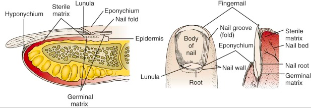
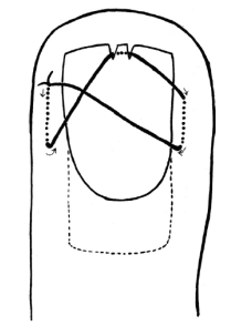
If the headache is occipital and radiates forward at all, it could be occipital neuralgia, which often responds quite well to occipital nerve blocks. The diagnosis is not very scientific, but the potential treatment is very simple and, if nothing else, just takes a few minutes of my time and a little pain for the patient, which might provide them hours of relief. I recommen a bupivicaine occipital nerve block to any patient who I think could have occipital neuralgia, review the anatomy, make sure I'm not in the artery, and put in a nice dose of bupivicaine.

No opioids

Opioids don’t work for headache. I never* give opioids for headache. Don’t give opioids for headache.

That being said, the evidence base is surprisingly weak (see REF). My personal experience resonates here; I’ve had a handful of classic migraines, and some were when I was studying abroad in Australia where codeine is OTC. It made me sleepy-ish but didn’t help the headache at all, for whatever that’s worth.

*Rarely, if I see a patient with a pain contract that includes opioids for headaches from a reputable source, I don’t die on that hill.

2nd round: magnesium + (metoclopramide or haloperidol)

If the patient need a second round (or if their headache was terrible to begin with) I throw some IV mag at them. The evidence is weak at best, but it might help and is pretty safe, so why not.

I usually re-dose metoclopramide at this point, but if their current or prior headaches are generally refractory, I often switch to haloperidol (2.5mg IV or IM).

Dispo

Key points in communicating with the patient and evaluating them for discharge:

I don’t have a silver bullet to fix their headache. My goal is to make sure we’re not worried that there is something dangerous going on (“good news! we’re not”) – it’s safe to go home and we have a good, safe plan for follow up (we’re not just kicking them out). While I can’t make the headache go away completely, “my other goal is to get you to the point where you can be miserable here or miserable at home” and we can safely discharge with return instructions and follow-up; I consider Neuro or headache specialist referral if it seems appropriate.

I really think that articulating a lot of these steps is helpful. Much of what we do implicitly is not clear to the patient – our across-the-room gestalt, our assessment and thought processes. Patients want to be listened to, they want to know what to expect, they want to know what to do, and they want a doctor who cares.

Screen Shot 2018-04-27 at 6.00.21 PM.png


Seth Trueger, MD, MPH

Assistant Professor, Northwestern Emergency Medicine


How to cite this post

[Peer-Reviewed, Web Publication]  Eswaran V,   Miller D  (2018, April 30 ). The Migraine Cocktail: Emergency Department Management of Headaches.  [NUEM Blog. Expert Commentary by Trueger, S ]. Retrieved from http://www.nuemblog.com/blog/headache


Posts you may also enjoy


Posted on April 30, 2018 and filed under Neurology.

The ED Guide to Neuroimaging: Part 1

Emergency Department Neuroimaging

Written by: Justin Seltzer, MD (NUEM PGY-1) Edited by: Andrew Cunningham, MD, (NUEM PGY-3) Expert commentary by:  David Rusinak, MD


Neuroimaging, mainly using CT, has become an indispensable part of our emergency diagnostic process, but, all too often we rely on radiologists to interpret what we ordered. The goal of this multi-part blog is as follows:

  • To cover the basics of how to look at a CT brain and quickly identify life threat

  • Review the literature supporting the major ED indications

  • Discuss special considerations, such as when to use contrast, angiography, or MRI instead.


Systematic Reading of a CT Brain

The first portion of this blog will focus on how to read a CT brain quickly with a focus on life threats.

The classic mnemonic, “Blood Can Be Very Bad,” is a pathology oriented, step-wise method to look for blood, cistern changes, and alterations to the brain parenchyma, ventricle appearance, and bony anatomy.  Applying this approach to each image cut individually can help reveal subtle findings that would otherwise be easily missed by quick scrolling.

Blood:

Blood can collect both intra-axially (parenchymal) and extra-axially (outside the parenchyma).

Figure 1

  • Classically, spontaneous intra-axial bleeding originates in deep structures such as the basal ganglia and thalamus (Figure 1).

    • In the setting of trauma, intra-axial bleeding is often ipsilateral or directly contralateral to the injury site (coup-contrecoup) but can be anywhere

    • Inferior frontal and anterior temporal lobes are high risk for traumatic contusions due to close proximity to bone (Figure 2)


Figure 2

 
  • Extra-axial bleeding is defined by location: mainly subdural, epidural, subarachnoid, and intraventricular hemorrhages. The patterns for these are well known and readily identified, however below are some key points on extra-axial bleeding.

    • Finding chronic subdural hematomas can be difficult as older blood and grey matter are similar appearing

    • Mass effect, abnormal appearing brain folds on that side, and use of coronal reconstructions can help identify

    • Subarachnoid hemorrhage becomes difficult to see within hours to days but acutely is often observed well in the cisterns (see below)

    • Be careful not to mistake choroid or pineal calcifications for hemorrhage

    • Don’t forget about scalp hematomas


Cisterns:

The cisterns are not ventricles but rather outpouchings of the subarachnoid space. When evaluating a CT brain the following, certain cisterns have clinical relevance for potential herniation syndromes, layering of subarachnoid blood, and/or the significant structures that run through them. Figures 3-5 show the locations of the major cisterns described below.

Figure 3

Figure 4

  • Suprasellar: Located in the area of the sella turcica, forms a pentagon/star shape

    • Classic location of subarachnoid hemorrhage due to proximity to circle of Willis

    • Obliteration associated with downward transtentorial (i.e. uncal) herniation or due to severe elevated ICP

  • Perimesencephalic cistern: A group of interconnected basal cisterns surrounding the midbrain (mesencephalon), important location of subarachnoid hemorrhage, may see effacement (reduction or loss) with tonsillar herniation

    • Interpeduncular: Located in the area of the cerebral peduncles

    • Quadrigeminal: Classically forms a W shape, obliteration associated with upward herniation

    • Ambient and crural: Connections between quadrigeminal and interpeduncular cisterns

  • Cerebellopontine: Located between anterior cerebellum and lateral pons, synonymous with area of cerebellopontine angle

  • Cisterna magna: Located between the cerebellum and medulla, receives fourth ventricular CSF outflow (Figure 4)

  • Prepontine: Located at the anterior aspect of the pons

Figure 5

Brain parenchyma:

CT allows for gross evaluation of the major structures as well as a differentiation of grey and white matter by Hounsfield units. The focus here is major parenchymal disruptions.

  • Mass lesions, mass effect, midline shift: Because of the fixed nature of the skull, mass lesions of any type easily exert pressure on the surrounding tissue (mass effect) that can result in increased ICP, midline shift, and herniation.

    • Midline shift is measured in millimeters of displacement of the septum pellucidum at the level of the foramen of Monro from the midline of the skull

  • Ischemic changes: depending on the size of the involved territory and duration, may be subtle or obvious density or architectural changes.

    • Early signs of infarction: reduced grey-white matter distinction and loss of insular hyperdensity

Ventricles:

The ventricular system is where CSF is produced and the route by which it travels into the subarachnoid space. The lateral ventricles drain via the foramina of Monro to the third ventricle, which then drains via the cerebral aqueduct (aqueduct of Sylvius) to the fourth ventricle and then to the cisterna magna and the rest of the subarachnoid space via the median and lateral apertures (foramina of Magendie and Lushka, respectively). Figures 3-5 also show the locations of the major ventricles.

  • Interruption of ventricular CSF flow will cause proximal ventricular dilation that helps localize the level of obstruction

  • If unsure between hydrocephalus and atrophy, dilation of temporal horns of the lateral ventricles can be helpful as it occurs in hydrocephalus involving the lateral ventricles but not with hydrocephalus ex vacuo


Bones:

Intimate knowledge of bony anatomy is not essential fracture evaluation. However, it is crucial that the bony anatomy be viewed with a dedicated bone window. Skull and facial fractures can be subtle and the presence of blood, especially an epidural hematoma, may help localize them. As noted above, soft tissue findings such as scalp hematomas are important to rule out as well.


Key Learning Points and Conclusions

  • A systematic approach is essential to avoid missing significant findings, especially with complex neuroimaging—remember “Blood Can Be Very Bad”

  • Immediately look for: blood anywhere (don’t forget the scalp!), effacement of major cisterns, mass effect/midline shift, enlarged ventricles (temporal horns), skull fractures

  • Older blood, such as a chronic subdural hematoma, can be hard to find and may require different cuts or inference from mass effect or effaced sulci

  • Signs of infarction may be subtle (more on this later)


In the next installation, we will discuss the major indications for CT brain and the utility of CT for these indications.


Expert Commentary

Overall, this is a very nice approach to head CT interpretation.  The classic mnemonic, “Blood Can Be Very Bad,” is not something I’ve heard of before, but it works.  Let’s take each search item in turn.


Blood

A helpful way to think about intracranial hemorrhage is to consider the causes of hemorrhage and the most common location for each pathology.  Common causes include trauma, stroke (hypertensive or hemorrhagic conversion of a venous or arterial infarct), neoplasm (primary or secondary), vascular (aneurysm, AVM, dural AV fistula), and spontaneous (anticoagulation, amyloid angiopathy, vasculitis).  If you consider the location of each of these pathologies, the hemorrhage will typically be primarily in this location.  A tumor, for example, will cause a parenchymal bleed, a ruptured aneurysm will cause subarachnoid hemorrhage, an AVM will result in a parenchymal bleed, etc.  Often with parenchymal bleeds additional imaging, vascular and MRI, as well as follow up imaging will be necessary to determine the underlying cause.

A correction is that subacute hemorrhage, not chronic, has a density similar to gray matter.  Chronic subdural hemorrhages are usually very hypodense and easy to detect on CT. So, from a practical perspective, a patient experiencing headaches from subarachnoid hemorrhage that is greater than 3 or 4 days old may be occult by CT.  This underscores the role of lumbar puncture and vascular imaging in working up patients with headaches.

Another important concept to keep in mind is window and level when interpreting CTs. Different substances (air, metal, bone, blood, fat, etc) have different and defined densities.  The pathologies associated with each of these substances (fractures, edema in the setting of stroke, etc) can be better seen by adjusting the window and level settings.  This can be done manually or, typically, PACS viewers have preset brain, bone, lung and soft tissue windows that can be displayed by pressing different numbers on the keypad.  Subtle subdural hemorrhages are often only seen with the appropriate window and level that allows distinction of the hemorrhage from the overlying calvarium.


Cisterns

The blood vessels course through the cisterns, so these must be scrutinized for the presence of hemorrhage secondary to a ruptured aneurysm in a patient presenting with an atraumatic headache.  The cisterns are also effaced in the setting of mass effect. Mass effect may be from a space occupying lesion; such as a tumor, abscess, or hemorrhage; or from diffuse cerebral edema with generalized brain swelling.  Often the absence of something (i.e. patent basal cisterns) can be harder to detect than the presence of something, like hemorrhage.  It is, therefore, important to examine the basal cisterns on each case to get comfortable with their normal variation of appearance so that their absence, such as in diffuse cerebral edema, is not missed.


Brain parenchyma

Subtle changes in parenchymal density can be difficult to detect.  It is important to get acquainted with ideal window and level settings to uncover subtle parenchymal changes.  Also comparison with prior imaging, if available, is necessary to determine the chronicity of parenchymal findings.  Understanding where a physical exam finding localizes intracranially can also be very useful- aphasia or left upper extremity weakness localize to very different locations, for example.  Lastly, always look at the vessels in the subarachnoid space to identify hyperdense thrombus in the setting of a suspected stroke.


Ventricles

Distinguishing volume loss from ventricular dilatation takes experience to understand the variation of normal across the entire age spectrum.  If hydrocephalus is suspected, determining if it is obstructive or communicating can help to understand the underlying cause. The temporal horns are the most elastic portion of the ventricles and dilate first in the setting of hydrocephalus. 


Bones

Depressed skull fractures and easy to see on routine bone windows.  Things get complicated when subtle non-displaced fractures mimic normal sutures or if the fracture involves the skull base/temporal bones.  It is probably not within the normal ED physician’s scope of practice to have a detailed knowledge of skull base anatomy, but if a skull base fracture is suspected (loss of hearing, hemorrhage in the external auditory canal, facial nerve paralysis, etc) it is important or order the proper test for further evaluation, like a temporal bone CT.  A helpful tip is to look for subtle foci of intracranial air and soft tissue swelling which may direct you to a subtle fracture.    

Screen Shot 2018-04-20 at 1.09.34 PM.png

David Rusinak, MD

Assistant Professor of Radiology, Northwestern Medicine


 How to cite this post

[Peer-Reviewed, Web Publication]  Whipple T,   Gappmeier V (2018, April 23 ). Demystifying the Hand Exam.  [NUEM Blog. Expert Commentary by Rusinak D ]. Retrieved from http://www.nuemblog.com/blog/neuroimaging


Posts you may also enjoy


References

1. Shaw AS, Prokop M. Computed Tomography. In Grainger & Allison's Diagnostic Radiology, 6th Edition (2016).

2. McKetty MH. The AAPM/RSNA physics tutorial for residents. X-ray attenuation. RadioGraphics, 1998; 18(1):151-163

3. Cadogan M. CT Head Scan. Life in the Fast Lane. Retrieved from https://lifeinthefastlane.com/investigations/ct-head-scan/.

4. Nadgir R, Yousem DM. Approach and Pitfalls in Neuroimaging. In Neuroradiology: The Requisites, 4th Edition (2017).

5. Mehta A, Jones BP. Neurovascular Diseases. In Grainger & Allison's Diagnostic Radiology, 6th Edition (2016). Chapter 62, 1456-1496.

6. Jones J. Subarachnoid Cisterns. Radiopaedia. Retrieved from https://radiopaedia.org/articles/subarachnoid-cisterns.

7. Nadgir R, Yousem DM. Cranial Anatomy. In Neuroradiology: The Requisites, 4th Edition (2017).

8. Skalski M, Dawes L. Cerebral herniation. Radiopaedia. Retrieved from  https://radiopaedia.org/articles/cerebral-herniation.

9. Baron EM, Jallo JI. TBI: Pathology, Pathophysiology, Acute Care and Surgical Management, Critical Care Principles, and Outcomes. In Brain Injury Medicine: Principles and Practice, 2nd Edition (2012). Chapter 18: 265-282.

10. Nadgir R, Yousem DM. Head Trauma. In Neuroradiology: The Requisites, 4th Edition (2017).

11. Waxman SG. Ventricles and Coverings of the Brain. In Clinical Neuroanatomy, 28th Edition (2013).

12. Nadgir R, Yousem DM. Neurodegenerative Diseases and Hydrocephalus. In Neuroradiology: The Requisites, 4th Edition (2017).

13. Galliard F, Jones J. Intraventricular haemorrhage. Radiopaedia. Retrieved from https://radiopaedia.org/articles/intraventricular-haemorrhage


Demystifying the Hand Exam

Screen Shot 2018-03-05 at 8.31.59 AM (1).png

Written by: Terese Whipple, MD (NUEM PGY-2) Edited by: Victor Gappmaier, MD, (NUEM PGY-4) Expert commentary by:  Aviram Gialdi, MD, MS


The human hand is a fascinatingly intricate arrangement of pulleys, tendons, muscles, and nerves that work together in a complex system to perform daily tasks. It is often difficult to visualize the various paths that the tendons and muscles take.  It can also make a thorough hand exam difficult to perform with proficiency. This post will review the clinically relevant anatomy of the hand, and apply it to both a screening exam and detailed exam with maneuvers used in the diagnosis of common hand injuries.


This screening exam can be used in the case of a fracture/dislocation at or proximal to the wrist, or in a general trauma to ensure that there has not been a nerve injury – from the cervical spine, through the brachial plexus, and into the extremity.

Basic Screening Exam:  

Vascular

To examine the vascular supply of the hand the examiner should palpate the radial pulse and check digital capillary refill.  Using a finger pulse oximeter is a useful adjunct for evaluating perfusion; anything below 95 in a traumatized limb/digit raises concern.

Neuro

The radial, median, and ulnar nerves each have sensory and motor functions that should be evaluated. 

 Radial (C5-C8):

Img 1. Sensory innervation of hand

  • Motor: Extend the wrist. If too painful due to injury, then extension of the thumb IP joint may be substituted.
  • Sensory: Test the dorsal webspace between the thumb and index finger

Median (C5-T1):

  • Motor:
    •  Recurrent motor branch of the median nerve: Have the patient attempt opposition (bringing the thumb tip across to the small finger tip)
    • Anterior interosseus branch of the median nerve:  Make an OK sign by having the patient touch the tip of the thumb to the tip of the index finger

Img 2. Correct OK Sign

Img 3. Incorrect OK Sign

  •  Sensory: Palmar surface of the index finger or thumb

Ulnar (C8-T1):

  • Motor: Test by having patient spread fingers against resistance
  • Sensory: Palmar aspect of the little finger


Check the individual digital sensory nerves to any finger by testing the radial and ulnar sides of each digit

If the patient can perform each of the above functions and has intact sensation, as well as good cap refill and pulses, they have passed the basic screening exam and are “neurovascularly intact.”


Now for a more detailed exam, which should be used when a patient comes in with a specific hand complaint or if there is concern for muscle or tendon injury.

A thorough musculoskeletal (MSK) exam should include:

  • Inspection
  • Palpation
  • Range of Motion (ROM)
  • Nerve/Vascular assessment
  • Muscle/tendon exam
  • Specific maneuvers


Detailed Hand Exam:

Inspection

Inspect the hand for evidence of:

Img 3. Mallet finger

  • Asymmetry
  • Lacerations/abrasions: Any skin break over a joint (eg : fight bite) may look innocent, but actually provides a route for inoculation of the joint with infection and can be serious.
  • Inflammation:  Can be acute from recent injury/infection or chronic from inflammatory states such as RA.
  • Atrophy: Think critically about the location of the atrophy, is it diffuse or does it fit one nerve distribution? For example, carpal tunnel syndrome may produce atrophy in the thenar muscles supplied by the median nerve. Ulnar nerve entrapment at the elbow (Cubital tunnel syndrome) could cause hypothenar muscle wasting and intrinsic wasting (most visible at first dorsal interosseous, along dorsal radial border of the index metacarpal).
  • Any evidence of traumatic deformity such as unusual angulation or rotation. You should always check alignment of the fingers in flexion and extension. Sometimes abnormal rotation will only be visible when making a fist, when one finger crosses over/under the next
    • Any alteration to the normal cascade of the fingers (one finger that is not flexed/extended to match the position of the others) may represent a tendon injury
    • Mallet finger: A flexed DIP with inability to actively extend due to rupture of the terminal extensor tendon of the digit. (Img. 3)
    • Boxer’s fracture: May have a “dropped knuckle sign” where the fracture of the metacarpal shaft causes a “disappearance” of the metacarpal head  (Img. 4)
 

Img 4. Dropped knuckle sign


Palpation

Img 5. Scaphoid Tubercle

It can be difficult to visualize all of the bones in the hand and wrist in order to palpate them correctly. However, there are a few that emergency medical providers should know in order to catch the most common and consequential injuries.

The scaphoid is technically part of the wrist, however it is usually part of a screening hand exam for anyone with a fall onto a hand. It can be palpated in 3 places:

Img 6. Anatomic snuffbox

  • Scaphoid tubercle   
  • The waist of the scaphoid can be palpated in the anatomic snuffbox 
  • The proximal scaphoid can be palpated on the dorsal wrist in the soft spot between the tendons of the 3rd and 4th compartment of the wrist, just distal to Lister’s tubercle 

Img 7. Lister's Tubercle

Img 8. Proximal Scaphoid

Previous studies have demonstrated that tenderness at the scaphoid tubercle is actually more sensitive than the anatomic snuffbox (95% v. 85%) in diagnosing scaphoid fracture. When palpating the anatomic snuffbox you can maximize the surface area that you are palpating by having the patient move their hand into ulnar deviation and thumb abduction.

Range of Motion

Test range of motion both passively and actively in each joint. Passive ROM gives you information about the joint.  You may feel clicking, catching or crepitance. Active ROM provides information about nerve function, muscle strength, joint congruity/stability, and tendon integrity.

[Insert aforementioned neurovascular exam here]

Muscle/tendon exam

A full muscle/tendon exam doesn’t need to be a part of every exam in the Emergency Department, we don’t have the time. However, if there is an injury that makes you concerned about the integrity of deep structures in the hand, wrist, or forearm, knowing the course and function of each muscle and tendon is useful.  Theoretical cases have been included to provide context.

Case 1:

A patient sustained a deep laceration to his right volar forearm from a glass bottle during an altercation at a bar. The sensory exam in the hand is normal, but function is abnormal.  In addition to the usual laceration care, you want to ensure all of the underlying tendons from the extrinsic muscles are intact. You need to check the finger and wrist flexors. Most of these are innervated by the median nerve, with the exception being the Flexor Carpi Ulnaris and the Flexor Digitorum Profundus to the small and ring fingers, which are innervated by the ulnar nerve.

Img 9. Flexors of forearm

  • Flexor Pollicis Longus (FPL): Test by asking patient to flex thumb at the IP joint (AIN)
  • Flexor Digitorum Profundus (FDP): Test by asking patient to flex DIP joint of index or middle finger while stabilizing PIP of the same digit
  • Flexor Digitorum Superficialis (FDS): Test by asking patient to flex PIP while examiner holds all the other digits in extension (this blocks FDP and completely isolates the FDS)
  • Flexor Carpi Ulnaris and Flexor Carpi Radialis: Test by asking patient to flex the wrist and palpate tendon/muscular contraction


In summary, to test the extrinsic flexors:

  • Flex thumb IP joint
  • Stabilize PIP and have patient flex each DIP in succession
  • Hold remainder of fingers in extension, ask patient to flex each PIP in succession
  • Volar flex wrist


Case 2:

The same patient presents again after a bar fight, this time sustaining a deep laceration to his dorsal forearm.  You want to ensure all of the underlying tendons from the extrinsic muscles are intact. You need to check the extensors.  These muscles are all innervated by the radial nerve and are separated into six compartments.

First Dorsal Wrist Compartment

  • Abductor Pollicis Longus and Extensor Pollicis Brevis: Ask the patient to bring their thumb out to the side (abduct) and palpate the tendons along the radial border of the wrist

Img 10. Extensors of forearm

2nd Dorsal Wrist Compartment

  • Extensor Carpi Radialis Longus (ECRL) and Extensor Carpi Radialis Brevis (ECRB): Have the patient make a fist and extend against resistance

3rd Dorsal Wrist Compartment

  • Extensor Pollicis Longus: Place hand flat on table and lift thumb off the table

4th Dorsal Wrist Compartment (the MCP joint extensors of the fingers)

  • Extensor Digitorum Communis and Extensor Indicis Proprius (EIP): Test by straightening individual fingers at the MCP. The EIP can be isolated by extending index finger with the rest of the fingers closed in a fist

Img 11. Extensors of forearm 

5th Dorsal Wrist Compartment

  • Extensor Digiti Minimi: Extend small finger with the rest of the fingers closed in a fist

6th Dorsal Wrist Compartment

  • Extensor Carpi Ulnaris: Extend and ulnar deviate wrist

In summary, to test the finger extensors:

  • Abduct the thumb, then place on table and lift thumb off
  • Extend fingers against resistance at MCP
  • Make fist and extend wrist against resistance
  • Ulnar deviate fist
  • Extend index finger from closed fist
  • Extend small finger from closed fist

Case 3:

The same unfortunate patient returns after yet another bar fight. His other two lacerations are well healed, but now he has sustained a deep laceration to his right palm. This time you need to check the intrinsic muscles and tendons of the hand. These are innervated by the median and ulnar nerves and are also separated into compartments.

Thenar muscles: both median and ulnar nerve innervation

Img 12. Intrinsic muscles of hand

  • Abductor Pollicis Brevis, Opponens Pollicis, Flexor Pollicis Brevis (median n.): Ask the patient to touch thumb and small finger tips together so the nails are parallel
  • Adductor pollicis (ulnar n.): Have the patient hold paper between thumb base and radial side of 1st finger.  Try to pull the paper away and see if they can hold it.  When the adductor muscle is weak the thumb flexes at the IP joint to grab the paper (Froment’s sign)


Interosseus and Lumbrical: ulnar nerve innervation

Interosseus testing

  • Lumbricals: Flex MCP and straighten IP
  • Interosseus: Adduct and abduct the fingers.  Place the hand flat on table to eliminate interference by extrinsic extensors, hyperextend middle MCP, and move finger from side to side.


Hypothenar muscles: ulnar nerve innervation (difficult to isolate, especially in an injured patient)

  • Abductor Digiti Minimi: Test by abducting small finger
  • Opponens Digiti Minimi: Function to bring small finger towards thumb

In summary, to test the intrinsic muscles of the hand:

  • Touch small finger to the thumb so the nails are parallel
  • Pinch paper between thumb and radial side of index finger in the first webspace
  • Flex MCP and straighten PIP
  • Place hand flat on table, hyperextend at MCP, adduct and abduct each finger
  • Spread fingers against resistance, (also abducts the 5th finger and tests the hypothenar muscles)


-------------------------------------------------------------------------------------------------------------------

Special tests:

There are several tests that can be used to examine for common and important injuries.

Case 4

You are working at a ski clinic in Lake Tahoe as part of an elective rotation. A patient presents after a fall backwards onto his R hand while holding his ski pole.  He has pain in his thumb, especially on the ulnar aspect of the MCP joint.

  • Most likely diagnosis: Skier’s thumb/Gamekeeper's thumb, a rupture of the ulnar collateral ligament (UCL)
  •  Evaluation: Test UCL integrity. Hold thumb metacarpal with one hand, and fully extend thumb MCP and apply gentle radial deviation force to see if there is laxity or pain. Test again at 30 degrees of MCP flexion. Test other thumb as a reference (people vary widely in baseline joint laxity).


Case 5

A patient reports that he was playing pickup basketball, got a finger snagged on the opposing player’s shirt, and felt pain when the player pulled away suddenly.  Now he has difficulty flexing the fingertip.

  • Most likely diagnosis: Jersey finger, a rupture of the FDP tendon from the distal phalanx.
  • Special test:  Hold the patient's MCP and PIP in full extension and ask patient to flex at the DIP. If the FDP is intact the patient will be able to flex at the DIP.  The PIP must be held in full extension to isolate FDP function. 

Case 6

The same patient presents 6 months later, again playing pickup basketball, but this time he got his finger jammed on the ball going up for a rebound.  Now he cannot fully extend it at the tip.

  • Most likely diagnosis: Mallet finger, an avulsion of the extensor digitorum from the distal phalanx.
  • Special test: Hold the middle phalanx of affected finger to isolate DIP and ask patient to actively straighten DIP. If the patient cannot, then the test is positive for Extensor Digitorum injury.  You can also passively extend the tip and see if patient is able to hold it there or if it returns to the flexed position.


Case 7

A patient presents with a deep laceration to the dorsum of his 3rd finger, over the middle phalanx. He appears to be able to extend and flex the finger easily, however as an astute ED physician, you are concerned about occult tendon injury.

Screen Shot 2018-04-14 at 3.40.35 PM.png
  • Most likely diagnosis: Central slip injury, a rupture of the central band of the extensor mechanism causes the lateral bands to slide ventrally, preventing extension of the PIP and extension of the DIP.
  • Special Test: Elston’s test. Passively flex the PIP to 90 degrees to relax the lateral bands. Have patient try to extend the finger and provide counter force on middle phalanx. When the patient tries to extend the PIP test the tension at the DIP: If DIP is floppy the central slip is intact, but if the DIP becomes taut then central slip is injured.

Expert Commentary

Evaluating hand trauma requires understanding the anatomy and the functions associated with that anatomy.  Having a systematic approach helps, as pain, bleeding, intoxication, and fear can affect the upper extremity exam.   It is good practice to start by evaluating for deformity, color change, and wounds.  Ask the patient to make a fist and then open it, which can help direct you to the problem area.  Test wrist flexion and extension.  Evaluate extension of each finger.  Evaluate flexion and extension of the thumb IP.  Evaluate FDP and FDS of each finger.  Test OK sign, fingers crossed (index and middle), spread fingers wide and hold them out against resistance.  Test gross sensation in each fingertip, and on the back of the hand.  If you do this every time, you are unlikely to miss a substantial injury. From the findings of the general hand exam you can then focally test any trouble areas.

Determining adequate perfusion, often via clinical exam (color, temperature, turgor, etc) is critical.  Using a pulse oximeter on an injured finger can help identify threatened digits before the clinical ischemia or venous congestion becomes obvious.  Doppler exam of each digital vessel is another useful evaluation tool. 

Always consider the proximal to distal nature of the anatomy, and tailor the focal exam based on the level of injury.  For example, if a patient presents with an injury at the wrist, testing finger flexion will not give any information about the median nerve.  The level of injury helps guide what additional components of the exam you need to perform to get the full picture, as laid out in the different discussions between cases 1 and 3 above.

The sensory exam is often challenging, especially in traumatized fingers.  Pain can be distracting, and edema can cause sensory changes.  Gentle sharp sensation (pin-prick) testing is a useful adjunct to the digital sensory exam, especially if only one side is injured and you are trying to clarify whether the digital nerve is intact.  Also, if a patient had a tourniquet placed in the field, they may present with an abnormal sensory exam (or even functional exam, depending on duration of ischemia) even if all structures are intact.

And, although this may be an obvious reminder, always document a thorough sensory exam before ever administering local anesthesia. 


Screen Shot 2018-04-14 at 3.45.33 PM.png


Aviram Giladi, MD, MS

The Curtis National Hand Center, MedStar Union Memorial Hospital


 How to cite this post

[Peer-Reviewed, Web Publication]  Whipple T,   Gappmeier V (2018, April 16). Demystifying the Hand Exam.  [NUEM Blog. Expert Commentary by Giladi A ]. Retrieved from http://www.nuemblog.com/blog/hand-exam


Posts you may also enjoy


References

• Ghane, MR et al. How trustworthy are clinical examinations and plain radiographs for diagnosis of scaphoid fractures. Trauma Mon Nov 2016. 21(5): e23345.

• Giuglae, J et al. The palpable scaphoid surface area in various wrist positions. Journal of Hand Surgery. 1 Oct 2015. 40(1): 2039-2044. 

• Netters Orthopedic Clinical Exam. Ed: Cleland, Joshua A., PT, DPT, PhD; Koppenhaver, Shane, PT, PhD; Su, Jonathan, PT, DPT, LMT. Third Edition. Copyright 2017

• Lin, M.  Quick Tip: Elston’s Test for the Finger.  Jul 29 2013. ALiEM.

• Bookman, A. A., von Schroeder, H. P., & Pham, A. G. (2010). The Wrist and Hand. In Fam’s Muskuloskeletal Exam and Joint Injection Techniques (pp. 29–43). Mosby.

• Seiler, JG. (2002). Essentials of Hand Surgery (pp 23-48). Lippincott Williams & Wilkins.


Approach to Nail Trauma

Screen Shot 2018-03-05 at 8.26.14 AM.png

Written by: Gabby Alzadeh, MD (NUEM PGY-3) Edited by: Jim Kenny, MD, (NUEM PGY-4) Expert commentary by:  Matt Levine, MD


Why nails are important

  • Nail injuries may have significant associated functional and cosmetic morbidity
  • The nail bed provides adherence and support for the nail
 
Nail_anatomy diagram 2.png
 

Nail Anatomy

  •  Nail bed: overlies the cortex of the distal phalanx and lies directly beneath the nail plate
  •  Eponychium: the skin that covers the proximal end
  •  Hyponychium: the skin edge at the distal nail margin
  • Cuticle: an outgrowth of the eponychium that provides a seal between the proximal nail fold and nail plate
  • Germinal matrix: the proximal portion of the nail bed responsible for nail formation and begins 7 to 8 mm under the eponychium; the distal end of the germinal matrix is the lunula


Subungual Hematoma

  • A simple subungual hematoma is not an indication to remove the nail. Trephination is not indicated if the hematoma encompasses only 25%, there is no significant pain, or if injury was over 24 hours ago, as the blood likely clotted and will not flow out.
  • Blood under the cuticle proximal to the nail is a clue that there is a deeper injury and usually the nail should be removed if there is significant pain.
  • There is controversy regarding treatment of subungual hematomas and whether simple trephination is enough or whether inspection of the nail bed for injury is required.
  • It was suggested that for subungual hematomas involving more than 50% of the nail bed, the nail should be removed given the risk of nail bed laceration.
    • This was based on an initial study in 1987 that found that 16/27 patients with hematomas >50% had associated nail bed lacerations that required repair
    • This study did not follow up with patients and did not have a control group, so long term outcomes are unknown.
    • However, subsequent studies have shown that if there is no other significant finger tip injury, treatment by trephinating alone provides a similar good cosmetic and functional result.
  •  If you don’t have a trephinator, what else can you use?
    •  Heated paper clip
    • 23-gauge 1-inch needle: Hold the needle over the hematoma, avoiding the lunula, twist and rotate the needle back and forth like a drill; no pressure needed.
    •  Number 11 scalpel (slower, more painful, larger hole and better drainage)
    •  Insulin syringe needle (29-gauge)
  •  What if there is a fracture underneath?
    • Though there is a risk of turning the fracture into an open fracture, consider still performing the procedure if the injury is painful.
    •  You can consider antibiotics if trephination is pursued, though there is no data.
    •  It is always important to obtain an x-ray with any traumatic injury.

Nail Bed Repair

Suture the nail bed if a large subungual hematoma is associated with an unstable or avulsed nail. Good outcome depends on maintaining the space under the cuticle where the new nail will grow out from (the germinal matrix). If this area scars down a new nail will not grow. 

Figure 1

Figure 1

  •  If the nail is only partially avulsed or loose, especially at the base, lift the nail slightly to assess the nail bed.
  • If the nail is completely transected, it is best to remove the entire nail to suture the nail bed. In this case, suture the proximal and lateral nail folds first for better approximation prior to repairing the actual nail bed.
  • A sturdy needle (3-0 or 4-0) is needed to suture the nail back in place. Before replacing the nail and suturing it back in place, you can poke a hole through so the needle and suture can pass more easily.
  • A study in 2008 used dermabond for nail bed laceration repair showed similar follow up cosmetic and functional outcomes; using dermabond took about 1/3 of the time. It was a small study with only 40 patients and repair was done by orthopedic residents, but definitely a consideration
    •  The key to success is achieving hemostasis and making sure you have a dry field before dermabond application
  • Another method to secure the nail in place is the figure 8 stitch (Figure 1, 2) proposed by hand surgeons 
 

Figure 2


Protecting the exposed nail bed is essential, which can be done with the nail itself (wash well beforehand with normal saline), with the sterile aluminum foil from the suture pack, or with a piece of vaseline gauze. The nail should be reinserted under the eponychium to protect the open space for nail growth. Consider a hand surgeon consult if the nail bed is extensively lacerated or if part of the nail bed is lost, as the patient my need a matrix graft.


Discharge Instructions:

  • Tell the patient to return for a wound check 3-5 days post repair. Replace any non-adherent material that was inserted into the proximal nail fold. Afterwards, the patient should perform dressing changes every 3-5 days.
  • Sutures that were used to reattach the nail should be removed in 2 weeks.
  • Nails grow at a rate of 0.1 mm/day and it takes approximately 6 months for a new nail to grow.
  • Instruct the patient to avoid any trauma or chemical irritants to the healing nail.

Tips:

Figure 3

  • Always use absorbable suture to repair the nail 
  • Use a large suture and sturdy needle when suturing the nail back in place; consider dermabond as an option
  • Use a finger tourniquet to maintain a bloodless field (Figure 3)
  • Digital blocks are key
  • Clean the nail bed prior to repair; clean the nail very well before replacement
  • If possible use the avulsed nail to protect the exposed nail bed and maintain the space for a new nail to grow
  • Repair the proximal and lateral nail folds first

Expert Commentary

Dr Ahlzadeh has presented a nice review of the approach to nail trauma and some useful techniques.  While there are seldom formal lectures during residency training dedicated to nail trauma, it is something we regularly see and treat in the ED, so it is important that we do this well.  It is well within the scope of practice of Emergency Medicine to be the primary providers for most nail injuries.

The subungual hematoma is probably the most common nail injury encountered in Emergency Medicine.  Traditional dogma directed the provider to remove the nail to repair underlying injuries in the presence of >25-50% hematoma.  This recommendation left many of us scratching our heads. We would go through the elaborate procedure of removing the nail by dissecting the nail away from the underlying nailbed, meticulously repairing a nailbed laceration if required, and then stenting the eponychial fold open, hoping that a normal nail would regrow without deformity despite the trauma from the injury and the procedure itself.  It seemed like we were introducing a lot of trauma to the nail bed and eponychial fold for a theoretical (non evidence based) benefit.  The more we did this, the more many of us would ask ourselves, “What if we just left this alone?”

Today it seems the tide has turned.  It is now well accepted practice to leave a nail in place in the setting of a subungual hematoma as long as the nail is intact and laying flat on the nailbed, regardless of the percentage size of the hematoma.  This makes intuitive sense.  An anatomically intact nail lying flat upon the nailbed should lead to the flat healing of an underlying laceration.  A common practice to ensure that the nail remains flat after discharge is to trephinate nail so that pressure from the underlying hematoma does not elevate the nail off of the nailbed after discharge.

Nail trephination is one of the more rewarding procedures in Emergency Medicine.  Patients present with throbbing pressure from the tense subungual hematoma and typically get immediate improvement once the nail is trephinated.  Many techniques have been described. Dr Ahlzadeh even refers to a “trephinator”.  I am not actually sure what this device even is.  Regardless, this procedure should be quick and easy.  No anesthesia is necessary.  I suggest keeping it simple.  Rather than heating a needle or paper clip, use a cautery device.  In our ED we have a cordless disposable plastic cautery device that is the size of a magic marker.  It has a single button and the tip becomes orange with heat when the button is pressed so you know it is hot and ready to go.  Warn the patient that they will feel some heat and possibly see or smell a bit of smoke but it will be brief and not painful (unless you are too forceful, in which case it will be hot and painful!).  Pick the location in the center of the hematoma.  Repeatedly very lightly touch the hot cautery tip to the nail over the center of the hematoma until you are through the nail.  You will know you are through when a drop of blood comes out of the hole you have trephinated.  At that point you can stop and gently express whatever blood you can.  That’s the whole procedure.  It should take mere seconds. When the patient gets over the initial anxiety of the procedure they typically realize they feel better. 

Nails that are deformed or elevated off of the nail bed are not good candidates for trephination. These nails should be removed for nailbed repair so that the nailbed heals flat.  Don’t even bother unless your digital block is highly effective.  We do not have commercial finger tourniquets in our ED so I like to make my own professional looking tourniquet.  I get a sterile glove is about the size (or a half size smaller) than would fit the patient.  I cut the very tip off the finger of the glove that corresponds with the patient’s injured finger.  Then I put the glove on the patient and roll the cut glove finger proximally and voila, you have both a tourniquet and a clean field.  Now you are ready to get comfy and repair the nail bed according to Dr Ahlzadeh’s techniques!  While a sturdy needle is needed to puncture through a nail, I still find the nail often deforms the needle. So instead of piercing the nail with my needle, I once again use my trusty cautery device to make holes in the nail that my needle will easily pass through without deformity.

Screen Shot 2018-04-08 at 5.24.00 PM.png


Matt Levine, MD

Assistant Professor, Northwestern Emergency Medicine


 How to cite this post

[Peer-Reviewed, Web Publication]  Alzadeh G,  Kenny J  (2018, April 8). Nail Trauma.  [NUEM Blog. Expert Commentary by Levine M ]. Retrieved from http://www.nuemblog.com/blog/nail-trauma. 


References

  • Batrick N. Treatment of uncomplicated subungual hematoma. Emerg Med J 2003;20:65.
  • Bowen WT, Slaven EM. Evidence-based management of acute hand injuries in the emergency department. Emergency Medicine Practice  EB Medicine. 2014;16(12):1-28. http://www.ebmedicine.net/media_library/files/1214%20Hand%20Injuries
  • Guthrie, Kane. “Minor Injuries 001.” Life in the Fastlane. <http://lifeinthefastlane.com/minor-injuries-001/>.
  • Hedges, Jerris, James Robers. “Methods of Wound Closure.” Clinical Procedures in Emergency Medicine, 6th ed. Philadelphia: Elsevier/Saunders, 2014.
  • Roser SE, Gellman H. Comparison of nail bed repair versus nail trephination for subungual hematomas in children. J Hand Surg [Am]1999;24:1166–70.
  • Strauss E, Weil W, Jordan C, Paksima N. A prospective, randomized, controlled trial of 2-octylcyanoacrylate versus suture repair for nail bed injuries. J Hand Surg Am. 2008;33(2):250-253.


Ocular Ultrasound: From Floaters to Fogginess

Screen Shot 2018-01-25 at 10.42.52 AM (3).png

Written by: Steve Chukwulebe, MD (NUEM PGY-3) Edited by: Michael Macias, MD, (NUEM Graduate 2017, US Fellow UC San Diego) Expert review by:  John Bailitz, MD 


The Case

A 60 year old male with a history of hypertension presents to the emergency department with three days of intermittent floaters in his right eye.  Concurrently, he also notes that vision in the right eye has become progressively blurred, first starting at the base of his visual field and now advancing up towards the center of his vision. He denies any trauma to the area as well as any other neurological complaints.


Ocular Exam

Motor: PERRL, EOMI,

Superficial Exam: Clear conjuctiva, left eye cataract

Visual Acuity: OD 20/25, OS 20/40 values corrected for a distance of 10 feet from the chart

Visual Fields: Decreased in the lower quadrants of the R eye to confrontation


The Differential Diagnosis for Floaters

  • Retinal detachment (RD)

  • Posterior vitreous detachment (PVD)

  • Vitreous hemorrhage

  • Intraocular foreign body

  • Posterior uveitis/vitreous inflammation

  • Migraine w/ aura

A bedside ultrasound obtained the following image:

 
eye.gif


The patient was found to have a retinal detachment and was admitted for definitive management under ophthalmology.


 Acute Vision Loss and Floaters

Floaters, often described by patients as lines, circles, dots, cobwebs, and other shapes, are common as part of the degenerative process of the vitreous body.  While in the chronic setting they are thought to be related to condensation of the vitreous collagen fibers, new onset floaters in patients 50 years or older have been related to PVD in 95% of cases. Of patients with vitreous floaters and/or flashes as a consequence of PVD, the incidence of RD is 14% [1]. If the PVD is complicated by vitreous hemorrhage, the incidence of RD rises to 70%.

From a population perspective, the incidence of RD ranges between 6.2-17.9 per 100,000 people with the highest rates occurring in the age group 60-69 [2].  Additionally, patients with history of myopia or uncomplicated cataract surgery have a significantly increased risk of developing RD compared to the general population.  It is also important to note that there is a 3-33% chance of bilateral involvement [3].


Using the Ultrasound for Detection of Retinal Detachment

On ultrasound, RD appears as a hyperechoic rippled (or undulating) line/membrane in the posterior to lateral globe.  A recent paper in Annals of Emergency Medicine reviewed 78 articles and ultimately included three studies (or 201 eyes) in a meta-analysis to evaluate the performance of emergency physicians at identifying RD through ultrasound [4].  Though the 95% confidence intervals for sensitivity and specificity range from 60%-100% in the three studies, each study boasted high accuracy to diagnosing RD.  Furthermore the receiver operating characteristic curve for the three studies had an excellent summary area of 0.97, suggesting that bedside ocular ultrasonography is an accurate tool in an emergency physician’s arsenal when a fundoscopic exam is technically challenging.


Keys to Successful Evaluation for Retinal Detachment with Ultrasound
 

Differentiating between RD and PVD - Keep the gain down:

Since the eye is usually a homogeneous fluid filled structure, it provides a great acoustic window for ultrasonography.  Too much posterior retrobulbar acoustic enhancement decreases the observer’s ability to visualize pathology in the vitreous body.  It can be difficult to see a difference between RD and PVD on ultrasound since both may present as a wavy membrane in the posterior orbit.  In this case, it is important to remember that the retina is a highly reflective surface and should still be seen as a thick or stiff undulating membrane with reduced ultrasonographic gain [5]. Another trick is when asking the patient to move their eye, the PVD membrane appears to be more mobile and dynamic, as if it is moving with the motions of the vitreous, while a RD membrane tends to retain its slow oscillation.


Ultrasound both eyes not just the affected one:

Though RD commonly occurs with the classic story of painless monocular vision loss, described as if a “curtain” or a “shade” is being pulled over their eye, in this case the patient actually presented with better visual acuity in his affected eye.  Remember that there is a 3-33% likelihood that a RD can be developing in the other eye.


Artifacts in the vitreous:

Artifact appears as bright echogenic material in the vitreous body that disappear when the patient is asked to move their eye.  However, if a persistent hyperechoic object is seen in the body, or there is shadowing or reverberations associated with the object, this is concerning for a foreign body or vitreous hemorrhage.


Using Tegaderm bandage over the eye:

This technique provides a few advantages.  One, it allows the ultrasonographer to apply as much gel as needed over the eye without the worry of getting it into the patient’s eye.  It is important to note that any pressure on the eye should be avoided if there is any suspicion for globe rupture or foreign body.  By applying a generous amount of gel over the eye and stabilizing the hand by placing a finger on the forehead or bridge of the nose, it is possible to stabilize the probe without having it make any contact with the eyelid.  This technique also allows for easier clean up after the exam, again focusing on preventing any pressure to the eye.


Conclusion

Bedside ultrasound is a useful tool for rapidly diagnosing RD and getting the patient seen by an ophthalmologist emergently.  However, if there is a high enough clinical suspicion for RD and ultrasound is negative, it is still important for the patient to receive a dilated fundoscopic exam by an ophthalmologist in a timely manner.


Expert Commentary:

Thank you for sharing this outstanding case describing one of the most straightforward and useful clinical ultrasound (CUS) applications. Few ED shifts go by when I do not need to perform an Ocular US exam. As mentioned, CUS is helpful for the evaluation of patients with suspected PVD, RD, Vitreous Hemorrhage, and Intraocular Foreign Body, but also for the assessment of patients with other ocular injuries or increased intracranial pressure. New clinicians may sometimes not perform an ocular US due to the perceived difficulty of preparing for, and safely performing the exam.

Pro-Tips on Preparing and Performing Ocular CUS Quickly and Safely

A physical examination of any sensitive structure such as the eye begins by first simply explaining the exam thoroughly to the patient. The explanation also provides a nice review for junior trainees in the room and even for the clinician sonographer! So take the time to explain the ocular ultrasound just as you would a Tonopen measurement or Slit Lamp exam.

Tegaderms are important for any patient who may not be entirely reliable or with suspected traumatic injury. However, for the experienced clinician sonographer with a reliable non-trauma patient, covering the orbit with a Tegaderm is not always necessary. Getting every artifact producing air bubble out of the way during the Tegaderm application may prove difficult. And patients may not appreciate the eye lash and brow waxing, as well as sticky residue left behind by the Tegaderm. As long as the patient agrees to keep their eyes completely until all gel is removed, the likelihood of gel getting into the eye is low. If cooperation is at all an issue, then I ask the patient their preference. From having the technique performed without a Tegaderm on myself countless times by students, and performing clinically on hundreds of patients, gel contamination is rare, and minor eye irritation even less so.

Next, recline the patient to a 45 degrees or even completely supine position. Then place a rim of gel on the clean linear probe. Ask the patient to close both eyes completely but not forcefully. Perform the exam from the head of the bed, first stabilizing your hand on the patient’s forehead, then gently placing the probe over the closed lid. With any soft tissue or musculoskeletal exam, always start with the normal side first to relearn your anatomy and optimize your settings. In particular, according to the As Low As Reasonably Achievable (ALARA) Principle, utilize the lowest Mechanical Index (MI) possible when performing ocular ultrasound. This minimizes the theoretical risk of damage to the delicate retinae from excess ultrasound energy. If you do not know how to adjust MI, then just select the Ocular preset on your machine. If you have no ocular preset, but a patient who desperately needs the ultrasound, at the very least be sure to minimize the ultrasound exam duration.

As you finish examining each eye, remind the patient to keep both eyes closed until all gel is removed. Have an assistant gently wipe any gel from the eyelid using gauze to lift the gel completely from the lid and lashes. For most CUS applications, white cotton napkins found in just about every ED work the best for gel removal. Paper towels simply smear gel, chucks are too expensive, and clothe towels hard to locate in a busy ED. For the ocular exam stick with gauze to lift all the gel away and give the patient one after you are finished just in case. With brief but adequate preparation and explanation, ocular CUS is a safe and effective technique to rapidly rule in emergent ocular pathology!


Screen Shot 2018-03-05 at 10.12.42 AM.png

John Bailitz, MD

Associate Professor of Emergency Medicine

Program Director, Northwestern Emergency Medicine


FOAMed Resources

1. Just in Time Learning

         a. Jacob Avilla’s 5 Minute Sono; http://5minsono.com/vids/

         b. ACEP Sonoguide https://www.acep.org/sonoguide/smparts_ocular.html

 2. Detailed Learning:

         a. Introduction to Bedside Ultrasound iBook Volume 2 Chapter 16. https://itunes.apple.com/us/book/introduction-to-bedside-ultrasound-volume-2/id647356692?mt=11

          b. Ultrasound of the Week: Ocular Ultrasound https://www.ultrasoundoftheweek.com/tag/ocular/


Posts you may also enjoy


 How to cite this post

[Peer-Reviewed, Web Publication]  Chukwulebe S,  Macias M  (2018, March 19). From Floaters to Fogginess.  [NUEM Blog. Expert Review by Bailitz J ]. Retrieved from http://www.nuemblog.com/blog/ultrasound-in-RD. 


References

  1. Lumi X, Hawlina M, Glavač D et al. Ageing of the vitreous: From acute onset floaters and flashes to retinal detachment. Ageing Research Reviews. 21:71-77. 2015.

  2. Mitry D, Charteris DG, Fleck BW, Campbell H, Singh J. The epidemiology of rhegmatogenous retinal detachment: geographical variation and clinical associations. British Journal of Ophthalmology. 94(6):678-684. 2009.

  3. Gupta OP, Benson WE. The risk of fellow eyes in patients with rhegmatogenous retinal detachment. Current opinion in ophthalmology. 16(3):175-8. 2005.

  4. Vrablik ME, Snead GR, Minnigan HJ, Kirschner JM, Emmett TW, Seupaul RA. The diagnostic accuracy of bedside ocular ultrasonography for the diagnosis of retinal detachment: a systematic review and meta-analysis. Annals of emergency medicine. 65(2):199-203.e1. 2015.

  5. Schott ML, Pierog JE, Williams SR. Pitfalls in the Use of Ocular Ultrasound for Evaluation of Acute Vision Loss. The Journal of Emergency Medicine. 44(6):1136-1139. 2013.


5 Palliative Care Myths DEBUNKED!

Written by: Hashim Zaidi, MD (NUEM PGY-3) Edited by: Alison Marshall, MD, (NUEM Graduate 2017) Expert review by:  Eashwar B. Chandrasekaran, MD MSc


1) “Palliative care is only for people close to death”

Image Credit: “Reconceptualizing palliative care as a continuum of support”Wang, David H., MD. Published March 31, 2017. Volume 69, Issue 4. Pages 437-443.

End-of-life care is only one of the many components of palliative care. Palliative care consists of interdisciplinary efforts to decrease the physical, spiritual, and psychological stress related to an incurable or chronic illness. Sound familiar? We do this daily in the emergency department (ED) for a variety of patients with acute on chronic conditions, many of whom would benefit from early palliative care involvement. Despite multiple studies illustrating the benefit of early palliative intervention, palliative care has traditionally been offered late in one’s disease course, thus creating the misperception that palliative care is synonymous with hospice, or end-of-life care. Specifically, hospice is a benefit and program for patients whom providers believe are prognostically likely to live less than 6 months if the disease is to run its usual course and are philosophically inclined with not pursuing further life-prolonging interventions. Many conditions and disease states could benefit from the model proposed below, with palliative care offered in tandem with life-prolonging care in order to ease symptom burden and improve quality of life. As someone’s chronic illness starts to worsen with concern that their time may be getting shorter, palliative care can then transition to planning for end-of-life, including discussions about enrolling in hospice. Within the landscape of modern disease shifting towards long term management of chronic disease, emergency medicine and palliative care are beginning to share increasing common ground. Many of these patients present to the ED on a regular basis, making the emergency department a unique access point for initiation of early palliative services.

2) “Palliative care discussions are really just about establishing code status.”

            As mentioned previously,  palliative care accomplishes much more that just what happens when a patient has cardio-respiratory compromise. The exact actions performed at the very end of life are not as significant as identifying the values that inform patients and surrogate decision making. It is better to avoid jumping into a goals of care discussion with questions such as “Would your loved one like everything done?” or more gore filled specifics of “Do you really want us beating on their chest or shocking them with electricity to try and keep them alive?”  No one would like to approach the context of their goals for treatment and care framed as a series of end of life checkboxes and procedures. Rather, palliative care providers stress the importance of identifying what’s important to patients and families with the worsening of their disease progression, in order to fill the gap between expectations and reality.

“‘Hello I’m Dr. Emergency please fill out this checklist of invasive procedures that you think sound appealing for your loved one.”

While it is important for us as ED providers to understand how a patients’ wishes relate to medical interventions, we must remember that collaboration and the development of mutual trust are needed to provide excellent care. Trust is achieved through verbal communication style and through non-verbal cues such as sitting with the patient and/or family and giving them time to communicate their wishes without haste or decision-steering conversation. Attempts should be made to identify broad goals that are important to the patient and family. Building on those principles, providers should then propose appropriate goals of treatment based upon expectations.

Screen Shot 2018-01-26 at 10.38.05 AM.png


Once trust and rapport are developed and the goals of care identified, providers should elaborate on a plan as well as timeline just as you do with all ED patients. If your patient's goals are more aligned with comfort and aggressive symptom control at the end of life, future discussions should establish code status as well as discussion of initiating hospice services be it as an inpatient or outside the hospital. . When speaking specifically of code status, a “Do Not Resuscitate” (DNR) specifically comes into play when the patient’s heart stops beating, even though it is often misconstrued as a theme for withholding treatment of any kind at the end of life. Thus, discussions for withholding interventions at the end of life should include discussion of not only code status, but also of discussion issues such as artificial nutrition/hydration and use of antibiotics. If appropriate, most emergency departments have a case-management based approach to initiate a hospice referral. Often, this is done in concert with the admitting team and/or palliative care providers to guarantee a smooth transition and often may require the patient to be admitted for setting up these services. Future directions will look to establish formalized care patterns for the initiation of hospice services in the ED to reduce hospitalizations for individuals at the end of life.

If the patient’s or family’s goals align with ensuring they live as long as possible or continue for hope of curative treatments, then it would be prudent still to offer your perspective of prognosis and document this discussion for future providers that may come in communication with the patient in the future. This certainly does not preclude trying to get palliative care involved with the patient as they can continue to follow the patient even if he is pursuing life-prolonging therapy in anticipation of a change in disease progression in the future.

3) “A patient who is DNR or DNR/DNI should not be admitted to the ICU”

The rough translation of this statement is “if we can’t offer everything in terms of intensive care then the only option is no critical care at all.”

“Wait a minute- if I can’t intubate the patient then how do I get a plateau pressure? Where are we on the CVP monitoring?” - ICU MD

“Wait a minute- if I can’t intubate the patient then how do I get a plateau pressure? Where are we on the CVP monitoring?” - ICU MD

For the emergency physician, identifying the broad goals of care in a criticallyi ill patient helps to delineate what specific interventions should and should not be done in the ED and furthermore during their inpatient stay. Identifying that CPR and intubation aren’t in line with the patient’s goals is critical first step if that intervention could happen in the ED. ICU level of care is not determined by this, but rather based upon the amount of intensive nursing care which will be required for that patient as well as frequency with which medications must be administered. Often times if patients are pursuing life-prolonging therapy, a short ICU stay is necessary to further discussions with inpatient ICU and palliative care providers as they de-escalate use of technology at the end of life and transition to aggressive symptom control. In the end, most ICU doctors have an informal fellowship themselves in palliative care and feel comfortable with admitting these patients knowing that their goals and their disease trajectory may change acutely.  It is important however to be explicit with specific discussions that have already taken place and what decisions may have been made based upon these discussions as to help the accepting service provide better care for these patients.

4) “Having a productive goals of care conversation takes hours; I don’t have that kind of time in the ED”

Meaningful discussions happen between patients and providers every day in the ED on a minute to minute basis. Having a productive and efficient goals of care conversation should be no different. These are critical communication skills that allow us as providers to understand the values of our patients and ensure that they understand their trajectory of care.  ACEP’s Simple Palliative Toolkit includes a framework for a 5-minute goals of care conversation in order to facilitate  meaningful dialogue in a time sensitive manner. Like any procedure, it requires patience, preparation and practice to accomplish well. The initial conversation should focus on defining the current situation and establishing the values of the patient. We’ve found the following question stems useful (shout out to Dr Justin Morgenstern who taught us this and so much more during his EM:RAP segment on Dyspnea in the Palliative Care Patient):

  1. What do you understand about your condition?
  2. How do you normally spend your days?
  3. How much has that changed over the last month?
  4.  What is most important to you (your loved one) right now?

Validate the patient and their loved ones continuously; acknowledge their challenges to date and what they have accomplished to get to that point. Utilizing this value framework will allow you to consider treatment options from the patient’s perspective in the current medical context. Discuss these options and then recommend a course of action based on both your medical knowledge and your understanding of the patient’s values. Finally, remember that emergency medicine is a team sport--get your colleagues from social work and pastoral care involved early, so that they can assist in the established care pathway.

5) “When discussing goals of care for a patient with a dire prognosis, I should avoid partiality in treatment options.”

This myth goes back to the 5 minute goals of care conversation. After establishing wishes and mapping out care goals broadly, it is imperative care providers take the role of adviser rather than host in terms of care. It is key to understand that this does not mean to be irreverent or negligent of a patient or family's wishes. Rather, when the goals of care and treatment have been established and the rapport developed, this is the time you give your professional opinion about the best course of action. If, with the appropriate patient with an incurable disease, you feel the patient would benefit from increased palliative measures and less invasive care you need to communicate that to the patient and family. Connecting their broad reaching goals with actionable palliative interventions such as pain control, dyspnea control, and secretion control will help build the sense that the providers are not “doing nothing” simply. With concerns and care issues validated you can build on issues and also discuss critical invasive interventions that would not help the overall prognosis and lead to prolonged suffering such as intubation, mechanical ventilation, and central line placement. As an ED provider, it is imperative we have discussions about interventions which could directly change the patients trajectory in the hospice such as the procedures detailed above, but more chronic life-prolonging therapy is best guided by the patient’s primary oncologist or in concert with their primary care physician. My recommendation is to make sure you have done due diligence to try to get these individuals in discussion if possible as to have appropriate follow up for future conversations.


Regardless of the patients chronic healthcare conditions, each visit to the ED is an opportunity to assess for palliative care needs

Regardless of the patients chronic healthcare conditions, each visit to the ED is an opportunity to assess for palliative care needs

Talking to the oncologist first often reveals a foundation of previous care goals that can be built upon. Instead of saying, “I’m going to talk to your oncologist about stopping chemotherapy and getting you the palliative care that I think is better for you at this stage,” this would be better communicated by saying, “I know you talked about what care and comfort issues were most important to you as this disease progresses with Dr. X. I talked to them as well. I wanted to revisit this issue and talk more about working to make you actively comfortable moving forward with or without cancer therapies.”


While these “myths” being debunked likely didn’t leave any physicians in shock or awe, it’s important to realize many of the underlying concepts get lost in the chaos that is the emergency department. Remember the ED sets the pace and tone for a lot of care delivered in a hospital system and establishes a patients trajectory through the greater hospital system. By taking a step back and remembering the principles and benefits of early palliative care, a substantial impact can be imparted on patients and their families  that will increase patient satisfaction, and provide better resources that align with their wishes.

Screen Shot 2018-01-26 at 11.14.52 AM.png

Expert Review by Eashwar B Chandrasekaran MD MSc

Assistant Clinical Professor Indiana University SOM Dept of EM

Associate Hospice Medical Direct IUH Ball Memorial Hospital


 How to cite this post

[Peer-Reviewed, Web Publication]  Zaidi H,  Marshall A  (2018, March 12). 5 Palliative Care Myths Debunked.  [NUEM Blog. Expert Review by Chandrasekaran E ]. Retrieved from http://www.nuemblog.com/blog/palliative-care. 


Posted on March 5, 2018 and filed under Palliative Care.

Open Globe Injury

Screen Shot 2018-01-25 at 9.31.41 AM (1).png

Written by: Justine Ko, MD (NUEM PGY-1) Edited by: Hashim Zaidi, MD (NUEM PGY-3)  Expert commentary by:  Rehan Hussain, MD


00001.jpg

Expert Commentary

Hi Drs. Ko & Zaidi,

Thank you for this great review on emergency management of open globe injuries. The presentation of open globe injuries can vary widely depending on the mechanism of injury, and so can the final visual outcome. For obvious ruptured globes with peaked pupils or extruding intraocular contents, I am glad you emphasized that it is best to avoid any manipulation of the eye to prevent further extrusion of contents or increase the risk of infection. Cover the eye with a shield, start topical and systemic antibiotics, and consult ophthalmology so they can arrange surgery in a timely manner. The patient must be kept NPO to avoid delaying surgery unnecessarily. Definitely don’t check the eye pressure if you already know it is a ruptured globe.

For the less obvious ruptured globes, which may sometimes mimic a corneal or conjunctival abrasion, it is imperative to perform a Seidel test carefully. This involves placing a wet fluorescein strip (not the pre-made drop) over the suspected entry site, looking at it under the cobalt blue light of the slit lamp, and checking to see if a stream of aqueous fluid is coming out of the injury site (it can be either quick or a slow stream). Fluorescein staining in absence of a stream of fluid indicates that only an abrasion is present. When in doubt, consult ophthalmology for confirmation. Other slit lamp findings that may be present include subconjunctival hemorrhage, chemosis, corneal abrasion, hyphema, flat anterior chamber, iris defects, foreign body in the anterior chamber, or traumatic cataract.

For imaging workup, CT of the ORBITS is preferable, as it allows for 1 mm sections that increase the likelihood of detecting small intraocular foreign bodies (IOFB) that might be missed with a standard head CT. MRI should be avoided as it is slow, costly, and can cause movement of metallic IOFBs, causing further damage to the eye. Gentle ultrasound over the closed eyelids could be used in absence of availability of CT, but I prefer to avoid it since puts pressure on the eye.

The patient and family members are often times distraught over the injury, and appropriate counseling is an essential part of the encounter. They frequently ask if they will be permanently blind. I avoid making any predictions on the final visual outcome, as it is difficult to predict and can lead to either unmet expectations or unnecessary anxiety. I say that the goal is to save the eye at this time, and we will follow closely to see how the vision turns out. Sometimes patients do very well if the extent of injury is not severe, but on other occasions patients require multiple surgeries to correct associated retinal detachment, vitreous hemorrhage, or lens dislocation. If appropriate antibiotic therapy is not initiated, there is an increased risk of developing endophthalmitis, which portends a poor visual prognosis.

Screen Shot 2018-01-26 at 11.43.21 AM.png

Rehan Hussain, MD

Vitreoretinal Surgery Fellow,  Bascom Palmer Eye Institute, Univeristy of Miami


Posts You May Also Enjoy


How to cite this post

[Peer-Reviewed, Web Publication]  Ko J,  Zaidi H  (2018, Feb 26). Open Globe Injury.  [NUEM Blog. Expert Commentary By Hussain R]. Retrieved from http://www.nuemblog.com/blog/globe-rupture. 


Resources

  1. Alteveer, J and Lahmann B. 2010. “An Evidence-Based Approach To Traumatic Ocular Emergencies.” Emergency Medicine Practice, 12(5): 1-24. 2017.

  2. “Emergency Management of Traumatic Eye Injuries | 2001-07-01 | AHC Media: Continuing Medical Education Publishing.” 2017. Accessed October 6. https://www.ahcmedia.com/articles/71714-emergency-management-of-traumatic-eye-injuries.

  3. Guluma K, Lee JE. Ophthalmology. In: Gausche-Hill M, Hockberger R, Walls R, eds. Rosen’s Emergency Medicine: Concepts and Clinical Practice. 9th ed. Philadelphia, Pa: Elsevier, Inc. 2018:790-819.e3.

  4. Bord, Sharon P., and Judith Linden. 2008. “Trauma to the Globe and Orbit.” Emergency Medicine Clinics of North America, Ophthalmologic Emergencies, 26 (1): 97–123. doi:10.1016/j.emc.2007.11.006.

  5. “Trauma: Open-Globe Injuries.” 2015. American Academy of Ophthalmology. November 4. https://www.aao.org/pediatric-center-detail/open-globe-injuries.

  6. Harlan JB, Pieramici DJ. Evaluation of patients with ocular trauma. Ophthalmol Clin N Am. 2002;15:153-161. (Review article)

  7. Ahmed, Y, A M Schimel, A Pathengay, M H Colyer, and H W Flynn. 2012. “Endophthalmitis Following Open-Globe Injuries.” Eye 26 (2): 212–17. doi:10.1038/eye.2011.313.

  8. Arey, Mark L., V. Vinod Mootha, Anthony R. Whittemore, David P. Chason, and Preston H. Blomquist. 2007. “Computed Tomography in the Diagnosis of Occult Open-Globe Injuries.” Ophthalmology 114 (8): 1448–52. doi:10.1016/j.ophtha.2006.10.051.

Post Partum Hemorrhage in the ED

Screen Shot 2018-01-24 at 6.14.19 PM.png

Written by: Spenser Lang, MD (NUEM PGY-4) Edited by: Michael Macias, MD, (NUEM Graduate 2017)  Expert commentary by:  Annie Dude, MD


Introduction

A 26-year-old female G3P3 arrives via ambulance with heavy vaginal bleeding after having a precipitous home delivery of her third child. EMS reports a “pool of blood,” and en route to your facility she continued to bleed briskly.

Post partum hemorrhage (PPH) is a common and dangerous complication of child birth. According to CDC estimates, hemorrhage is the most common cause of maternal death in both developed and developing countries. About 2 out of every 100 births occur either at home, pre-arrival to the hospital or in the ED. With a trend towards home births and free standing delivery centers increasing dramatically in recent years, emergency physicians need to be able to recognize and treat this life threat.

The official diagnosis of PPH is volume-based, however this information is not easily obtainable in the emergency department.  A more reasonable approach is to treat the PPH patient the same way you would a traumatic hemorrhage. Allow the patient’s vitals and visualized hemorrhaging to guide the aggressiveness of your resuscitation. Remember, a pregnant woman has ~40% extra circulating blood volume and can cope with a higher amount of blood loss than her non-pregnant counterpart.

Following an algorithmic approach as detailed below is essential to management of these patients.


Management

1. Notify an obstetrician.

  • Having the obstetrician on board early will allow for mobilization of definitive treatment such as trans-arterial embolization and/or laparotomy should physical maneuvers, tamponade & uterotonics fail

 2. Resuscitate.

  • As with any resuscitation, begin with the simple ABC algorithm, addressing any issues as they are identified. Patients should be placed on 15 L of oxygen via face mask regardless of their saturation (if hemorrhage is significant, this will dramatically increase their blood oxygen levels via dissolved O2). If a patient is hemodynamically unstable, early administration of blood products should be considered over large volume crystalloid.

3. Obtain adequate access.

  • Two large bore IVs will be necessary if aggressive resuscitation is needed. Consider an intraosseous line early if difficulty obtaining access. Send type and screen, CBC, coagulation panel and fibrinogen. Keep coagulopathy on your differential (Thrombin). This should be done in concert with step 2 described above.

4. Source control

  • By far, the most common cause of PPH is uterine atony (Tone).  Therefore the first action taken should be physical maneuvers to improve tone. A bimanual uterine massage can be useful in stimulating uterine contractions. At the same time, one can evaluate for retained products of conception (Tissue). If tissue is felt, try to sweep out as much as possible while taking care to avoid uterine perforation. Note that this should be done with an empty bladder therefore a foley catheter should be placed prior to attempting massage.


  • If the patient continues to bleed briskly, an effort can be made via balloon tamponade of the uterus, with a foley catheter (or, if available, a Bakri) with ~ 150 ml normal saline injected into the balloon. If the uterus is firm and bleeding continues it is reasonable to assess the genital tract for lacerations of the vaginal wall or cervix (Trauma). Cervical lacerations, ideally, should be repaired by an experienced obstetrician as this can have implications on future fertility. However, a vaginal laceration can be repaired just as a perineal laceration repair, taking care to approximate anatomy with absorbable sutures.

5. Administer uterotonics

  • It is reasonable to begin uterotonic therapy in conjunction with uterine massage in a briskly bleeding patient. Oxytocin is first line and 10 U can be given immediately IM or as an infusion at 10 - 40 mU/minute to achieve and maintain uterine contractions. If hemorrhage is refractory to massage and oxytocin, continue pharmacotherapy for ongoing bleeding with Carboprost (Hemabate - 0.25 mg IM) and Misoprostol (cytotec – 1000 mcg per rectum). 

Summary

Hemodynamically unstable PPH patients should be resuscitated like any other severely hemorrhaging patient. Utilizing a step wise approach as described above will help you maintain control of the situation. Notify an obstetrician. Resuscitate. Obtain adequate access. Source control. Administer uterotonics.

If the patient remains unstable, a myriad of other options exist with your interventional or obstetrical colleagues, including uterine artery embolization, ligation of uterine/internal iliac arteries, or hysterectomy.


Expert Commentary


This case outlines a common presentation of a postpartum hemorrhage in the ED.


A few points:

  1.  While the patient in this scenario had just delivered, postpartum hemorrhage can occur days or even weeks following delivery/hospital discharge. Delayed postpartum hemorrhages are often caused by infection or retained products of conception, so if a patient is stable enough to perform a bedside scan, looking for retained products (which will show as echogenic on ultrasound) can be helpful. Ultimately, a patient with retained products is likely going to need to go to the OR for a D &C, so this is another reason to call OB/Gyn early.
  2. In the case of delayed postpartum hemorrhage, realize a patient may have been sent home following delivery with a fairly low hemoglobin, and may not have much reserve even given that pregnant and recently postpartum women have higher circulating blood volumes. She may have also lost a lot of blood prior to presentation, either that day or slowly over the past days/weeks.
  3.  When interpreting DIC labs, remember that fibrinogen levels are higher in pregnant, as compared to nonpregnant, women. Thus, a ‘normal’ fibrinogen level may still represent a significant decrease. Most protocols for blood product resuscitation in the case of a postpartum hemorrhage involve replacement of clotting factors and fibrinogen along with packed red blood cells as DIC is fairly common with large volume blood losses.
  4. There are two goals of performing a bimanual exam: fundal massage and clot evacuation. Uterine atony will not improve if there is a large volume of clot in the uterus, so be aggressive about clearing these clots out.
  5. Uterine massage can be quite painful; if possible give either IV or IM narcotic (morphine, fentanyl, or dilaudid) prior to starting.
  6. Cervical lacerations and complex perineal lacerations often need to be repaired in the OR, either for better visualization or better pain control, often with either a spinal or epidural to help keep the patient comfortable and still. Packing the vagina with kerlix gauze while waiting for OB/Gyn can be one strategy to reduce bleeding.
  7.  A good way to determine ongoing blood loss is to have someone weigh the chux with the blood on it (1 gram = 1 ml).

One other note: the box with risk factors for postpartum hemorrhage also include parity (the more
babies a woman has had, the higher the risk
), a macrosomic fetus, and polyhydramnios.

Annie Dude, MD PhD

Maternal-Fetal Medicine Fellow, Northwestern Obstetrics & Gynecology


Posts You May Also Enjoy


How to cite this post

[Peer-Reviewed, Web Publication]  Lang S,  Macias M  (2018, Feb 12). Post Partum Hemorrhage in the ED.  [NUEM Blog. Expert Commentary By Dude A]. Retrieved from http://www.nuemblog.com/blog/post-partum-hemorrhage. 


References

  1. MacDorman, M. F., Mathews, T. J., & Declercq, E. (n.d.). Trends in out-of-hospital births in the United States, 1990-2012.
  2. MacDorman, M. F., Mathews, T. J., & Declercq, E. R. (2012). Home births in the United States, 1990-2009. Hyattsville, MD: U.S. Dept. of Health and Human Services, Centers for Disease Control and Prevention, National Center for Health Statistics
  3.  Marx, J. A., Hockberger, R. S., Walls, R. M., & Adams, J. (2002). Rosen's emergency medicine: Concepts and clinical practice. St. Louis: Mosby.
  4.  Prevention and management of postpartum haemorrhage - RCOG. (n.d.). Retrieved July 23, 2016, from https://www.rcog.org.uk/globalassets/documents/guidelines/gt52postpartumhaemorrhage0411.pdf
  5. Anderson, J. M., MD, & Etches, D., MD. (n.d.). Prevention and Management of Postpartum Hemorrhage. Retrieved July 27, 2016, from http://www.aafp.org/afp/2007/0315/p875.html

Journal Club: Do Emergency Physician Opioid Prescribing Practices Impact Long-Term Opioid Use?

Screen Shot 2018-01-25 at 10.11.06 AM.png

Written by: Jon Andereck, MD (NUEM PGY-3) Edited by: Rachel Haney, MD, (NUEM Graduate 2017) Expert
commentary by:  Seth Trueger, MD


Introduction

Figure 1. Opioid Pain Reliever Sales, Related Treatment Admissions, and Related Deaths from 1999-2010 (CDC).

Over the past two decades, rates of opioid prescribing in the United States have skyrocketed, with the total amount of opioids distributed quadrupling from  1999 to 2010 (see Fig. 1). [1]  Rates of opioid related unintentional overdoses and deaths have risen in concurrent fashion, from 4,030 in 1999 to 14,800 in 2008. Public health experts, policy makers, and physicians have slowly come around to acknowledging the epidemic of opioid abuse now facing the country.

Much like in other care settings, there has been an increasing push to reduce the volume of opioids prescribed from the ED. The exact role of Emergency Department (ED) opioid prescriptions in this epidemic has been difficult to quantify. Among people aged 10-29, EDs represented 12% of opioid prescriptions and ranked as the third most common setting for which opioids were prescribed. [2]

Indeed, nearly 4 in 5 heroin users reported prior exposure to non-medical prescription pain relievers, and prior exposure to narcotic pain medications carried a 19-fold increased risk of future heroin use. [3] A study from 2014 estimated that as many as 13.8% of patients discharged from ED's across the country in 2010 were written a prescription for opioid pain medications, up from 11% in 2005. [2] Still, a large amount of uncertainty persists about the true impact of prescribing habits of Emergency Physicians (EP's) on the incidence of narcotic abuse. This study helps shed some light on how EP prescribing practices impact long-term narcotic use.


Study

Barnett ML, Olenski AR, Jena AB. Opioid-Prescribing Patterns of Emergency Physicians and Risk of Long-Term Use. N Engl J Med. Feb 16 2017. 376(7): 663-673.

Study Design

Retrospective analysis.

Population

Medicare Beneficiaries who visited any ED in the US from Jan 1, 2008 – Dec 31, 2011 who had not received an opioid prescription in the preceding 6 months and who were not admitted to the hospital on the index ED visit studied. Patients with cancer or on hospice were also excluded.

Measurement Protocol

Using Medicare Part D data, the authors calculated the morphine equivalents dispensed both in the 7 days following the index ED visit and any further opioid prescriptions over the following 12 months.

Treating EPs were categorized as either high-intensity or low-intensity opioid prescribers based on comparison with their peers at the same hospital. The authors calculated the percentage of patients that filled an opioid prescription after seeing any provider in a given hospital, and then divided providers into quartiles of rates of opioid prescribing within their own hospital. Physicians in the top quartile were designated high-intensity prescribers; those in the bottom quartile were designated low-intensity prescribers.

Outcome Measures

The primary outcome of interest was rate of long-term opioid use among patients in the 12 months following a visit in which they were seen by either a low-intensity or high-intensity opioid prescriber. Long-term use was defined as at least 180 days of opioids supplied in the 12 months after the initial ED visit, excluding the first 30 days following the ED visit.

Secondary measures included rate of hospital encounters possibly related to the adverse effects of opioids in the 12 months following the index ED visit. The authors also measured repeat ED visits at 14 and 30 days for the same primary diagnosis to assess for possible undertreated pain.

Results

Long-term opioid use was significantly higher among those treated by high-intensity prescribers, with an odds ratio of 1.3 (p<0.001) and an absolute rate of 1.51% as compared to 1.16% in the low-intensity group. The authors calculate a number needed to harm of 48 patients receiving an opioid prescription to lead to one excess long-term opioid user.

Long-term opioid use increased in a stepwise fashion for patients treated by physicians in each quartile of prescribing intensity (Fig 1).

A total of 377,629 patients were included in this retrospective analysis; 215,678 were seen by low-intensity EPs and 161,951 were seen by high-intensity EPs. Characteristics of each patient population were similar, though several of these were significant given the large sample size (see Table 1).

Over three times as many patients seen by a high-intensity prescriber were discharged with an opioid prescription than those seen by a low-intensity prescriber (24.1% vs. 7.3%), though there was no difference between the two groups in the median dose of morphine equivalents per prescription.

Figure 1

Table 1

In the secondary analysis, the authors found a small but significant increase in rates of opioid-related encounters (OR 1.03, p = 0.02) as well as ED visits for fall or fracture (OR 1.07, p < 0.001) for patients treated by high-intensity prescribers. In contrast, there was no difference in rates of hospital encounters for non-opioid related complaints. Additionally, rates of short-term ED visits for the same chief complaint were no different at 14 or 30 days for patients treated by either low- or high-intensity prescribers (See Table 3).


Table 3


Interpretation

This study does an impressive job of looking at an important but poorly understood issue in the field of emergency medicine – how do prescribing practices of physicians affect long-term opioid abuse their patients?

This retrospective study design is limited in that it was not randomized-controlled, but it was the most logical design to answer the question at hand. The number of patients included was certainly adequate to detect a meaningful difference. The study was limited by the fact that only Medicare beneficiaries were studied, in part because this was the most accessible database for such a large retrospective study. However, it therefore excludes many in the 19-39 age range in which long-term abuse potential is highest. Designing a randomized-controlled trial to attempt to answer this question would be difficult as it would require standardization of discharge prescriptions; few physicians would be amenable to ceding their right to determine the analgesic plan for their own patients.

The primary outcome of interest – long-term opioid use among patients seen by either type of provider – demonstrated that there is a correlation between high-intensity prescribers and long-term opioid use among patients they see. The authors calculate a number needed to harm (NNH) of 48 among patients prescribed opioids on discharge. This means that for every 48 patients given a discharge prescription for an opioid analgesic by a high-intensity prescriber, one will go on to use opioids long-term (as defined by this study) that could have been avoided if the patient had been seen by a low-intensity prescriber.

One of the most interesting results from this study is not even the question the authors set out to answer, but is the difference in opioid prescribing rates between high-intensity and low-intensity prescribers. This difference was over three-fold (7.3% to 24.1%) and represents an extraordinarily wide practice variability that underscores the lack of standard practice for opioid use. However, this variation was only in the number of prescriptions written and not for the amount of morphine equivalents per prescription as demonstrated in Fig. S3 above.

The counter-measure of pain control adequacy is an important one. The authors attempt to address whether patients treated by low-intensity providers had inadequate analgesia at home. While this question was not directly answered by the study, a surrogate measure of return visits to the ED with the same chief complaint demonstrated no significant difference between the two groups, which suggests but does not prove that there was no meaningful difference in analgesia between the groups.

Is this article practice changing? Perhaps. It does provide evidence that there is a correlation, small though it may be, between prescribing practices of EPs and long-term opioid use of patients. There are also small but significant differences in complications from the opioids given by high-intensity prescribers. The study further highlights the profound variability among EPs regarding their opioid prescribing practice, which I argue is an area to target for improvement especially without any known deficiency of pain treatment by doing so. We can all strive to only prescribe opioids that are truly necessary to treat acute pain, and this article serves as further motivation that over-prescribing can in fact cause our patients direct harm.


Take Home Points

  •  Variability of opioid prescribing within departments is large
  •  Opioid prescribing patterns do have an impact on long-term opioid use
  •  Fewer opioids do not lead to worse pain control, at least as measured by the return rate to the ED

Expert Commentary

This is a great overview of an impactful article. While ultimately there will always be some variation in opioid prescribing (by chance, some physicians will likely see more patients with more painful conditions), this paper suggests that the prescriber variability is high and its not due to chance. Regardless, my takeaway here is that there does seem to be a dose-response to opioid prescribing in the ED and longer term opioid use.

Both the sheer scale and physicians’ role in the opioid epidemic is startling and a number of factors are at play. Years of focus on oligo-analgesia were likely a mix of genuine concern for undertreating pain but unfortunately also driven by those with specific financial interests. Similarly, the increasing focus on patient satisfaction/experience and even with the link to payments, I suspect physicians are too quick to shift blame to others and we need to prescribe more responsibly. While most emergency department opioid prescriptions are short (75% are for 20 pills or fewer [4]), as Barnett and others have shown, a startling fraction of ED patients receive opioid prescriptions. And to borrow from Lewis Nelson, everyone who is addicted to opioids had to have had a first exposure.

Of course there are no easy answers – plenty of patients we see in the emergency department are in substantial pain, and we do not have a lot of tools. But it is not hopeless. My approach to pain management is similar to patients with URIs and antibiotics: sometimes we cut corners (“they’re just here for a z pack / Norco prescription”) and underestimate how satisfied our patients can be being taken care of by a physician who cares and explains things. Some of the things I focus on:

  1.  Acknowledge the patient’s pain: just because I’m not writing for a ton of opioids doesn’t mean I don’t believe they’re not in pain
  2.  Set realistic goals: I don’t have a silver bullet to make pain go away. My goal is to make their pain manageable, not gone.
  3.  Emphasize our priorities: Our main goal in the ED is to make sure there isn’t anything dangerous causing the patient’s symptoms.
  4.  The door isn’t shut when I discharge the patient: “The good news is you don’t need an MRI now but you do need to follow up with your primary doctor over time who will keep an eye on your symptoms and help determine if you do eventually need more testing or to see a specialist.”
  5. Information, information, information: what concerning symptoms to look for at home, when to call your doctor, when to come back to the ED.
  6.  What to do for symptoms: What works well for this? I find a lot of patients are very happy to hear me thoughtfully say “what works really well for this is prescription-strength ibuprofen.” I’ve also had a lot of success with lidocaine patches. Anecdotally they seem to work well, but more importantly, I think it demonstrates to the patient that we’re paying attention and being thoughtful (especially as I need to explain that sometimes insurance doesn’t cover them well but there are over the counter versions so here is how to approach that…)

Incidentally I rarely prescribe or co-prescribe benzodiazepines as we have some data they aren’t very helpful (e.g. Friedman [5]) and that we vastly underestimate their harms, particularly when patients take both opioids and benzos (e.g. Sun [6]).

We have a tight needle to thread between oligoanalgesia and the opioid epidemic, but right now I think it’s clear that the pendulum has swung too far. I don’t think we can nor should stop using opioids altogether (yet) but we can be thoughtful and careful as we care for our patients.

Seth Trueger MD MPH

Assistant Professor of Emergency Medicine, NUEM


Posts You May Also Enjoy


How to cite this post

[Peer-Reviewed, Web Publication]  Andereck J,  Haney R  (2018, Jan 29). Journal Club:  Do Emergency Physician Opioid Prescribing Practices Impact Long-Term Opioid Use? [NUEM Blog. Expert Commentary By Trueger S]. Retrieved from http://www.nuemblog.com/blog/opioids. 


Resources

  1.  “Vital Signs: Overdoses of Prescription Opioid Pain Relievers --- United States, 1999-2008.” Morbidity and Mortality Weekly Report. Centers for Disease Control and Prevention. Nov 4, 2011. 60(43): 1487-1492. < https://www.cdc.gov/mmwr/preview/mmwrhtml/mm6043a4.htm#fig2>. 
  2.  Cantrill SV, Brown MD et al. Clinical Policy: Critical Issues in the Prescribing of Opioids for Adult Patients in the Emergency Department. From the American College of Emergency Physicians Opioid Guideline Writing Panel. Ann Emerg Med. 2012. 60:499-525.
  3.  Muhuri PK, Gfroerer JC, and Davies MC. Associations of Nonmedical Pain Reliever Use and Initiation of Heroin Use in the United States. Center For Behavioral Health Statistics and Quality. CBHSQ Data Review. August 2013.
  4. Hoppe JA, Nelson LS, Perrone J, Weiner SG. Opioid Prescribing in a Cross Section of US Emergency Departments. Ann Emerg Med. 2015 Sep;66(3):253-259.
  5. Friedman BW, Irizarry E, Solorzano C, Khankel N, Zapata J, Zias E, Gallagher EJ. Diazepam Is No Better Than Placebo When Added to Naproxen for Acute Low Back Pain. Ann Emerg Med. 2017 Aug;70(2):169-176.
  6. Sun EC, Dixit A, Humphreys K, Darnall BD, Baker LC, Mackey S. Association between concurrent use of prescription opioids and benzodiazepines and overdose: retrospective analysis. BMJ. 2017 Mar 14;356:j760. doi: 10.1136/bmj.j760.