Posts tagged #syncope

Canadian Syncope

Written by: Jonathan Hung, MD (NUEM ‘21) Edited by: Jon Anderek (NUEM ‘19) Expert Commentary by: Andrew Moore, MD, MS

Written by: Jonathan Hung, MD (NUEM ‘21) Edited by: Jon Anderek (NUEM ‘19) Expert Commentary by: Andrew Moore, MD, MS


Introduction

Syncope is defined as a brief loss of consciousness that is self-limited. [1] It is a commonly seen chief complaint in the emergency department (ED), consisting of up to 3% of ED visits. [2] There are both benign causes of syncope such as vasovagal syncope and more serious causes such as arrhythmias. By the time these patients present to the ED, they are often asymptomatic and hemodynamically stable. Part of the ED workup and disposition includes risk stratification of these patients that can vary by provider and hospital system. [3] For those who present with high-risk features, ED physicians often recommend admission to the hospital for telemetry monitoring and expedited evaluation with echocardiography. [4] Multiple decision rules, most notably the San Francisco Syncope Rule (SFSR), have been developed to identify syncope patients at risk for poor outcomes. The SFSR takes into account predictors such as a history of heart failure, an abnormal electrocardiogram (ECG), and hypotension to determine 7-day negative outcomes for patients presenting to the ED with syncope. [5] Another study called the Osservatorio Epidemiologico sulla Sincope nel Lazio (OESIL) includes age over 65 and syncope without prodrome in addition to a history of cardiovascular disease as part of their decision-making tool. [6] Lastly, the Risk Stratification of Syncope in the Emergency Department (ROSE) also takes lab results such as brain natriuretic peptide and hemoglobin into account. [7] Despite the numerous studies examining risk stratification in syncope, each one has limitations and ultimately lack adequate sensitivity and specificity for widespread clinical adoption. A new study published in Academic Emergency Medicine is one of the largest studies to develop a risk tool that identifies adult syncope patients at 30-day risk for serious adverse outcomes defined as a serious arrhythmia, need for intervention to correct arrhythmia, or death. [8]

Study

Thiruganasambandamoorthy V, Stiell IG, Sivilotti MLA, et al. Predicting Short-term Risk of Arrhythmia among Patients With Syncope: The Canadian Syncope Arrhythmia Risk Score. Baumann BM, ed. Acad Emerg Med. 2017;24(11):1315-1326.

Study Design

  • Multi-center, prospective, observational cohort study.

  • This was a derivation study used to define the parameters of the risk score.

Population

Inclusion criteria:

  • Syncope patients presenting within 24 hours of the event

  • Adults age ≥16

Exclusion criteria:

  • Prolonged loss of consciousness

  • Change in mental status from baseline

  • Witnessed seizure

  • Head trauma or other trauma requiring admission

  • Unable to provide history due to alcohol intoxication, illicit drug use or language barrier

  • Obvious arrhythmia or nonarrhythmic serious condition on presentation

Intervention protocol

ED physicians and emergency medicine residents were trained to assess standardized variables at the initial ED visit including time and date of syncope, event characteristics, personal and family history of cardiovascular disease, and final ED diagnosis. Other variables were obtained through chart review and included age, sex, vital signs, laboratory results and ECG variables. All ECGs were reviewed by a cardiologist, and abnormal variables were reviewed by a second cardiologist. Physician gestalt for dangerous etiology was also recorded for each patient. Multivariable logistic regression was used for the analysis.

Outcome Measures

Composite of death, arrhythmia, or procedural interventions to treat arrhythmias within 30 days of ED disposition

Results

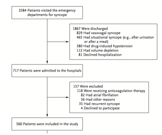
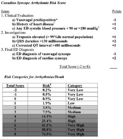
5,010 patients were enrolled in the study with 106 (2.1%) patients suffering arrhythmia or death within 30 days of ED presentation. Forty-five of the 106 patients suffered their adverse event outside of the hospital. The mean age of the study population was 53.4 (SD 23.0 years) and 54.8% were females. A total of 8 variables were included in the final model:

  1. Vasovagal predisposition

  2. History of heart disease (CAD, atrial fibrillation/flutter, CHF, valvular abnormalities)

  3. Systolic blood pressure <90 or >180 mm Hg at any point

  4. Troponin elevation

  5. QRS duration >130 msec

  6. QTc interval > 480 msec

  7. ED diagnosis of cardiac syncope

  8. ED diagnosis of vasovagal syncope

The Canadian Syncope Arrhythmia Risk Score had a sensitivity of 97.1% and specificity of 53.4% at a threshold score of 0 based on the study’s internal validation.

CS1.jpg
CS2.jpg

Interpretation

This study is the largest, multicenter study assessing predictors of short-term outcomes following initial ED presentation of syncope. The results are similar to previous studies that examined long-term outcomes. One interesting difference is that in prior studies, advanced age was a risk factor in arrhythmia or death, however it did not make the final model in this study. The strengths of this prospective study include the large patient population and that only 6.5% were lost to follow up. Furthermore, developing a simplified risk tool similar to the HEART score for chest pain, it can be easily utilized in the ED to help aid in decision making. Some limitations are that a large portion (54%) of patients did not have a troponin level measured and the study notes that these were usually younger patients with less comorbidities.

In practice, it may be difficult to use this tool if there is provider variation for when cardiac syncope is suspected and when a troponin level is measured. Whether or not the provider diagnoses vasovagal syncope or cardiac syncope is subjective as well, though may serve as a surrogate for “physician gestalt.” These results are helpful in risk stratifying syncope patients especially in regard to short-term outcomes, however this disease process is complex and cannot be oversimplified. Overall, this decision tool at the very least allows ED providers to have a shared decision-making conversation with more robust data to support the various options.

Take Home Points

  • The Canadian Syncope Arrhythmia Risk Score is a large, multicenter trial evaluating serious 30-day outcomes following an ED presentation for syncope

  • Emergency medicine physicians may consider using this tool to guide their clinical-decision making for syncope patients by offering risk percentages for 30-day adverse events

  • At the time this was written a validation study was underway


Expert Commentary

The management and disposition of syncope has been a conundrum for emergency physicians for decades. In fact, the last 20 years of syncope research have focused on development of a risk stratification score for the ED management of syncope. With the recent external validation of the Canadian Syncope Risk Stratification Score [9] (CSRSS) and the recent publication of the FAINT Score [10] for syncope in older adults, we now have two prospectively derived studies to support risk stratification of the syncope patient. The external validation of the CSRSS showed good sensitivity for low risk patients with a sensitivity of 97.8%.  None of the very low risk or low risk patients in the external validation died or suffered cardiac arrhythmia in 30 days. Based on this if your patient is very low risk or low risk you can safely discharge the patient home with primary care follow up.

In my practice, the CSRSS serves as an adjunct to clinician judgement. Using a risk stratification score is often the impetus for a shared decision-making discussion regarding risk and safe disposition. The results of the external validation study further support clinical use of the CSRSS. 

The FAINT score also shows promise for risk stratification in older patients with syncope and near syncope. This score has not been externally validated, but focuses on the older population that many emergency physicians reflexively admit for cardiac monitoring.

Regardless of which decision score you decide to use in personal practice, most of these patients with unexplained syncope can be safely admitted for a short observation stay.  It is safe to say that we have entered a golden age of syncope decision rules.

Andrew Moore.PNG

Andrew Moore, MD, MS

Emergency Physician and Emergency Care Researcher

Department of Emergency Medicine

Carilion Clinic


How To Cite This Post:

[Peer-Reviewed, Web Publication] Hung, J, Anderek, J. (2020, Sept 14). Canadian Syncope. [NUEM Blog. Expert Commentary by Moore, A]. Retrieved from http://www.nuemblog.com/blog/canadian-syncope.


Other Posts You May Enjoy

References

  1. Brignole M, Moya A, de Lange FJ, et al. 2018 ESC Guidelines for the diagnosis and management of syncope. European heart journal 2018;39:1883-948.

  2. Sun BC, Emond JA, Camargo CA, Jr. Characteristics and admission patterns of patients presenting with syncope to U.S. emergency departments, 1992-2000. Acad Emerg Med 2004;11:1029-34.

  3. Probst MA, Kanzaria HK, Gbedemah M, Richardson LD, Sun BC. National trends in resource utilization associated with ED visits for syncope. The American journal of emergency medicine 2015;33:998-1001.

  4. Cook OG, Mukarram MA, Rahman OM, et al. Reasons for Hospitalization Among Emergency Department Patients With Syncope. Acad Emerg Med 2016;23:1210-7.

  5. Quinn JV, Stiell IG, McDermott DA, Sellers KL, Kohn MA, Wells GA. Derivation of the San Francisco Syncope Rule to predict patients with short-term serious outcomes. Annals of emergency medicine 2004;43:224-32.

  6. Colivicchi F, Ammirati F, Melina D, Guido V, Imperoli G, Santini M. Development and prospective validation of a risk stratification system for patients with syncope in the emergency department: the OESIL risk score. European heart journal 2003;24:811-9.

  7. Reed MJ, Newby DE, Coull AJ, Prescott RJ, Jacques KG, Gray AJ. The ROSE (risk stratification of syncope in the emergency department) study. J Am Coll Cardiol 2010;55:713-21.

  8. Thiruganasambandamoorthy V, Kwong K, Wells GA, et al. Development of the Canadian Syncope Risk Score to predict serious adverse events after emergency department assessment of syncope. CMAJ : Canadian Medical Association journal = journal de l'Association medicale canadienne 2016;188:E289-98.

  9. Thiruganasambandamoorthy V, Sivilotti MLA, Le Sage N, et al. Multicenter Emergency Department Validation of the Canadian Syncope Risk Score. JAMA internal medicine 2020;180:1-8.

  10. Probst MA, Gibson T, Weiss RE, et al. Risk Stratification of Older Adults Who Present to the Emergency Department With Syncope: The FAINT Score. Annals of emergency medicine 2019.

Posted on September 14, 2020 and filed under Cardiovascular.

Management of Environmental Heat Injury in the ED

heat injury image.png

Written by: Sean Watts, MD (NUEM PGY-3) Edited by: Phil Jackson, MD (NUEM ‘20) Expert Commentary by: George Chiampas, DO, CAQSM, FACEP


Heat related illness has become an increasing source of morbidity and mortality due to environmental injuries from rising global temperatures and increased interest in outdoor activities. The National Oceanic and Atmospheric Administration reported that 2016 was the hottest year on record, and that temperatures were on average 3.2 F° higher than the 20th century averages.[1] Increasing temperatures have manifested in fatal heat waves such as one claiming the lives of 70,000 individuals living in Europe during 2003.[1] The population most subject to these heat waves include the extremes of age and athletes.

Human body temperature is normally set at 37 ° C, and is maintained via the preoptic nucleus of the anterior hypothalamus.[1,2] Hyperthermia results from exposure to an exogenous heat source without altering the hypothalamic set point. As core temperatures elevate during exertion and with exposure to heat, the posterior hypothalamic nucleus signals sympathetic pathways that result in vasodilation of peripheral vascular beds and shunting blood away from gastrointestinal vasculature in order to maximize heat dissipation. Additionally, eccrine sweat glands are cholinergically activated resulting in an evaporative cooling effect. When the duration and magnitude of heat exposure outpace these physiologic mechanisms, the symptoms of heat-related illness become evident and vary from mild heat cramps to severe heat stroke and death.[2]

Heat cramps result from both potassium wasting from persistent utilization of aldosterone in order to maintain a euvolemic state and sodium loss through sweat. Edema can result from increased hydrostatic pressure of the peripheral vasculature. Additionally, syncope and hypotension can manifest due to dehydration, orthostatic pooling of blood, peripheral vasodilation, and a subsequent decrease in cardiac output. Without appropriate treatment, heat exhaustion and the more extreme heat stroke can present.

Heat exhaustion is defined as a core temperature between 37 ° C and 40 ° C with signs and symptoms including intense thirst, weakness, discomfort, anxiety and dizziness.[1,2,6,8] Heat stroke, on the other hand, is defined as a core temperature greater than 40 C° with signs of central nervous system dysfunction. Heat stroke can be further categorized into exertional and non-exertional.[4] The demographic of exertional heat stroke includes athletes, military personal, or young individuals participating in prolonged exercise.[4,8] Non-exertional heat stroke includes the elderly, young children, or individuals with metabolic or cardiac comorbidities that engage in brisk to minor activity at elevated temperatures.[1,4] When the body reaches 40 C° denaturation of proteins, release of pro-inflammatory mediators, and direct activation of the coagulation cascade occurs.[1,2] This can ultimately result in disseminated intravascular coagulation, which is a common complication of heat stroke.[1,4,5] Disruption of the liver and the cerebellum from tissue ischemia, hypoxia, vascular dysfunction, secondary cascade inflammation manifest with elevated liver function tests and ataxia dysmetria, and coma.[1,6]

Summary of the Pathophysiology of Heat Stroke [1]

Summary of the Pathophysiology of Heat Stroke [1]

Treatment of heat related illness in the emergency department rests on appropriate recognition of the severity of disease. For heat syncope and heat cramps, isotonic or hypotonic electrolyte solutions may be administered in addition to actively flexing leg muscles to prevent peripheral pooling of blood.[7,8] Ice packs or cold towels around the neck, axillae and groin can also be used for comfort measures 6. In general, these heat illnesses are self-limiting.

For heat exhaustion and heat stroke, treatments become more aggressive and should be initiated within 30 minutes of recognition of the signs/symptoms.[1,4] These patients often present critically ill and rapid assessment of the patient’s airway, breathing, and circulation is paramount. Caregivers should obtain good IV access, as well as intubate the patient if they are obtunded or in danger of loss of airway protection.[1,6] Broad spectrum critical care labs should be obtained, as well as a CK to assess for evidence of rhabdomyolysis.[5]  Additionally, obtaining an accurate core body temperature is a crucial first step to determine the severity of illness.[1,2,4,5,6] This is best performed through continuous rectal probe monitoring. Rehydration should then be performed, preferably with 1 to 2 L of isotonic fluids.[1,4] Care should be taken to not over-correct hypovolemia as the aforementioned pathophysiology makes this population vulnerable to pulmonary edema.[1] Additionally, care should be taken not to over bolus hypotonic or isotonic solutions as this population, especially those involved in long distance endurance sports like triathlons or marathons, are particularly prone to hyponatremia.[9] If these patients are given too much of these solutions, this can actually exacerbate the hyponatremia. Patients with profound hyponatremia will actually require IV hypertonic solutions or salt tabs.[9]

 Clinicians should next focus on cooling core body temperature. The best treatment for exertional heat stroke is cold-water immersion therapy—where the patient gets placed in a cold body of water.[5,7] This method takes advantage of the high thermal conductivity of water and is most effective when the patient’s clothing is removed. Studies have demonstrated that immersion in an ice-water slurry at 2°C generated cooling rates of 0.35°C/min.[4] Comparatively, allowing hyperthermic subjects to rest in air-conditioned or temperature-controlled rooms only resulted in cooling rates of only 0.03°–0.06°C/min.[4] Evidence regarding an optimal temperature to halt cooling is still under debate, but is thought to be somewhere between 38°C to 39°C, with the fear that overcooling may result in cardiac arrhythmias, especially in the elderly suffering from non-exertional heat stroke.[1,4]

Subject in a cold water-immersion bath after heat- stroke [4]

Subject in a cold water-immersion bath after heat- stroke [4]

The use of cold-water immersion therapy in non-exertional heat stroke is still under debate, but the limited evidence shows that evaporative and convective cooling by a combination of cool water spray with continual airflow over the body may be superior, especially in the elderly suffering from non-exertional heat stroke.[4] In many emergency departments, complete cold water immersion therapy may not be readily  available and limited by the placement of cardiac leads, intubation, and IV access, so evaporative and convective cooling methods become first-line for both exertional and non-exertional heat stroke in the emergency department setting should cold water immersion be unavailable.[1,4,5] Should shivering become problematic, benzodiazepines are considered first line therapy.[1,6] In severe or refractory cases the patient may benefit from ECMO.[6]

Evaporative and conductive cooling methods--note the placement of ice packs in axilla, groin as well as the cooling fan overhead [4]

Evaporative and conductive cooling methods--note the placement of ice packs in axilla, groin as well as the cooling fan overhead [4]

With the rapid increase in heat-related injuries, and projected increase in global warming, researchers are continually seeking new and efficacious treatments. For example, recombinant activated protein C is currently being explored to manage the disseminated intravascular coagulation that may result from heat stroke.[2] Additionally, application of cold packs versus other methods of rapid cooling has been explored. An experimental study published in the journal of Wilderness and Environmental Medicine found that the use of ice packs provided a significantly higher enthalpy change over cold packs—suggesting that ice packs are more efficacious than cold packs when managing heat-injury.[3] Additionally, the study found that application of cold packs or ice packs to locations high in AV anastomoses provided superior cooling rates.[3] Evaporative plus convective cooling units are also under study as an alternative means to cold water immersion for the treatment of non-exertional heat stroke.[5]


Key Points and Summary

  • Heat Injury continues to be a major cause of environmental morbidity and mortality, and will likely increase due to rising global temperatures

  • Heat Injury exists on a continuum, with heat cramps/syncope on one end and heat exhaustion/stroke on the other end

  • Obtain a rectal temperature if you suspect heat exhaustion/stroke, assess ABC’s, get good IV access, and be careful not to over bolus isotonic/hypotonic solutions due to the risk of worsening hyponatremia in athletes

  • If feasible, cold water immersion is superior for exertional heat stroke, in the ED setting evaporative and conductive cooling with ice packs can be used

  • In severe or resistant cases cardiopulmonary bypass can be effective

table of cooling methods [6]

table of cooling methods [6]


Expert Commentary

A great review and reminders of what is a preventable death especially in exertional heatstroke. Unfortunately, still in the United States there are still approximately fifteen to twenty heat-related deaths in athletes annually, mostly seen in august. While there is a spectrum of illness, preventative measures, a high index of concern and management can all mitigate negative outcomes.

In non-exertional heat illness, removal from the environment, addressing the medical condition and or removing any contributing factors is key. Cooling methods and the aggressiveness of cooling are determined by the patient’s mental status and stability. As highlighted, Heat exhaustion presents with headaches, nausea, dizziness, and weakness. Using cooling blankets and cold packs to the groin axilla and circulating fans all are measures in passive cooling. One key element to address as typically a patient presents undifferentiated is to obtain a rectal temperature in a timely fashion as highlighted. Temperatures and glucose in altered mental status patients are critical for efficient management and positive outcomes. There are key studies that highlight that time and duration above 42C lead to higher morbidity including death. 

In athletics, the death of Minnesota football player Korey Stringer in August of 2001 shed greater light on the risks of exertional heatstroke. Since his death, more work and research has been done including best practices in sport to mitigate these outcomes. Across many sports, including Marathons, best practices as outlined in the blog are being implemented pre-hospital. These measures are comparable to the recent out of hospital cardiac arrest best practices of on-sight CPR and utilization of an AED and transport second mantra. In heatstroke “cooling” on sight with ice tub submersion is the current thread being communicated. This messaging is evidenced by a recent EMS consensus paper  that highlights to first-responders the importance of recognizing but also cooling on-sight prior to transport. The delay of cooling and transport times to delay of recognition and cooling in emergency departments may lead to not initiating life-saving rapid cooling beyond the thirty minutes highlighted in the blog.

As you accurately highlighted patients can present differently, however, the key is altered mental status (AMS). Based on experience this can have the forms of patients collapse and obtunded, seizing, irritable and combative to just being confused. Rapid assessments in the right environment with excluding other AMS possibilities will allow the practitioner to respond and manage in a timely fashion. At Northwestern, both Dr. Malik and Dr. Chiampas have published the attached “collapse algorithm” (below) which allows for a quick assessment and possible differential diagnoses. Lastly obtaining a rectal temperature, which at times may be challenging with the combative patient, allows the staff in the Emergency room to objectively determine when to cease cooling. I will share that some of these patients based on their presentation would traditionally be intubated upon arrival. I would caution and remind the practitioner that if you have prepared in advance and can rapidly cool the symptoms are reversible within 10-15 minutes of ice submersion.

Lastly for emergency departments, where out-door events (sporting, festivals or concerts) with the possibility of stimulant use, preparedness is key. At Northwestern, we have secured 100-gallon ice tubs, implemented the collapse algorithm in our trauma bay and on when high-risk events take place to trigger necessary resources. For the Chicago Marathon, Triathlon or major concerts such as Lolla Palooza we order ice to the ER, towels, and prep the tub while educating our staff of the likelihood of these conditions. As we head towards the summer ahead with all of the environmental concerns of climate change and increased temperatures, this blog provides key reminders of the emergency department’s role.

Collapse_Algorithm[1] (1) (1).png
chiampas.png
 

George Chiampas DO CAQSM

Assistant Professor Northwestern University, Feinberg School of Medicine

Departments of Emergency and Orthopedic Surgery

Chief Medical Officer U.S. Soccer

Chief Medical and Safety Officer Bank of America Chicago Marathon

Team Physician Chicago Blackhawks


How To Cite This Post

[Peer-Reviewed, Web Publication] Watts S, Jackson P. (2020, July 6). Management of Environmental Heat Injury in the ED [NUEM Blog. Expert Commentary by Chiampas G. Retrieved from http://www.nuemblog.com/blog/environmental-heat-injury


Other Posts You May Enjoy


References 

  1. Heat-Related Illness. Walter F. Atha, MD. Emerg Med Clin N Am 31 (2013) 1097–1108. http://dx.doi.org/10.1016/j.emc.2013.07.012

  2. Wilderness Medical Society Practice Guidelines for the Prevention and Treatment of Heat-Related Illness: 2014 Update. Grant S. Lipman, MD; Kurt P. Eifling, MD; Mark A. Ellis, MD; Flavio G. Gaudio, MD; Edward M. Otten, MD; Colin K. Grissom, MD. WILDERNESS & ENVIRONMENTAL MEDICINE, 25, S55–S65 (2014)

  3. Chemical Cold Packs May Provide Insufficient Enthalpy Change for Treatment of Hyperthermia. Samson Phan, MS; John Lissoway, MD; Grant S. Lipman, MD. WILDERNESS & ENVIRONMENTAL MEDICINE, 24, 37–41 (2013)

  4. Cooling Methods in Heat Stroke Flavio G.Gaudio MD∗Colin K.Grissom MD†The Journal of Emergency Medicine, Volume 50, Issue 4, April 2016, Pages 607-616

  5. Heat Stroke. Alan N. Peiris, MD, PhD, FRCP(London); Sarah Jaroudi, BS; Rabiya Noor, BS. JAMA. 2017;318(24):2503. doi:10.1001/jama.2017.18780

  6. Tintinalli’s Emergency Medicine: A Comprehensive Study Guide, 8e. Judith E. Tintinalli, J. Stephan Stapczynski, O. John Ma, Donald M. Yealy, Garth D. Meckler, David M. Cline. Section 16, Chapter 210: Heat Emergencies. http://accessmedicine.mhmedical.com.ezproxy.galter.northwestern.edu/content.aspx?bookid=1658&sectionid=109384117. Accessed June 10, 2019.

  7. Heat-Related Illness in Athletes Allyson S. Howe MD, Barry P. Boden, MD First Published August 1, 2007 https://doi-org.ezproxy.galter.northwestern.edu/10.1177/0363546507305013

  8. Heat-Related Illnesses. ROBERT GAUER, MD, Womack Army Medical Center, Fort Bragg, North Carolina. BRYCE K. MEYERS, DO, MPH, 82nd Airborne Division, Fort Bragg, North Carolina. Am Fam Physician. 2019 Apr 15;99(8):482-489.

  9. Hyponatremia among runners in the Boston Marathon. Almond CS, Shin AY, Fortescue EB, Mannix RC, Wypij D, Binstadt BA, Duncan CN, Olson DP, Salerno AE, Newburger JW, Greenes DS. N Engl J Med. 2005 Apr 14;352(15):1550-6.


Posted on July 6, 2020 and filed under Environmental.

The PESIT Trial: Do All Syncope Patients Need a PE Workup?

pesitgraphic.png

Written by: Alex Ireland, MD (NUEM PGY-2) Edited by: Josh Zimmerman, MD,  (NUEM class of 2017) Expert Commentary by: D. Mark Courtney, MD


Syncope is defined as a transient loss of consciousness and postural tone caused by cerebral hypoperfusion. This chief complaint is familiar to most emergency physicians given it accounts for more than 1-2 million patient visits in the US annually.[1]  The differential diagnosis is broad and crosses multiple organ systems, including, but not limited to, cardiovascular, neurovascular,  and hemorrhagic/hematologic causes.  Pulmonary embolism (PE) is one cardiovascular cause that is considered to be infrequent. This is because when associated with syncope, it is often made more clinically apparent by signs and symptoms such as dyspnea, chest pain, tachycardia, hypotension, and hypoxemia.  The American Heart Association consensus statement on the evaluation of syncope gives little attention to diagnostics for PE, instead focusing on arrhythmia, ischemia, and structural heart disease. [2]  We know from a San Francisco Syncope Rule validation trial that when adverse events occur in the minority of patients with generalized syncope, they are related mostly to cardiac arrhythmias, followed by MI- not PE. [3] Previous studies have cited the prevalence of pulmonary embolism as a cause of syncope in hospitalized patients as low as 1.6%. [4]  But are we underestimating and under-diagnosing this potentially lethal condition? The PESIT study sought to systematically assess the prevalence of pulmonary embolism in patients admitted for syncope. [5] 


Prevalence of Pulmonary Embolism among Patients Hospitalized for Syncope. Paolo Prandoni, M.D., Ph.D., Anthonie W.A. Lensing, M.D., Ph.D., Martin H. Prins, M.D., Ph.D., Maurizio Ciammaichella, M.D., Marica Perlati, M.D., Nicola Mumoli, M.D., Eugenio Bucherini, M.D., Adriana Visonà, M.D., Carlo Bova, M.D., Davide Imberti, M.D., Stefano Campostrini, Ph.D., and Sofia Barbar, M.D., for the PESIT Investigators*. N Engl J Med 2016; 375:1524-1531. October 20, 2016

What was the PESIT trial? 

Figure 1. Inclusion and Exclusion Flowchart

Figure 1. Inclusion and Exclusion Flowchart

    The PESIT trial was a cross-sectional study of patients older than 18 years of age who were hospitalized for a first episode of syncope. This was a multicenter trial, taking place in 11 hospitals (2 academic, 9 nonacademic) in Italy. There were several important exclusion criteria to this study including patients on anticoagulation, pregnant patients, and those with previous syncopal episodes.   Most importantly, however, the study excluded all patients discharged and solely focused on those admitted for an inpatient evaluation (Figure 1). 


    All included patients were subjected to a standardized history aimed at suggesting the cause of syncope and identifying risk factors for pulmonary embolism (Figure 2). They then underwent mandatory chest radiography, electrocardiography, arterial blood gas testing, and routine blood testing, including a D-dimer assay. Further diagnostic workup for causes other than pulmonary embolism was not standardized and varied between patients based on attending physician discretion.

Figure 2. Standardized History Questions

    The presence or absence of PE was assessed with a validated algorithm based on pretest clinical probability and the result of the D-dimer assay. The pretest clinical probability was defined according to the simplified Wells score, which classifies PE as being “likely” or “unlikely” (Table 1). In patients who had a high pretest clinical probability, a positive D-dimer assay, or both, computed tomographic pulmonary angiography or ventilation-perfusion lung scanning was performed.


Outcomes

    The primary outcome of the PESIT trial was to measure the prevalence of pulmonary embolism in patients admitted to the hospital for syncope. Secondarily, the thrombotic burden was assessed by measuring the most proximal location of embolus or the percentage of perfusion defect area.

    In 58.9% (339/560) of patients, PE was ruled out by low pre-test probability and a negative D-dimer. Of the remaining patients, 42.2% (97/229) had a pulmonary embolism. In the entire cohort, the prevalence of PE was 17.3% (97/560). Thus, the authors concluded that nearly one of every six patients hospitalized for a first episode of syncope has a pulmonary embolism (Figure 3, Table 2).

Figure 3. Results Flowchart

Figure 3. Results Flowchart


Strengths and Weaknesses of the Study

    The major strength of the PESIT trial is the systematic approach with which PE's were diagnosed. ALL admitted patients underwent consideration of and testing for PE based on risk factors and pretest probability. Even if, for example, the initial ECG suggested an arrhythmia as the cause of syncope, if they did not rule out for PE, they went on to get a scan. This ensured a complete and thorough investigation. Furthermore, their results were internally valid, as the prevalence of PE was consistently 15-20% across all 11 centers.

    Additionally, the population studied is also fairly representative of those who most emergency medicine physicians would admit to the hospital with syncope. Reasons for admission included trauma, severe comorbidities, failure to identify a cause in the ED, and a high probability of cardiac syncope. While we don’t commonly site the EGSYS score that they used, the components such as palpitations, heart disease, and an exertional component to syncope, are all considerations that factor into our clinical gestalt for admission.

    A glaring problem with this study is the significant variation in diagnostic workup beyond PE. Other potential explanations for syncope were much more likely to be undetermined in those with confirmed embolism (see table 2). Because further workup was left to the discretion of individual physicians, alternative and perhaps more causative causes of syncope may have been under-diagnosed or underreported.

    Furthermore, this study has limited external validity when used to assess our ED population as a whole, given the exclusion criteria of all patients discharged from the ED. When recalculating the prevalence based on all patients who presented to the ED, only 1 in 26 (97/2584 = 3.8%) patients were diagnosed with a PE, far fewer than their conclusive 1 in 6. A major contributing factor is likely age. As expected, the mean age of patients discharged was much younger at 54 years (range 16 to 79). These patients are much less likely to develop PE based on currently available decision rules such as the PERC Rule.

    We commonly use tools such as the PERC Rule or the Wells score to quantify our pretest probability because they include characteristics known to be associated with PE. As seen in the clinical characteristics described in table 2, patients diagnosed with pulmonary embolism had a high prevalence of symptoms commonly associated with PE, such as hypoxemia and tachycardia, or clinical manifestations of DVT. They were also far more likely to have a history of previous venous thromboembolism or to have active cancer. A large proportion of PESIT patients diagnosed with PE presented exactly as we would expect patients with a PE to present. These  data strengthen support for current clinical practice, which for most physicians means only entering the diagnostic pathway for pulmonary embolism when history and physical exam suggests it as a potential diagnosis.

    Lastly, the PESIT trial spends significant effort quantifying radiographic burden of pulmonary embolism across all their positive cases. As mentioned earlier, all patients were evaluated for PE, regardless of clinical gestalt. However, we must remember that degree of radiographic filling defect does not necessarily correlate with clinically significant pulmonary embolism. In 40% of patients with PE, the extent of pulmonary vascular obstruction was at the segmental or subesegmental level or was less than 25% of total lung area. Some of these may have been clinically insignificant, not likely to have caused the syncopal event, and perhaps were discovered incidentally. A better predictor for the severity of PE might have incorporated factors such as heart rate, systolic blood pressure < 100 mmHg, elevated BNP, and elevated troponins, which are used in the simplified PE Severity Index and a subsequently developed composite score to predict mortality from PE. [6,7}


Take Home Message

    In summary, Pulmonary embolism is an important “must-not miss” cause of syncope, but it is likely an uncommon diagnosis in patients who pass out, recover, and appear well – the main way that most patients in the US with syncope present. Overall, 1 in 26 patients who present to the ED with first time syncope may have a PE. A large portion of these may be clinically insignificant and not causative of syncope. Those with symptoms and signs of PE are more likely to have a significant PE and should be evaluated as such. Thus, we should not change our current clinical practice based on the results of the PESIT trial.


Expert Commentary

Internal validity:

Dr. Ireland’s review of the Prandoni study reviews it’s methods and results quite well.  He also points out the important ways in which we as consumers of literature should address a study.  The first is with respect to internal validity (how well the authors measured what they in fact intended to measure).  This takes into account potential for bias, or systematic ways in which error could be introduced into the measurement.  Dr. Ireland does not seem to identify many problems with this study from that standpoint.   In general I agree with this, if the goal of measurement of this study was to exhaustively test all patients who are admitted largely at the discretion of their doctors after a first event of syncope.  If so, then the methods of this paper seem to have resulted in reasonable internal validity.  However if the goal of the paper is to identify the prevalence of symptomatically significant PE among ALL patients with first time PE, then it is quite possible that there is bias due to the methods of inclusion.  Specifically, the fact that only admitted patients were included, and those admitted had a fairly large degree of comorbid conditions may have resulted in a measurement of PE that is higher than what would be expected for in general “syncope.”

External validity:

The second key question for a consumer of medical literature to ask is, simplistically, “are these patients like mine?”  The answer to this is probably no.  The patients included are different than a typical US ED "undifferentiated passing out patient" not just because they are Italian, but also because they had a high prevalence of symptoms to suggest PE such as history of PE/DVT,  or history of malignancy. In the US, we would probably not consider many of these patients to be “undifferentiated syncope.” Rather, we would consider them to be possible symptomatic PE patients and simply test them.  Dr. Ireland points this out in his evaluation of table 2.  It is not a novel finding that many of the patients with these comorbidities, signs, and symptoms went on to have PE when tested (Courtney Annals of EM Annals of Emergency Medicine 2010;55(4):307–315).

Another way to examine generalizability is to examine the course of these syncope patients and ask if this is similar to the course that would be taken in the US. Of the 2584 ED syncope patients in this study, 1867 were discharged. Are we discharging 72% of our syncope patients?  Whether or not we should be is another question.  However,  it is likely that the US ED environment has a much lower threshold for admission for syncope than the Italian setting, similar to the way that the US ED environment has a much lower threshold for testing for PE than the European setting.  Therefore, it is highly likely that, at least to some extent, these Italian syncope patients are more ill than the average US ED syncope patient.  This is supported by the elderly median age in the Prandoni study of 80….meaning half of all their patients were over 80 years of age!  Also note that of the 717 remaining patients not discharged, a further 157 were excluded.  So this sample really is a unique selected group…..making it difficult to generalize this study to the average US ED patient with syncope.  

Dr. Jeff Kline and I explored the possible external generalizability of this report in a re-examination of the PERC dataset which included 7940 patients from 12 US emergency departments, all of whom had  symptoms prompting testing for PE.  Among 466 PE positive patients, 6.6% reported syncope, while among 7474 PE negative patients 6.0% reported syncope (95% CI for difference, -1.7 - 3.0).  This suggested syncope was not a predictor of PE. We also noted in the Prandoni study a mean age of 75 and a high prevalence of main pulmonary artery clot (42%), something we have not found in US studies of undifferentiated PE patients where median percent obstruction was 9%.

Bottom line: 

In elderly syncope patients with some combination of tachycardia, tachypnea, hypotension, active cancer, and perhaps especially those without a clear suspected cause of syncope, PE should be a consideration that warrants testing.  Perhaps this should be considered even when patients do not have the more traditional symptoms of PE such as dyspnea or chest pain.  However, we would caution clinicians NOT to interpret this study as rationale for widespread testing on all or nearly all US ED syncope patients.  The outcome of such a simplistic interpretation of this study would undoubtedly result in further radiation and contrast burden and harms for our patients.

d-mark.png


D. Mark Courtney, MD

Associate Professor of Emergency Medicine and Medical Social Sciences, Northwestern Emergency Medicine


Posts You May Also Enjoy


How to cite this post

[Peer-Reviewed, Web Publication] Ireland A,  Zimmerman J (2017, Sep 27). The PESIT Trial: Do All Patients Need a Syncope Workup?  [NUEM Blog. Expert Commentary By Courtney DM]. Retrieved from http://www.nuemblog.com/blog/PESIT


Resources

1. Syncope Evaluation in the Emergency Department Study (SEEDS): A Multidisciplinary Approach to Syncope Management. Win K. Shen, Wyatt W. Decker, Peter A. Smars, Deepi G. Goyal, Ann E. Walker, David O. Hodge, Jane M. Trusty, Karen M. Brekke, Arshad Jahangir, Peter A. Brady, Thomas M. Munger, Bernard J. Gersh, Stephen C. Hammill and Robert L. Frye. Circulation. 2004;110:3636-3645.

2. AHA/ACCF Scientific Statement on the evaluation of syncope: from the American Heart Association Councils on Clinical Cardiology, Cardiovascular Nursing, Cardiovascular Disease in the Young, and Stroke, and the Quality of Care and Outcomes Research Interdisciplinary Working Group; and the American College of Cardiology Foundation: in collaboration with the Heart Rhythm Society: endorsed by the American Autonomic Society. Strickberger SA, et. Al. American Heart Association Councils on Clinical Cardiology, Cardiovascular Nursing, Cardiovascular Disease in the Young, and Stroke; Quality of Care and Outcomes Research Interdisciplinary Working Group. American College of Cardiology Foundation.; Heart Rhythm Society.; American Autonomic Society. Circulation. 2006 Jan 17;113(2):316-27. No abstract available. Erratum in: Circulation. 2006 Apr 11;113(14):e697.

3. Prospective validation of the San Francisco Syncope Rule to predict patients with serious outcomes. Quinn J, McDermott D, Stiell I, Kohn M, Wells G.. Ann Emerg Med. 2006 May;47(5):448-54.

4. Etiology of syncope in hospitalized patients. Saravi M, Ahmadi Ahangar A, Hojati MM, Valinejad E, Senaat A, Sohrabnejad R, Khosoosi Niaki MR. Caspian J Intern Med. 2015 Fall;6(4):233-7.

5. Prevalence of Pulmonary Embolism among Patients Hospitalized for Syncope. Prandoni P, Lensing AW, Prins MH, Ciammaichella M, Perlati M, Mumoli N, Bucherini E, Visonà A, Bova C, Imberti D, Campostrini S, Barbar S; PESIT Investigators. N Engl J Med. 2016 Oct 20;375(16):1524-1531.

6. Jiménez D, Aujesky D, Moores L, et al. Simplification of the pulmonary embolism severity index for prognostication in patients with acute symptomatic pulmonary embolism. Arch Intern Med 2010; 170:1383.

7. Jiménez D, Kopecna D, Tapson V, et al. Derivation and validation of multimarker prognostication for normotensive patients with acute symptomatic pulmonary embolism. Am J Respir Crit Care Med 2014; 189:718.


Posted on September 26, 2017 and filed under Cardiovascular.

To Admit or Not to Admit: Initial Results from the Intermediate-Risk Syncope (IRiS) Study

There is not clear guidance on which patients presenting with syncope to the emergency department should be admitted. This week we discuss new evidence from the IRiS trial that may help with determining which patients may be safe for outpatient work up. 

Methadone Induced Torsades

Torsades de Pointes (Tdp) is a term that is often used synonymously with polymorphic ventricular tachycardia (PVT) but it is important to understand the differences. This week we take a deep dive into an interesting case of Tdp with expert commentary by Amal Mattu!