Posts tagged #risk stratification

How to Talk Like a Neurologist

Written by: Saabir Kaskar, MD (NUEM ‘23) Edited by: Nick Wleklinski (NUEM ‘22)
Expert Commentary by: Fan Caprio, MD


Neurology Scores: LVO, NIHSS, and ICH

As first line providers, being able to effectively communicate with ancillary services and specialties is key to advancing patient care within the emergency department. When patients present with symptoms concerning for ischemic or hemorrhagic stroke, there are a variety of clinical decision tools available to help direct interventions and predict patient outcomes.  Having a basic understanding of these scoring systems helps ED providers communicate more effectively with our neurology colleagues. This post highlights indications, strengths, and limitations of common stroke assessment scales used in the prehospital and hospital setting.  

Cincinnati Prehospital Stroke Scale (CPSS)

The Cincinnati Prehospital Stroke Scale is a simple, easy to teach, three-part evaluation and is the most cited scale in statewide EMS protocols. Patients with one of these three findings, as a new event, will have 72% probability of ischemic stroke. If they have three of these deficits, that probability increases to 85%. Further, those scoring higher on this scale are more likely to have a large vessel occlusion (LVO) and warrant transfer to a comprehensive stroke center. One major limitation is that the CPSS does not identify features of posterior circulation strokes.

Figure 1: Cincinnati Prehospital Stroke Scale components

Predicting Large Vessel Occlusion 

There are many stroke severity scales that are useful in predicting large vessel occlusion (LVO) in the pre-hospital setting. Early LVO detection is useful as these patients have better outcomes if transported to comprehensive stroke centers (CSCs) which have endovascular interventions, such as thrombectomy, readily available. Such interventions are not available at primary stroke centers (PSC). LVO screening tools include the Rapid Arterial Occlusion Evaluation Scale (RACE), the Cincinnati Prehospital Stroke Severity Scale (CP-SSS/C-STAT), the Los Angeles Motor Scale (LAMS), and the Emergent Large Vessel Occlusion Scale (ELVO). While these scales are good, none have achieved an optimal sensitivity/specificity combination which is why there is no “gold standard” test per the most recent 2019 AHA guidelines (Powers et al. Guidelines for Early Mgmt of Patients with AIS. Stroke 2019). 

The Rapid Arterial Occlusion Evaluation Scale (RACE), for example, is one of these severity scales that predicts stroke caused by large vessel occlusion. It is based on the NIHSS but provides quicker assessment in the pre-hospital environment. It focuses on facial palsy, extremity motor function, head deviation, gaze deviation and aphasia or agnosia. The scale ranges from 0-9 with scores ≥ 5 being associated with detection of an LVO. RACE has a sensitivity of 85% and specificity of 68% for LVO at scores ≥ 5. 

Another example of a LVO screening tool is the Cincinnati Prehospital Stroke Severity Scale (CP-SSS/CSTAT) which is important to differentiate from the CPSS outlined above.  CSTAT focuses on gaze deviation, level of consciousness and arm weakness. Both RACE and CSTAT are validated in the prehospital setting and with external data sets. However, CSTAT is more convenient with fewer items to score. 

EMS protocol in Chicago (Region XI), utilizes a two-tier system that first involves the Cincinnati Stroke Scale and finger to nose test. If either aspect is abnormal, then stroke severity is assessed with the 3-Item Stroke Scale (3I-SS) which assesses level of consciousness, gaze preference and motor function, scored from 0-6. If the 3I-SS score is ≥4 and the last known normal is ≤6 hours ago then the patient is transported to the closest CSC instead of the closest primary stroke center (PSC), as long as the added transport time is not >15 minutes.  

National Institutes of Health Stroke Scale (NIHSS)

The NIHSS is a 11-part scoring tool and is the gold standard when assessing stroke patients in hospital (figure 3). Higher scores indicate a more severe stroke and usually correlate with infarct size on CT and MRI. Taken within the first 48 hours of acute stroke, the NIHSS helps predict three month and one-year clinical outcomes. For example, patients with a NIHSS of 1-4 have a high likelihood of functional independence and favorable outcome regardless of treatment. The NIHSS does not serve as the primary clinical guide in determining tPA administration. However, given that higher scores correlate with larger infarct size, caution is advised when considering tPA in patients with a NIHSS >22 as there is a higher risk of hemorrhagic conversion (see figure 2 for full tPA exclusion criteria). Analysis from subjects of the NINDS trials show that a NIHSS of >20 was associated with a 17% rate of intracranial hemorrhage with tPA when compared to 3% hemorrhage rate in patients with a score of <10.

Figure 2: Contraindications for tPA administration

Overall, the NIHSS is a reliable scoring tool to quicky assess the effects of stroke. Medical providers and nurses have been shown to have similar levels of accuracy when trained. Limitations include assessing posterior circulation stroke that involve gait abnormality, dizziness, or diplopia.

Figure 3: NIHSS, adopted from the American Stroke Association

Intracerebral Hemorrhage Score (ICH Score)

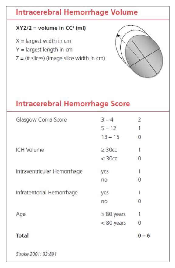
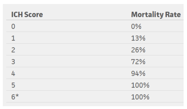
The ICH score is an important tool when evaluating a hemorrhagic stroke. This score was developed to standardize clinical grading of ICH and to improve communication between providers. This five-component scoring system (Figure 4) helps quantify ICH severity and subsequently 30-day mortality  (Figure 6) with a sensitivity of 66%. It is not used to determine treatment modality. This score helps universalize the grading of ICH severity, providing a standardized language that can be used between EM providers, neurologists, and neurosurgeons. Further, this score can help providers guide goals of care conversations with patient’s families and determine appropriate level of care or transfer.

Figure 4: ICH score, adapted from the American Stroke Association

Figure 5: Mortality rates based on ICH score

*No patients in the study scored 6, but estimated 100% mortality

Conclusion

In summary, it is important to understand how to utilize these scoring tools for ischemic and hemorrhagic stroke. Knowing how to interpret pre-hospital stroke scores and how to calculate a NIHSS score accurately and quickly is helpful in not only quantifying severity but also in improving communication between providers. Improved understanding and effective use of these tools can help better advance care of our stroke patients efficiently. These tools can also remind us of the severity of the neurologic deficit we observe on clinical exam. Subsequently, this can be helpful in guiding discussions with patients and their families regarding the severity of their condition.

References

Adams HP Jr, Davis PH, Leira EC, et al. Baseline NIH Stroke Scale score strongly predicts outcome after stroke: A report of the Trial of Org 10172 in Acute Stroke Treatment (TOAST). Neurology 1999; 53:126.

Goldstein, L. (2019). Use and utility of stroke scales and grading systems. Up To Date

Goldstein L, Bertels C, Davis JN. Interrater reliability of the NIH stroke scale. Arch Neurol 1989; 46:660.

Generalized efficacy of t-PA for acute stroke. Subgroup analysis of the NINDS t-PA Stroke Trial. Stroke 1997; 28:2119.

Hemphill JC 3rd, Bonovich DC, Besmertis L, Manley GT, Johnston SC. The ICH score: a simple, reliable grading scale for intracerebral hemorrhage. Stroke. 2001 Apr;32(4):891-7. PubMed PMID: 11283388.

Kothari RU, Pancioli A, Liu T, et al. Cincinnati Prehospital Stroke Scale: reproducibility and validity. Ann Emerg Med 1999; 33:373.

Pérez de la Ossa N, Carrera D, Gorchs M, et al. Design and validation of a prehospital stroke scale to predict large arterial occlusion: the rapid arterial occlusion evaluation scale. Stroke 2014; 45:87.

Schlemm L, Ebinger M, Nolte CH, Endres M. Impact of Prehospital Triage Scales to Detect Large Vessel Occlusion on Resource Utilization and Time to Treatment. Stroke 2018; 49:439.


Expert Commentary

Thanks for writing this comprehensive summary of common screening tools used in stroke patients. Having a good handle on these tools will allow you to quickly and effectively communicate with comanaging care providers. It is also important to understand how and why each scale was developed, so they can be used in the appropriate setting to expedite care in extremely time-sensitive neurologic emergencies.

Keep in mind that scales are merely screening tools and are not meant to give a definitive diagnosis. No scale is perfect, but you have highlighted some that yield the highest sensitivity and specificity for identifying a potential stroke patient. In addition to leaning on these scales as decision support tools, always use your clinical judgement. A few things to remember in addition to the neurologic symptoms:

* Strokes are potentially intervenable within the first 24 hours:

1. Up to 4.5 hours – IV-TPA / tenecteplase.

2. Up to 6 hours – Thrombectomy with LVO on vessel imaging.

3. Up to 24 hours – Thrombectomy with LVO + favorable penumbra on perfusion imaging.

* Last known normal (LKN) starts the timer to when stroke patients are eligible for intervention (not to be confused with time of symptom discovery!)

* Strokes typically cause a sudden loss of function (in contrast to positive phenomena such as convulsive movements, tingling sensation, sparkling vision, which can point away from a stroke diagnosis)

* In patients with prior deficits, ask which symptoms are new or different in comparison to their baseline.

The NIHSS is widely accepted as THE stroke severity scale, and it has many strengths and some pitfalls. The NIHSS was initially developed to be used in research, and, as mentioned here, was designed to be reproducible between various groups – physicians, nurses, research staff. Higher scores correlate with bigger infarct volume. The NIHSS is not an accurate scale in that it does not necessarily capture each patient’s deficits, omitting brain functions such as gait, distal limb dexterity, and cognition. It also scores higher for dominant (L) hemispheric functions as many points depend on language function.

When screening for large vessel occlusion, remember key brain structures and functions from the L MCA, R MCA, and posterior circulation. Looking for cortical signs can be very helpful to identify larger stroke syndromes: aphasia, neglect, gaze deviation, visual field deficit.

Last but not least, keep in mind that hemorrhagic strokes (intracerebral hemorrhage, subarachnoid hemorrhage) account for about 15% of all strokes. The same screening tools for acute neurologic symptoms can be used to identify these patients, though they more often have concurrent headache or LOC than ischemic strokes (due to increased ICP and irritation from blood products). For SAH, two scales are commonly used to describe the clinical and radiographic severities: Hunt-Hess (surgical risk index) and modified Fisher scales (risk index for developing vasospasm).

Figure 1: Hunt-Hess Scale

Figure 2: Modified Fisher Scale

Fan Caprio, MD

Assistant Professor of Neurology (Stroke)

Department of Neurology

Northwestern Memorial Hospital


How To Cite This Post:

[Peer-Reviewed, Web Publication] Kaskar, S. Wleklinski, N. (2021, Oct 25). How to Talk Like a Neurologist. [NUEM Blog. Expert Commentary by Caprio, F]. Retrieved from http://www.nuemblog.com/blog/neuro-scores


Other Posts You May Enjoy

Canadian Syncope

Written by: Jonathan Hung, MD (NUEM ‘21) Edited by: Jon Anderek (NUEM ‘19) Expert Commentary by: Andrew Moore, MD, MS

Written by: Jonathan Hung, MD (NUEM ‘21) Edited by: Jon Anderek (NUEM ‘19) Expert Commentary by: Andrew Moore, MD, MS


Introduction

Syncope is defined as a brief loss of consciousness that is self-limited. [1] It is a commonly seen chief complaint in the emergency department (ED), consisting of up to 3% of ED visits. [2] There are both benign causes of syncope such as vasovagal syncope and more serious causes such as arrhythmias. By the time these patients present to the ED, they are often asymptomatic and hemodynamically stable. Part of the ED workup and disposition includes risk stratification of these patients that can vary by provider and hospital system. [3] For those who present with high-risk features, ED physicians often recommend admission to the hospital for telemetry monitoring and expedited evaluation with echocardiography. [4] Multiple decision rules, most notably the San Francisco Syncope Rule (SFSR), have been developed to identify syncope patients at risk for poor outcomes. The SFSR takes into account predictors such as a history of heart failure, an abnormal electrocardiogram (ECG), and hypotension to determine 7-day negative outcomes for patients presenting to the ED with syncope. [5] Another study called the Osservatorio Epidemiologico sulla Sincope nel Lazio (OESIL) includes age over 65 and syncope without prodrome in addition to a history of cardiovascular disease as part of their decision-making tool. [6] Lastly, the Risk Stratification of Syncope in the Emergency Department (ROSE) also takes lab results such as brain natriuretic peptide and hemoglobin into account. [7] Despite the numerous studies examining risk stratification in syncope, each one has limitations and ultimately lack adequate sensitivity and specificity for widespread clinical adoption. A new study published in Academic Emergency Medicine is one of the largest studies to develop a risk tool that identifies adult syncope patients at 30-day risk for serious adverse outcomes defined as a serious arrhythmia, need for intervention to correct arrhythmia, or death. [8]

Study

Thiruganasambandamoorthy V, Stiell IG, Sivilotti MLA, et al. Predicting Short-term Risk of Arrhythmia among Patients With Syncope: The Canadian Syncope Arrhythmia Risk Score. Baumann BM, ed. Acad Emerg Med. 2017;24(11):1315-1326.

Study Design

  • Multi-center, prospective, observational cohort study.

  • This was a derivation study used to define the parameters of the risk score.

Population

Inclusion criteria:

  • Syncope patients presenting within 24 hours of the event

  • Adults age ≥16

Exclusion criteria:

  • Prolonged loss of consciousness

  • Change in mental status from baseline

  • Witnessed seizure

  • Head trauma or other trauma requiring admission

  • Unable to provide history due to alcohol intoxication, illicit drug use or language barrier

  • Obvious arrhythmia or nonarrhythmic serious condition on presentation

Intervention protocol

ED physicians and emergency medicine residents were trained to assess standardized variables at the initial ED visit including time and date of syncope, event characteristics, personal and family history of cardiovascular disease, and final ED diagnosis. Other variables were obtained through chart review and included age, sex, vital signs, laboratory results and ECG variables. All ECGs were reviewed by a cardiologist, and abnormal variables were reviewed by a second cardiologist. Physician gestalt for dangerous etiology was also recorded for each patient. Multivariable logistic regression was used for the analysis.

Outcome Measures

Composite of death, arrhythmia, or procedural interventions to treat arrhythmias within 30 days of ED disposition

Results

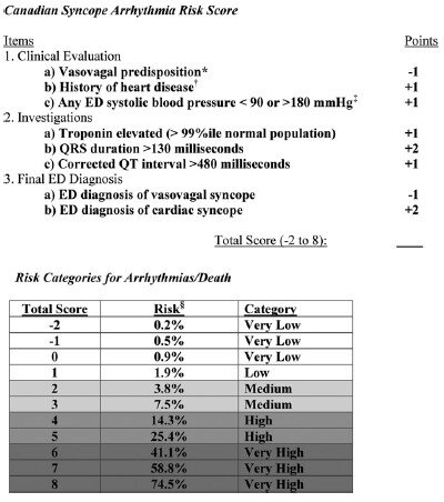
5,010 patients were enrolled in the study with 106 (2.1%) patients suffering arrhythmia or death within 30 days of ED presentation. Forty-five of the 106 patients suffered their adverse event outside of the hospital. The mean age of the study population was 53.4 (SD 23.0 years) and 54.8% were females. A total of 8 variables were included in the final model:

  1. Vasovagal predisposition

  2. History of heart disease (CAD, atrial fibrillation/flutter, CHF, valvular abnormalities)

  3. Systolic blood pressure <90 or >180 mm Hg at any point

  4. Troponin elevation

  5. QRS duration >130 msec

  6. QTc interval > 480 msec

  7. ED diagnosis of cardiac syncope

  8. ED diagnosis of vasovagal syncope

The Canadian Syncope Arrhythmia Risk Score had a sensitivity of 97.1% and specificity of 53.4% at a threshold score of 0 based on the study’s internal validation.

CS1.jpg
CS2.jpg

Interpretation

This study is the largest, multicenter study assessing predictors of short-term outcomes following initial ED presentation of syncope. The results are similar to previous studies that examined long-term outcomes. One interesting difference is that in prior studies, advanced age was a risk factor in arrhythmia or death, however it did not make the final model in this study. The strengths of this prospective study include the large patient population and that only 6.5% were lost to follow up. Furthermore, developing a simplified risk tool similar to the HEART score for chest pain, it can be easily utilized in the ED to help aid in decision making. Some limitations are that a large portion (54%) of patients did not have a troponin level measured and the study notes that these were usually younger patients with less comorbidities.

In practice, it may be difficult to use this tool if there is provider variation for when cardiac syncope is suspected and when a troponin level is measured. Whether or not the provider diagnoses vasovagal syncope or cardiac syncope is subjective as well, though may serve as a surrogate for “physician gestalt.” These results are helpful in risk stratifying syncope patients especially in regard to short-term outcomes, however this disease process is complex and cannot be oversimplified. Overall, this decision tool at the very least allows ED providers to have a shared decision-making conversation with more robust data to support the various options.

Take Home Points

  • The Canadian Syncope Arrhythmia Risk Score is a large, multicenter trial evaluating serious 30-day outcomes following an ED presentation for syncope

  • Emergency medicine physicians may consider using this tool to guide their clinical-decision making for syncope patients by offering risk percentages for 30-day adverse events

  • At the time this was written a validation study was underway


Expert Commentary

The management and disposition of syncope has been a conundrum for emergency physicians for decades. In fact, the last 20 years of syncope research have focused on development of a risk stratification score for the ED management of syncope. With the recent external validation of the Canadian Syncope Risk Stratification Score [9] (CSRSS) and the recent publication of the FAINT Score [10] for syncope in older adults, we now have two prospectively derived studies to support risk stratification of the syncope patient. The external validation of the CSRSS showed good sensitivity for low risk patients with a sensitivity of 97.8%.  None of the very low risk or low risk patients in the external validation died or suffered cardiac arrhythmia in 30 days. Based on this if your patient is very low risk or low risk you can safely discharge the patient home with primary care follow up.

In my practice, the CSRSS serves as an adjunct to clinician judgement. Using a risk stratification score is often the impetus for a shared decision-making discussion regarding risk and safe disposition. The results of the external validation study further support clinical use of the CSRSS. 

The FAINT score also shows promise for risk stratification in older patients with syncope and near syncope. This score has not been externally validated, but focuses on the older population that many emergency physicians reflexively admit for cardiac monitoring.

Regardless of which decision score you decide to use in personal practice, most of these patients with unexplained syncope can be safely admitted for a short observation stay.  It is safe to say that we have entered a golden age of syncope decision rules.

Andrew Moore.PNG

Andrew Moore, MD, MS

Emergency Physician and Emergency Care Researcher

Department of Emergency Medicine

Carilion Clinic


How To Cite This Post:

[Peer-Reviewed, Web Publication] Hung, J, Anderek, J. (2020, Sept 14). Canadian Syncope. [NUEM Blog. Expert Commentary by Moore, A]. Retrieved from http://www.nuemblog.com/blog/canadian-syncope.


Other Posts You May Enjoy

References

  1. Brignole M, Moya A, de Lange FJ, et al. 2018 ESC Guidelines for the diagnosis and management of syncope. European heart journal 2018;39:1883-948.

  2. Sun BC, Emond JA, Camargo CA, Jr. Characteristics and admission patterns of patients presenting with syncope to U.S. emergency departments, 1992-2000. Acad Emerg Med 2004;11:1029-34.

  3. Probst MA, Kanzaria HK, Gbedemah M, Richardson LD, Sun BC. National trends in resource utilization associated with ED visits for syncope. The American journal of emergency medicine 2015;33:998-1001.

  4. Cook OG, Mukarram MA, Rahman OM, et al. Reasons for Hospitalization Among Emergency Department Patients With Syncope. Acad Emerg Med 2016;23:1210-7.

  5. Quinn JV, Stiell IG, McDermott DA, Sellers KL, Kohn MA, Wells GA. Derivation of the San Francisco Syncope Rule to predict patients with short-term serious outcomes. Annals of emergency medicine 2004;43:224-32.

  6. Colivicchi F, Ammirati F, Melina D, Guido V, Imperoli G, Santini M. Development and prospective validation of a risk stratification system for patients with syncope in the emergency department: the OESIL risk score. European heart journal 2003;24:811-9.

  7. Reed MJ, Newby DE, Coull AJ, Prescott RJ, Jacques KG, Gray AJ. The ROSE (risk stratification of syncope in the emergency department) study. J Am Coll Cardiol 2010;55:713-21.

  8. Thiruganasambandamoorthy V, Kwong K, Wells GA, et al. Development of the Canadian Syncope Risk Score to predict serious adverse events after emergency department assessment of syncope. CMAJ : Canadian Medical Association journal = journal de l'Association medicale canadienne 2016;188:E289-98.

  9. Thiruganasambandamoorthy V, Sivilotti MLA, Le Sage N, et al. Multicenter Emergency Department Validation of the Canadian Syncope Risk Score. JAMA internal medicine 2020;180:1-8.

  10. Probst MA, Gibson T, Weiss RE, et al. Risk Stratification of Older Adults Who Present to the Emergency Department With Syncope: The FAINT Score. Annals of emergency medicine 2019.

Posted on September 14, 2020 and filed under Cardiovascular.

Assessment of the Suicidal Patient

Assessment of the suicidal patient img.png

Written by:  Kaitlin Ray, MD (NUEM PGY-4) Edited by: Matt Klein, MD (NUEM ‘18) Expert commentary by: Julie Cooper, MD (NUEM ‘11)


Approach to Assessing Suicidal Ideation in the Emergency Department

As the 10th leading cause of death in the United States, suicide has become a pervasive public health issue taking over 44,000 lives annually [1]. Each year, over 12 million emergency department (ED) visits are related to mental health and substance abuse issues, and over 650,000 patients are evaluated for suicide attempts [2]. An estimated 9.3 million American adults reported having suicidal thoughts in 2015, among which 2.7 million thought through a suicide plan. Of those adults who have thought through a plan, half 1.3 million actually attempted suicide [3]. Unfortunately, suicidal ideation is one of the most common psychiatric chief complaints encountered by emergency medicine physicians, and the ED is playing an increasingly critical role in providing acute psychiatric care [3]. 

The unfortunate reality is that many mental health programs and community initiatives have limited resources and are at maximum capacity [2]. As such, the ED is often the only available option for management of acute and subacute psychiatric illness [2]. In fact, the Joint Commission’s National Patient Safety Goal (NPSG) orders that general hospitals “conduct a risk assessment that identifies specific characteristics of the individual served and environmental features that may increase or decrease the risk for suicide”. Further, the National Action Alliance Clinical Care and Intervention Task Force specifies that suicide assessment “should be completed by a professional with appropriate and specific training in assessing for and evaluating suicide risk…and [the professional] must have the skills to engage patients in crisis and to elicit candid disclosures of suicide risk in a non-threatening environment” [4]. 

In an attempt to meet these goals and provide psychiatric care to those in need, EM physicians are faced with the unique expectation to execute an organized, efficient, and effective approach to suicide assessment that ensures patient and public safety. The process of eliciting the aforementioned ‘candid disclosure’ can be a daunting task during an emergent visit without a previously established relationship [2]. This problem is further complicated by the increasing reliance on the ED for acute psychiatric care which can exacerbate overcrowding, leading to decreased quality of care and increased likelihood of medical error. Further, mental health associated visits are 2.5x more likely to result in an admission requiring resource intensive care, which can negatively impact quality of care for other patients [5].

The continued emphasis on screening for suicidal ideation in the ED necessitates EM physicians to understand and perform a suicide risk assessment [6]. Of note, it is critical to differentiate suicide screening and suicide assessment. Screening refers to a standardized instrument or protocol that identifies individuals at risk for suicide; a process often performed in triage independent of chief complaint or presenting symptoms. Assessment refers to a comprehensive evaluation performed by a clinician to not only confirm suspected suicide risk, but also to estimate immediate danger to the patient and implement a treatment plan [4]. The focus of this piece is targeted toward the assessment and evaluation of a patient once already determined to be at risk for suicide per various screening methods (Mental Health Triage Scale, Behavioral Health Screening, Manchester Self-Harm Rule, ReACT Self-Harm Rule, P4 Screener, Beck Depression Scale, Geriatric Depression Scale) [7]. 

Suicide assessment and evaluation in the ED is an imperfect science with a limited evidence base to guide management [4]. Further, neither the American Psychiatric Association nor the American College of Emergency Physicians (ACEP) have issued guidelines addressing acute ED management of suicidal patients, leading to markedly varied practice patterns in hospitals across the United States [2]. While there are efforts to develop a quantitative method through which to identify those at highest risk of suicide, there is no universally accepted scoring system, and currently clinical judgment remains the most essential factor [6].

In the majority of emergency departments across the country, the EM physician is responsible for the assessment and disposition of patients with suicidal ideation. Multiple factors are taken into account when defining the role of an EM physician during this process including the following: 

  • Providing a safe environment: Take care to ensure the safety of both the patient and other health care providers. This process often requires taking patient’s clothing in exchange for a hospital gown, searching and withholding personal belongings, 1:1 observation, and physical or chemical restraints if deemed appropriate. Conduct the evaluation in a non-judgmental fashion, preferably in a private or semi-private setting utilizing open-ended questions [2]. 

  • Ruling out “reversible” causes of depression/suicidal ideation: Consider toxic ingestions, infectious processes, toxic-metabolic etiology, and trauma as possible causative factors when clinically indicated. ACEP issued a Level B recommendation regarding obtaining routine laboratory testing in alert, cooperative patients with normal vital signs and a non-focal history and physical. Routine urine drug screens (UDS) are a Level C recommendation and should not delay patient evaluation or transfer to more advanced psychiatric care [2]. Of note, one should insist on a clinically sober assessment, not based on BAC, given the disinhibiting effect of alcohol. 

  • Assessing the degree of imminent risk to the patient: Arguably the most challenging yet critical component of the process. The Suicide Prevention Resource Center (SPRC) has developed a 5-step process to guide the clinical assessment of patients with suicidal ideation and is one that can be implemented in the ED setting [8]. SAFE-T, the Suicide Assessment Five-Step Evaluation and Triage, is a simple methodical approach that focuses on identifying risk factors for suicide, identifying protective factors, conducting the suicide inquiry, determining the risk level of the patient, and finally documenting the clinical assessment [8]. Each component will be elaborated on separately.

Identifying risk factors for suicide:

  • Prior history of suicide attempts: The single strongest predictor of suicide with these patients being 6x more likely to make another attempt [2].

  • Current lethal plan: Highly predictive of future suicide attempt [6].

  • Older age: While younger patients typically have more attempts at suicide, older patients are more likely to succeed2. The highest rates of suicide are found among middle-aged populations between 45-64 years old [1].

  • Coexisting psychiatric disorder: Major Depressive Disorder, schizophrenia, personality disorder, borderline personality disorder, bipolar disorder, PTSD [2]

  • Recent psychosocial stressor: Ask about marital status, employment, social support, homelessness, financial stressors

  • Caucasian Race: Highest suicide rates among whites, specifically white males who account for 7/10 suicides in the US [1]. The 2nd highest suicide rate is among American Indians/Alaska Natives, where suicide is now the leading cause of death in those aged between 10-34 years of age [3].

  • EtOH/Drug Abuse: Chronic use elevates suicide risk long term, while acute intoxication disinhibits and impairs thought process, increasing suicide risk in a more immediate context [6].

  • Other factors to consider [6]:

    • Gender: Women attempt suicide 4x more frequently than men; however men are 3x as successful as women in completing suicide [2].

    • Access to firearms: Utilization of firearms accounts for 50% of all suicides in the US, with higher rates among men1. Poisoning is the most common method among females [3].

    • Impulsivity: Look for behaviors and statements from the patient that establish a pattern of impulsive behavior [2].

    • Family history of suicide/mental illness

    • History of childhood trauma

    • Chronic physical illness

Identifying protective factors for suicide

  • No past suicidal ideation: Denies feelings of hopelessness and depression [6]

  • Supportive family and social network [9]

  • Willingness to seek and accept help [9]

  • Strong personal relationships [9]

  • Female gender [9]

  • Ethical, moral, or religious suicide taboos [9]

  • Employment and financial stability [9]

  • Having dependents [9]

  • Positive self-esteem [9]

Conducting suicide inquiry [8]

  • Ideation: Frequency? Intensity? Duration? How often in past 48 hours? Past month?

  • Plan: Inquire about timing, location, lethality, availability, and any preparatory acts that may be involved

  • Behaviors: Past attempts? Aborted attempts? Any rehearsals—tying a noose? Loading a gun?

  • Intent: Evaluate the extent to which the patient intends to carry out the plan and believes the act to be lethal versus self-injurious. If possible discuss with patient their reasons to die vs. reasons to live.

Determining risk level and need for interventions

  • A patient’s risk level and subsequent treatment disposition is based on clinical judgment

  • Charted below is a general rule of thumb in guiding a patient’s disposition from the ED [8]:

Screen Shot 2019-09-29 at 10.07.16 AM.png

Documenting the clinical assessment

  • Documentation is the fifth and final component of a suicide assessment in the ED. Be clear to document the patient’s estimated risk level as well as the rationale for doing so. Specify the treatment plan that will address the patient’s current risk [8]. 

Perhaps the most challenging portion of assessing a suicidal psychiatric complaint is determining the patient’s disposition. In many facilities, a formal psychiatric assessment would require an inpatient hospitalization. Additionally, it is state (not federal) laws that govern conditions in which you may involuntarily hold a patient for an emergency psychiatric evaluation, with a hold >72 hours typically requiring a court order. Unfortunately there is no clear evidence to support the use of suicide contracts in the ED—i.e. written or verbal agreements between the physician and the patient in which the patient agrees to abstain from self-harm behaviors while in the ED and for a set amount of time thereafter. While psychotropic medications are rarely initiated in the emergency department, it may be reasonable to prescribe a short course of anxiolytics as a bridge to psychiatric follow up in a patient determined safe for discharge home. Patients determined safe for outpatient follow-up should be given strict return precautions in addition to resources that include emergency and crisis phone numbers. Finally, as with all other life threatening conditions that come through the ED, documentation regarding the risk assessment and disposition of the patient is critical [2].

Ultimately, until our mental health and community resources have the means to meet the growing psychiatric demands of our country, the emergency department will continue to be a resource to provide acute psychiatric care. Limited evidence-based recommendations and no official standardized guidelines exist to assist emergency physicians in assessing risk of suicidality; however, adhering to the basic process of identifying high risk features in addition to protective factors, while simultaneously asking direct questions regarding suicidal ideation, plan, behavior, and intent, can guide EM physicians towards making an appropriately and timely disposition for the suicidal patient.


Expert Commentary

Thanks so much for this excellent review of the approach to the patient with suicidal ideation. What a complex task to perform in our already complex practice, but also what a pleasure to care for someone when the major tool in our toolbox is taking the history!  This review correctly notes that the emergency physician “must have the skills to engage patients in crisis and to elicit candid disclosures of suicide risk in a non-threatening environment”. So, what exactly are those skills? 

First, we know that the typical ED environment is not always conducive to sensitive conversations. I once heard a resident walk up to a patient in the hallway and say “Hi! I heard you were suicidal!”. Whether the patient is mean, intoxicated, or has some kind of perceived secondary gain, observe your cognitive biases and overcome the urge to minimize their perceived risk.  Consider location bias when they are in a hallway, anchoring bias when staff tell you “they were just discharged yesterday”. Those all may be reasons a patient is destabilized and at high risk, so be on alert. Many of these patients wait for hours, have difficult lives and none of us went into medicine to be mean to vulnerable people. Get them the sandwich and a warm blanket, create some privacy and pull up a chair. 

Conversations surrounding mental health and suicidality can trigger intense feelings of shame or embarrassment (in both the patient and clinician), elicit anxiety surrounding the consequences of seeking help or conjure memories of negative experiences with the mental health system. Language really does matter when it comes to building trust and conveying empathy. I always start my history open ended with “how did you end up here today?” and assume no knowledge of the events that brought them in. The details of how a person actually came to be in the ED can shine a light on their risk. Did they come seeking help themselves? Did another person encourage them who may have important collateral information? Was law enforcement involved? If they are not forthcoming I might try “I heard …   is that correct?” 

If a patient doesn’t bring up suicidal thoughts on their own I often start with “It sounds like you have been feeling really badly leading up to today, were you worried about your safety?”. I’ll work up to “were you worried you might harm or kill yourself?” and try to tease out “how close” they might have gotten by asking “did you actually do something to try to harm yourself or was there something you were worried about doing?” For an attempt that doesn’t seem serious to me (a small over the counter ingestion or superficial self-injurious behavior like cutting) I will ask what they thought was going to happen when they did that. Often it might be a serious attempt in their eyes. When considering protective factors I always ask “what kept you from going through with it?”. This might bring up mitigating factors that reduce their suicide risk.  

The assessment of a suicidal patient can be an opportunity to switch gears during a shift and focus on the kind of communication that is fundamental to the practice of medicine.  If you’re looking to build your skills, consider seeking feedback from mental health professionals like psychiatrists, nurses or social workers in your department or observe them on shift to learn language you might integrate. That is how I picked up one of my favorite tools for an emotional patient encounter: expressing gratitude. If a patient acknowledges suicidal feelings, try “thank you so much for sharing that with me, I know it was hard and we are here to help.”

NO_NAME-30.png
 

Julie Cooper, MD


How To Cite This Post

[Peer-Reviewed, Web Publication] Ray K,  Klein M. (2019, Sept 30). Assessment of the Suicidal Patient. [NUEM Blog. Expert Commentary by Cooper J]. Retrieved from http://www.nuemblog.com/blog/assessment-SI.


Other Posts You May Enjoy

Resources:

1. American Foundation for Suicide Prevention. (2015). Suicide Statistics — AFSP. Retrieved from https://afsp.org/about-suicide/suicide-statistics/

2. Bernard, C., Gitlin, D., & Patel, R. (2011). The Depressed Patient and Suicidal Patient in the Emergency Department: Evidence based management and treatment strategies. Emergency Medicine Practice, 13(9). Retrieved from ebmedicine.net

3. Suicide Facts at a Glance 2015. Retrieved from http://www.cdc.gov/violenceprevention

4. Suicide Prevention Resource Center. (2014, September 1). Suicide Screening and Assessment. Retrieved from http://www.sprc.org/sites/default/files/migrate/library/RS_ suicide%20screening_91814%20final.pdf

5. Owens, P., Mutter, R., & Stocks, C. (2010). Mental health and substance abuse-related emergency department visits among adults, 2007 (92). Agency for Healthcare Research and Quality.

6. Ronquillo, L., Minassian, A., Vilke, G. M., & Wilson, M. P. (2012). Literature-based Recommendations for Suicide Assessment in the Emergency Department: A Review. The Journal of Emergency Medicine, 43(5), 836-842. doi:10.1016/j.jemermed.2012.08.015

7. Brim, C., Lindauer, C., Halpern, J., & Storer, A. (2012). Clinical Practice Guideline: Suicide Risk Assessment. Institute for Emergency Nursing Research. Retrieved from https://www.ena.org

8. Jacobs M.D., Douglas. National Suicide Prevention Lifeline. (2017, January 14). SAFE-T: Suicide Assessment Five-step Evaluation and Triage for Mental Health Professionals. Retrieved from http://www.stopsuicide.org

9. Simon, Robert I. "Assessing protective factors against suicide: questioning assumptions." Psychiatric Times, Aug. 2011, p. 35. Academic OneFile, go.galegroup.com.ezproxy.galter.northwestern.edu/ps/i.do?p=AONE&sw=w&u=

northwestern&v=2.1&it=r&id=GALE%7CA264271238&asid=e996d06fc529b9a60a6d5306fb8c8fd4. Accessed 1 Feb. 2017.

Posted on September 30, 2019 and filed under Psychiatry.

Journal Club: Coronary CT Angiography Versus Traditional Care

Coronary CT Angiography (CCTA) has shown promise in three major randomized controlled trials evaluating its safety in risk-stratifying low- to intermediate-risk patients. Additionally, CCTA-based screening was shown to increase rate of discharge directly from the ED, and decreased length of stay. The study discussed in this Journal Club Review contributes to the growing body of evidence regarding use of  CCTA and its role in screening patients with lower risk chest pain.