Posts tagged #pregnancy

Molar Pregnancy


Written by: Conner Morton, MD (NUEM ‘26) Edited by: August Grace (NUEM ‘24)

Expert Commentary by: Dana Loke, MD, MS


Expert Commentary

Thank you to Drs. Morton and Grace for this excellent infographic highlighting molar pregnancy and its relevance to Emergency Medicine providers. While rarer than other pregnancy issues seen in the Emergency Department, molar pregnancy is an important diagnosis for Emergency Medicine providers to be able to recognize, understand, and treat. As listed in the above post, symptoms of molar pregnancy can be similar to viable pregnancy and its complications, so it is important to obtain a timely ultrasound in any patient with unknown pregnancy location or if considering molar pregnancy. While molar pregnancy is not a difficult diagnosis to make as long as an ultrasound is done, it is important to be wary of its complications and treat appropriately. This includes monitoring vitals, specifically in consideration of hemorrhage and pre-eclampsia, with resuscitation and transfusion as needed. Consultation with OB/GYN should occur in the Emergency Department for immediate next steps, which will include procedural management. When updating the patient about the diagnosis and next steps, make sure to sensitively explain that molar pregnancy is nonviable. Lastly, make sure to complete a comprehensive review of systems and physical exam; molar pregnancy is associated with choriocarcinoma, which is known to spread widely and aggressively throughout the body. 

Dana Loke, MD, MS

Assistant Professor

Emergency Medicine

University of Wisconsin-Madison


How To Cite This Post:

[Peer-Reviewed, Web Publication] Morton, C. Grace, A. (2024, Apr 1). Molar Pregnancy. [NUEM Blog. Expert Commentary by Loke, D. Retrieved from http://www.nuemblog.com/blog/molar-pregnancy


Other Posts You May Enjoy

Posted on April 1, 2024 and filed under Obstetrics & Gynecology.

Resuscitative Hysterotomy

Written by: Aldo Gonzalez, MD (NUEM ‘23) Edited by: Justine Ko, MD (NUEM ‘21)
Expert Commentary by: Paul Trinquero, MD (NUEM '19) & Pietro Bortoletto, MD


Introduction

Resuscitative hysterotomy (RH) is the new term for what was previously called perimortem cesarean delivery (PMCD). The new nomenclature is being adopted to highlight the importance of the procedure to a successful resuscitation during maternal cardiopulmonary arrest (MCPA). It is defined as the procedure of delivering a fetus from a gravid mother through an incision in the abdomen during or after MCPA. The goal of the procedure is to improve the survival of the mother and the neonate.

Physiology

There are physiologic changes that occur during pregnancy which reduce the probability of return of spontaneous circulation (ROSC) during cardiac arrest. Physiologic anemia of pregnancy reduces the oxygen carrying capacity of blood and results in decreased delivery of oxygen during resuscitation. The large gravid uterus elevates the diaphragm and reduces the lung’s functional reserve capacity (FRC),  which when combined with increased oxygen demand from the fetus results in decreased oxygen reserves and resultant risk for rapid oxygen desaturations. The size of a gravid uterus at 20 weeks results in aortocaval compression which reduces the amount of venous return from the inferior vena cava and reduces cardiac output during resuscitation. The theory behind resuscitative hysterotomy is to increase the probability of ROSC by reducing the impact of aortocaval compression.

Supporting Evidence 

A 2012 systematic review primarily investigated the neonatal and maternal survival rates after perimortem cesarean delivery and secondarily attempted to evaluate maternal and fetal neurological outcome and the ability to perform the procedure within the recommended time frame.

Inclusion Criteria

  • original articles, case series, case reports and letters to the editor, and reports from databases

  • had minimum of least five clinical details of the case (e.g. patient age, gravidity, parity, obstetric history, medical history, presenting rhythm, or location of arrest) 

    AND

  • the care administered (chest compression, ventilation, monitoring, drug administration)

    AND

  • maternal return of spontaneous circulation or survival to hospital discharge or fetal neonatal outcome

Exclusion Criteria

  • Post-delivery arrests

  • Studies without enough data to understand the details of the arrests

  • Studies with unclear maternal and fetal outcomes

Population

  • Pregnant woman that

    • (1) had a cardiac arrest or a non-perfusing rhythm 

    • (2) received chest compression and/or advanced life support medications and/or defibrillation

  • Average maternal age: 30.5±6.5 years (median 32, range 17–44, IQR, 26.5–35.5, n = 80)

  • Gravidity: 2.5±1.5 (median 2, range 1–7, IQR 1–4, n = 59)

  • Parity: 1.1±1.3 (median 1, range 0–6, IQR 0–2, n = 57)

  • Singleton Pregnancies: 90.4% (n = 85)

  • Average gestational age at arrest: 33±7 weeks (median 35, range 10–42, IQR 31–39, n = 85)

Results

  • for cases undergoing PMCD, earlier time from arrest to delivery was associated with increased survival (p < 0.001, 95%CI 6.9–18.2)

    • surviving mothers: 27/57; 10.0±7.2 min (median 9, range 1–37)

    • non-surviving mother: 30/57; 22.6±13.3 min (median 20, range 4–60)]

  • for neonates delivered by PMCD/RH earlier time from arrest to delivery was associated with increased survival (p = 0.016)

    • surviving neonates: 14±11 min (median = 10, range = 1–47)

    • non-survivor neonates: 22±13 min (median = 20, range = 4–60) 

  • Only 4 cases met the timeframe of less than minutes

Take-Aways: Performing a PMCD/RH in the 4-5 minutes time frame is difficult. However, PMCD/RH beyond the proposed time is still beneficial and earlier time to delivery from arrest is associated with better outcomes

Guideline Recommendations

Perform basic life support (BLS) in the same way as non-pregnant patients

  • Place patient in supine position

    • Left lateral decubitus (left lateral tilt) positioning is no longer recommended during compressions because of reduced efficacy of chest compressions

  • No modification of Chest compressions 

    • Rate: 100-120 per minute

    • Depth: at least 2 inches (5 cm)

    • Allow for full chest recoil between compressions

    • Avoid interruptions as much as possible

  • No modification of Ventilation

    • Use bag-ventilation 

    • Compression to breath ratio: 30:2 before advanced airway

Perform advanced cardiac life support (ACLS) as in non-pregnant women

  • No modification of Ventilation

    • Once breath every 6 seconds (10 BPM) with advanced airway

  • No modification of medications

    • Use 1 mg Epinephrine of epinephrine every 3-5 minutes

  • No modification to defibrillation

    • Use adhesive pads on patient

    • Place in anterolateral position 

      • Lateral pad should be placed under breast tissue

    • Defibrillate for Ventricular fibrillation or Ventricular tachycardia

    • Use usual Voltages

      • Biphasic: 120-200 Joules

    • Resume compressions after shock is delivered

Special considerations during resuscitation

  • Obtain access above the diaphragm to minimize the effect of aortocaval compression on the administration of drugs

  • Perform left uterine deviation during resuscitation to reduce aortocaval compression

  • If a gravid patient suffers a cardiac arrest mobilize resources to prepare for the need for resuscitative hysterotomy and the resuscitation of the fetus early

  • Palpate the size of the gravid uterus

    • If above the height of the umbilicus then patient is most likely greater than 20 weeks gravid and a candidate for RH

  • Strongly consider performing RH (PMCD) if the patient does not achieve ROSC by the 4-minute mark and qualified staff to perform the procedure are present

  • Aim to have the procedure done by the 5-minute mark

  • Consider performing RH (PMCD) sooner if maternal prognosis is poor or prolonged period of pulselessness

  • RH should be performed at the site of the resuscitation

  • Do not delay procedure to prepare abdomen

    • May pour iodine solution over abdomen prior to incision

  • Do not delay procedure for surgical equipment if scalpel is available

  • Continue performing LUD while performing RH

Figure 1: One-handed left uterine deviation technique

Figure 2: Two-handed left uterine deviation technique

Steps for Resuscitative Hysterectomy

Pre-procedure

  • Gather supplies to perform RH

    • Personal Protective Equipment

      • Gloves

      • Face mask

      • Apron/gown

    • Resuscitative Hysterotomy Equipment

      • Scalpel(the minimum equipment to perform procedure)

      • Blunted Scissors

      • Clamps/Hemostats

      • Gauze

      • Suction

      • Large absorbable sutures

      • Needle Holder

      • Antiseptic Solution

    • Neonatal resuscitation equipment

      • Dry Linens

      • Neonatal Bag Valve Mask

      • Neonatal Airway supplies

      • Suction

      • Umbilical venous access equipment

      • Neonatal resuscitation drugs

      • Baby Warmer

      • Plastic Bag

  • Form teams to perform Resuscitative Hysterotomy

    • Resuscitative Team

    • Resuscitative Hysterotomy Team

    • Neonatal Resuscitation Team

Procedure

  • Maintain patient in supine position and continue compressions

  • Continue Left Uterine Deviation until the start of incision 

  • Quickly prepare the skin with antiseptic solution (do not delay for skin prep)

  • Perform midline vertical Incision with scalpel on the abdomen from pubic symphysis to umbilicus and cut through skin and subcutaneous tissue until fascia is reached

  • Use fingers to bluntly dissect the rectus muscle fascia access the peritoneum (can use scalpel or blunt scissors)

  • Locate the uterus and differentiate it from the bladder (bladder yellow and enveloped in fatty tissue)

  • Make a vertical incision from the lower uterus to the fundus with scalpel (can use blunt scissors)

  • If the placenta is encountered while entering the uterus, cut through it

  • Use a cupped hand to locate the fetal part closest to pelvis

  • Elevate the located fetal part and pass through uterine incision while applying transabdominal pressure with other hand

  • Use traction and transabdominal pressure to deliver the rest of the baby

  • Clamp the cord at two spots and cut in between both clamps

  • Hand the baby to the neonatal team

  • Deliver placenta with gentle traction

Post-procedure

  • Continue performing compressions

  • Consider stopping if ROSC not achieved after several rounds and  depending on the cause of PMCA

  • Give medications to promote uterine contraction

  • Analgesia and sedation may be required if patient achieves ROSC

  • Bleeding will be worse if ROSC achieved and may require pharmacologic and nonpharmacologic interventions

  • Closure will depend on whether the patient achieves ROSC and may necessitate careful closure to prevent further bleeding. Best performed by an obstetrician. If an obstetrician is unavailable, pack the uterus with gauze and clamps actively bleeding vessels to reduce bleeding. 

  • Administer prophylactic antibiotics

References

  1. Einav, S., et al. (2012). "Maternal cardiac arrest and perimortem caesarean delivery: evidence or expert-based?" Resuscitation 83(10): 1191-1200.

  2. Jeejeebhoy, F. M., et al. (2015). "Cardiac Arrest in Pregnancy: A Scientific Statement From the American Heart Association." Circulation 132(18): 1747-1773.

  3. Kikuchi, J. and S. Deering (2018). "Cardiac arrest in pregnancy." Semin Perinatol 42(1): 33-38.

  4. Parry, R., et al. (2016). "Perimortem caesarean section." Emerg Med J 33(3): 224-229.

  5. Rose, C. H., et al. (2015). "Challenging the 4- to 5-minute rule: from perimortem cesarean to resuscitative hysterotomy." Am J Obstet Gynecol 213(5): 653-656, 653 e651.

  6. Soskin, P. N. and J. Yu (2019). "Resuscitation of the Pregnant Patient." Emerg Med Clin North Am 37(2): 351-363.

  7. Walls, R. M., et al. (2018). Rosen's emergency medicine: concepts and clinical practice. Philadelphia, PA, Elsevier.


Expert Commentary

This is an excellent review of an extremely rare, but potentially life-saving procedure. It may seem daunting to perform (and it should), but the evidence would say that a resuscitative hysterotomy (RH), especially if performed promptly, drastically improves survival during the catastrophic scenario of maternal cardiac arrest. This is even more important because these patients are young (and often relatively healthy) and could potentially have decades of meaningful quality of life if they can survive the arrest. That being said, this procedure is so rare that most of us not only have never performed it, but often have never even seen it. Not only that, but unlike other rare lifesaving procedures (such as cricothyroidotomy or resuscitative thoracotomy), RH is extremely difficult to practice in cadaver labs due to the unavailability of pregnant cadavers. So, we are left with the next best thing: familiarizing ourselves with the anatomy, physiology, and simplified technique of the procedure and mentally rehearsing it so that when the time comes, we can be ready.

For these rare procedures, in addition to the excellent and thorough review above, it is also helpful to simplify and rehearse the fundamental steps. I’m not an obstetrician and certainly not an expert on this procedure, but I’ve mentally prepared myself for what I would do in the event that I am faced with this grave situation and categorized it into the following simplified five step plan. Also, prior to writing this commentary I got a curbside consult from a friend from med school and actual obstetrician and gynecologic surgeon, Dr. Pietro Bortoletto. 

First off, the indications-- basically, a pregnant woman estimated to be >20 weeks EGA who has suffered a cardiac arrest. Don’t worry about the 4 minutes, make the decision to perform a RH right away and start prepping. Delegate someone to call the appropriate resuscitation teams if available. Then start the procedure. 

Step 1: Setup. You probably don’t have a c section kit in your trauma bay, so instead open the thoracotomy tray and you’ll have most of what you need. Go ahead and set aside the finochietto rib spreaders so that you don’t have a panic attack trying to remember how to put those together with other people watching. But everything else you’ll need will be in that tray (basically a scalpel, blunt scissors, and hemostats). 

Step 2: Cut into the Abdomen. Splash prep the abdomen with betadine. Then make your long vertical incision from the uterine fundus to the pubic symphysis. Cut through the skin and subcutaneous tissue then bluntly separate the rectus and enter the peritoneum with scalpel or blunt scissors. Extend the peritoneal incision with blunt scissors. 

Step 3: (carefully) Cut into the Uterus. First, locate the uterus. Then, take a deep breath and remember that there is a fetus inside the uterus. With that terrifying thought in mind, cut vertically into the uterus, insert your fingers, and extend the incision upwards with blunt scissors and a steady hand. If you encounter an anterior placenta, cut right through it.

Step 4: Delivery. Deliver the fetus either by cupping the head and elevating it through the incision or by grabbing a leg, wiggling out the shoulders, and then flexing the head. Hand over the neonate to whoever is taking the lead on the neonatal resuscitation (will need to be warmed, stimulated, and potentially aggressively resuscitated). Clamp and cut the cord, leaving a long enough umbilical stump for an easy umbilical line if needed. Then using gentle traction, attempt delivery of the placenta. If it isn’t coming easily, leave it alone so as not to stir up more bleeding. 

Step 5: Extra credit. If you’ve made it this far as an emergency physician and there is still no obstetrician in sight, you can continue resuscitation, focusing on stopping the uterine bleeding. While you don’t need to close the fascia or skin, it can be helpful to close the uterine incision to prevent additional blood loss. You can do this with a whip stitch using 0-0 vicryl (or if that seems like showing off, you can just pack it with sterile gauze. If you’ve got it handy, give 10 IU oxytocin to stimulate uterine contraction and further slow bleeding. Feel free to order some antibiotics as well. Otherwise, continue maternal resuscitation following typical ACLS.

The big picture here is that this is a heroic, potentially life-saving procedure that most of us will never do. But we can all take a few minutes to read an excellent review like the blog post above, watch a video, and mentally walk ourselves through the simplified steps. That preparation will afford us some much-needed confidence if we are ever faced with this terrifying scenario.

Paul Trinquero, MD

Medical Director

Department of Emergency Medicine

US Air Force Hospital - Langley

Pietro Bortoletto, MD

Clinical Fellow

Reproductive Endocrinology & Infertility

Weill Cornell Medical College


How To Cite This Post:

[Peer-Reviewed, Web Publication] Gonzalez, A. Ko, J. (2021, Dec 13). Resuscitative Hysterotomy. [NUEM Blog. Expert Commentary by Trinquero, P and Bortoletto, P]. Retrieved from http://www.nuemblog.com/blog/resuscitative-hysterotomy.


Other Posts You May Enjoy

Buprenorphine Use in the ED

Written by: Diana Halloran, MD (NUEM ‘24) Edited by: Sean Watts, MD (NUEM ‘22) Expert Commentary by: Quentin Reuter, MD (NUEM ‘18)

Written by: Diana Halloran, MD (NUEM ‘24) Edited by: Sean Watts, MD (NUEM ‘22) Expert Commentary by: Quentin Reuter, MD (NUEM ‘18)


The United States has been facing a debilitating opioid epidemic, which has been partially fueled by the over-prescription of these medications in the emergency department setting. In addition, the opioid epidemic has grown exponentially during the COVID-19 pandemic. More than 40 states have reported increases in opioid-related mortality, resulting in an increased burden on an already overstrained healthcare system. (1) Prescribing the medication Buprenorphine in the emergency department offers an opportunity to ameliorate these past faults and rising statistics.

The basics:

Buprenorphine, which goes by the trade name Subutex, works by acting as both a partial mu agonist and weak kappa antagonist on opiate receptors in the brain. (2) This mechanism of action enables buprenorphine to exert analgesic effects, as well as antagonistic effects when additional opiates are consumed. In addition, buprenorphine does not carry significant sedative effects, making respiratory depression extremely rare. (3) Buprenorphine is also safe in pregnancy – a 2016 meta-analysis found no difference in pregnant patients given methadone versus buprenorphine when assessing for congenital malformations. (4) The American College of Obstetrics & Gynecology has released a committee position statement, encouraging the use of buprenorphine in pregnant patients with opioid use disorder. (5)

How to prescribe:

While the DEA X-waiver is required to write a prescription for buprenorphine for addiction treatment, withdrawal, or detox, it is not required to order or administer a dose in the hospital or emergency department. (6) This exception, called the “three-day rule”, allows a patient to come to the emergency department for three consecutive days to obtain a dose of buprenorphine if found to be in opioid withdrawal. (7)

In order to dose buprenorphine in the emergency department, the patient must be in mild acute opioid withdrawal, with a Clinical Opiate Withdrawal Score (COWS) of at least 8. (8,9) Administration of buprenorphine should not occur if the patient does not appear to be clinically withdrawing, as administration in this setting could actually precipitate withdrawal.

Dosing: (10)

  • 4mg of sublingual buprenorphine can be given initially, allowing 20-40 minutes for resolution of withdrawal symptoms with repeat dosing every 1-2 hours as needed. (10)

  • On Day 2, the patient’s response to Day 1 should be assessed. If the patient’s opioid withdrawal symptoms were controlled, the same dose can be continued. If not, the dose should be increased by 2-4mg. (10)

  • On Day 3, the patient’s response to Day 2 should be assessed. Again, if the patient’s withdrawal symptoms are controlled then the same dose can be continued. If not, the dose can be increased by 2-4mg for Day 3. (10)

  • After 3 days this dose should be continued for 3-7 days until steady-state levels are achieved (10)

  • Doses should be decreased by 2mg if the patient experiences opioid intoxication (10)

Use in the emergency department:

While buprenorphine and long-term treatment of opioid use disorder may seem confined to primary care physicians and psychiatrists, emergency medicine physicians have been shown to be successful providers for initiating buprenorphine treatment versus brief intervention and referral with a result of decreased self-reported illicit opioid use. (11) In addition, Dr. Gail D’Onofrio, chair of the Department of Emergency Medicine at Yale, found that emergency department initiated buprenorphine treatment was associated with the increased self-reported engagement of addiction treatment and reduced illicit opioid use within a two-month interval. (12)  Increasing evidence demonstrates that the emergency department provides an opportunity to intervene on opioid use disorder, with more and more emergency medicine physicians becoming X-waiver certified.

References

  1. Issue brief: Reports of increases in opioid and other drug-related overdose and other concerns during COVID pandemic. American Medical Association. https://www.ama-assn.org/system/files/2020-12/issue-brief-increases-in-opioid-related-overdose.pdf. Published December 9, 2020.

  2. Wakhlu S. Buprenorphine: a review. J Opioid Manag. 2009 Jan-Feb;5(1):59-64. doi: 10.5055/jom.2009.0007.

  3. Walsh SL, Preston KL, Stitzer ML, Cone EJ, Bigelow GE. Clinical pharmacology of buprenorphine: ceiling effects at high doses. Clin Pharmacol Ther. 1994 May;55(5):569-80. doi: 10.1038/clpt.1994.71.

  4. Zedler BK, Mann AL, Kim MM, Amick HR, Joyce AR, Murrelle EL, Jones HE. Buprenorphine compared with methadone to treat pregnant women with opioid use disorder: a systematic review and meta-analysis of safety in the mother, fetus and child. Addiction. 2016 Dec;111(12):2115-2128. doi: 10.1111/add.13462.

  5. Committee Opinion No. 711 Summary: Opioid Use and Opioid Use Disorder in Pregnancy. Obstetrics & Gynecology. 2017;130(2):488-489. doi:10.1097/aog.0000000000002229

  6. Special Circumstances for Providing Buprenorphine. SAMHSA. https://www.samhsa.gov/medication-assisted-treatment/statutes-regulations-guidelines/special-circumstances. Published August 19, 2020.

  7. Nagel L. Emergency Narcotic Addiction Treatment. https://www.deadiversion.usdoj.gov/pubs/advisories/emerg_treat.htm.

  8. Wesson DR, Ling W. Clinical Opiate Withdrawal Scale. PsycTESTS Dataset. June 2003. doi:10.1037/t48752-000

  9. D'Onofrio G, O'Connor PG, Pantalon MV, et al. Emergency department-initiated buprenorphine/naloxone treatment for opioid dependence: a randomized clinical trial. JAMA. 2015;313(16):1636-1644. doi:10.1001/jama.2015.3474

  10. Dosing Guide For Optimal Management of Opioid Dependence. The National Alliance of Advocates for Buprenorphine Treatment.

  11. D’Onofrio G, O’Connor PG, Pantalon MV, et al. Emergency Department–Initiated Buprenorphine/Naloxone Treatment for Opioid Dependence: A Randomized Clinical Trial. JAMA. 2015;313(16):1636–1644. doi:10.1001/jama.2015.3474

  12. D'Onofrio G, Chawarski MC, O'Connor PG, Pantalon MV, Busch SH, Owens PH, Hawk K, Bernstein SL, Fiellin DA. Emergency Department-Initiated Buprenorphine for Opioid Dependence with Continuation in Primary Care: Outcomes During and After Intervention. J Gen Intern Med. 2017 Jun;32(6):660-666. doi: 10.1007/s11606-017-3993-2.


Expert Commentary

Thanks to Dr. Halloran and Watts for providing an informative discussion on buprenorphine prescribing from the ED. Buprenorphine continues to emerge as the state of the art treatment strategy for opioid use disorder (OUD) and thus, developing a working knowledge for when and how to use it is essential.

While there is little doubt that the medical field fueled the opioid epidemic through the prescribing of pain medications, EM is often given a disproportionate amount of blame for the current situation.  In 2012, EM prescriptions made up only 4.3% of all opioids in circulation (1). Furthermore, I anticipate our specialty will continue to lead the fight against the opioid epidemic as practices such as naloxone prescribing, education around safe injecting practices, reduction and optimization of opioid prescribing efforts, and buprenorphine initiation gain further traction in the ED.

Obtaining a DEA X is the first step to prescribing buprenorphine. In April of this year guidelines for the administration of buprenorphine were updated to allow practitioners to treat up to 30 patients at a time with no extra training (2). While these changes will likely expand buprenorphine prescribing from the ED, it is vital that we do not operate in a silo.

To effectively manage this complex patient cohort, a coherent system of addiction medicine services is vital.  EDs must partner with local community resources to make rapid addiction medicine appointments available. Our department utilizes specially trained addiction care coordinators, nurses with extensive training in addiction medicine to help evaluate OUD patients and navigate the fractured array of outpatient services.

Prior to the implementation of our Medication for Opioid Use Disorder (MOUD) program, our clinicians had relatively little to offer patients that directly addressed their underlying addiction.  While anecdotal, we believe that by utilizing MOUD, we have begun to rebuild trust between OUD patients and the medical system.  A once generally negative relationship between OUD patients and our ED staff has been replaced with a hopeful rapport, confident that recovery for these patients is a distinct possibility.  This therapeutic relationship continues to grow and we believe will lead to long-term sustained recovery for many of our OUD patients in the surrounding community. 

References

  1. Levy B, Paulozzi L, Mack KA, Jones CM. Trends in Opioid Analgesic-Prescribing Rates by Specialty, U.S., 2007-2012. Am J Prev Med 2015;49:409-13.

  2. Reuter Q, Smith G, McKinnon J, Varley J, Jouriles N, Seaberg D. Successful Medication for Opioid Use Disorder (MOUD) Program at a Community Hospital Emergency Department. Acad Emerg Med 2020.

quentin reuter.png

Quentin Reuter, MD

Emergency Medicine Physician

Core Faculty at Summa Health


How To Cite This Post:

[Peer-Reviewed, Web Publication] Halloran D., Watts S. (2021, Sept 13). Buprenorphine Use in the ED. [NUEM Blog. Expert Commentary by Reuter Q.]. Retrieved from http://www.nuemblog.com/blog/buprenorphine


Other Posts You May Enjoy

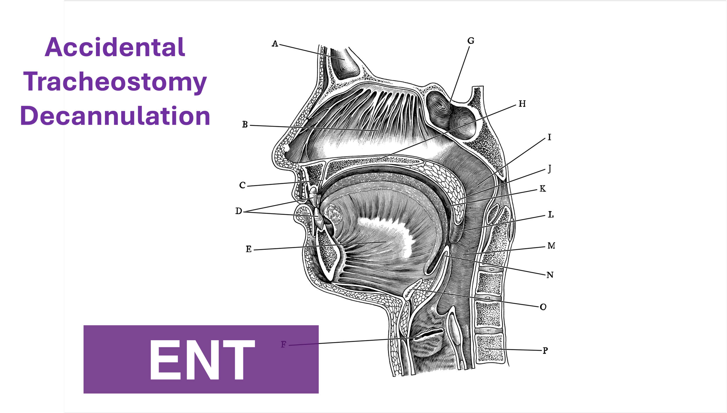
Intubating the Pregnant Patient in the ED

Written by: Priyanka Sista, MD (NUEM ‘20) Edited by: Steve Chukwulebe, MD (NUEM ‘19) Expert Commentary by: Samir Patel, MD

Written by: Priyanka Sista, MD (NUEM ‘20) Edited by: Steve Chukwulebe, MD (NUEM ‘19) Expert Commentary by: Samir Patel, MD


Expert Commentary

Tip for #1 - While 3-5 minutes of 100% oxygen is ideal to achieve denitrogenation, in an emergency 8 vital capacity breaths (maximal inhalation and exhalation) with a high FiO2 source is sufficient in a cooperative patient.

Tip for #2 - Airway edema is even worse in preeclamptic patients, and Mallampati scores acutely worsen DURING labor. Don’t bother with direct laryngoscopy and go straight to the video laryngoscope if it’s available.

Tip for #3 - In this scenario, the ideal LMA or supraglottic airway is one that includes a port for passage of an OG tube. Your pregnant patient in the ER with increased aspiration risk is not likely to be NPO for 8 hours like they are for anesthesiologists before surgery.

Tip for #4 - The rapid sequence dose of rocuronium is 1.2 mg/kg. You can immediately reverse rocuronium with sugammadex 16 mg/kg if necessary. For cost purposes, succinylcholine is still the best choice unless medically contraindicated.

Tip for #5 - According to ACOG, if cardiac arrest occurs in a woman greater than 23 weeks gestation, and there is no return of spontaneous circulation after 4 minutes of correctly performed CPR, a perimortem c-section should be performed with the goal of delivering the fetus by the fifth minute.

Samir Patel.PNG

Samir K. Patel, MD

Assistant Professor

Northwestern University Feinberg School of Medicine

Department of Anesthesiology


How To Cite This Post:

[Peer-Reviewed, Web Publication] Sista, P. Chukwulebe, S. (2021, Jan 18). Intubating the pregnant patient in the ED. [NUEM Blog. Expert Commentary by Patel, S]. Retrieved from http://www.nuemblog.com/blog/intubating-the-pregnant-patient.


Other Posts You May Enjoy

Posted on January 18, 2021 and filed under ENT, Airway.

Assisted Reproductive Technology

Written by: Jesus Trevino, MD (NUEM ‘19) Edited by: Keith Hemmert, MD (NUEM ‘18) Expert Commentary by: Lia Bernardi, MD

Written by: Jesus Trevino, MD (NUEM ‘19) Edited by: Keith Hemmert, MD (NUEM ‘18) Expert Commentary by: Lia Bernardi, MD

In 2015, assisted reproductive technology (ART) resulted in 72,913 live births, comprising approximately 1.6% of all infants born in the US (CDC 2017).  As ART is becoming increasingly common, it is important that emergency medicine providers are familiar with this treatment modality to appropriately diagnose and manage maternal complications.  This article will review the components and complications of ART that may present in the Emergency Department with a focus on the ovarian hyperstimulation syndrome (OHSS).

    ART encompasses therapies that address all causes of infertility, which span both the male factor (e.g., sperm motility) and female factor (e.g., mechanical, ovulatory).  In vitro fertilization is a common treatment strategy that overcomes reproductive barriers and involves: 1) controlled ovarian hyperstimulation, 2) oocyte retrieval, 3) oocyte fertilization and 4) oocyte implantation.  Below is an outline of these in vitro fertilization steps along with associated complications.

Controlled ovarian hyperstimulation

There are numerous protocols available to recruit ovarian follicles and they differ in the type and intensity of exogenous stimulation (Speroff).  Protocols are graded in order of increasing intensity (and success rate):

  • Natural cycle (i.e., no exogenous stimulation)

  • Minimal (i.e., clomiphene citrate)

  • Mild (i.e., clomiphene citrate + low-dose exogenous gonadotropins)

  • Aggressive (i.e., high-dose gonadotropins +/- gonadotropin-releasing hormone agonist or antagonist).

The most life-threatening complication of these strategies is ovarian hyperstimulation is OHSS (Adams):

  • Incidence - 0.5-5% of ART cycles; 0.1-2% involve severe presentations (Weinerman).

  • Pathophysiology - increased capillary permeability leads to edema, ascites, pleural and pericardial effusions; this inflammatory state may result in renal failure, respiratory failure and/or thromboembolism.

  • Timing - typically occurs within a week of exogenous HCG administration or in the peri-implantation period due to increases in endogenous HCG.

  • Risk factors - < 35 years, low BMI, gonadotropin-releasing hormone and/or analogues, hyperstimulation of ovarian follicles (detected via pelvic US), elevated estradiol levels

  • Presenting symptoms & signs - abdominal distention, rapid weight gain, peripheral edema, dyspnea, pulmonary edema/effusions, oliguria; minimize pelvic exams to avert ovarian cyst rupture and hemorrhage

  • Labs - +/- hyponatremia, AKI, estradiol > 3000 pg/mL (typically not practical in ED evaluation)

  • Management -

    • Criteria for outpatient management: normal VS, renal and hepatic labs, ovaries < 5 cm

    • Criteria for inpatient management: ovaries > 5 cm, ascites; admission is required for serial exams and pain control

    • Criteria for ICU: palpable ovaries, pleural effusions, ARDS, oliguria, hypotension, AKI, hepatic dysfunction; these patients require fluid resuscitation, +/- therapeutic thora/paracentesis, likely termination of ART cycle

    In addition, controlled ovarian hyperstimulation has an increased incidence of ovarian torsion - 0.08% without OHSS and 3% with OHSS (Weinerman).

Oocyte retrieval

This step is usually performed under conscious sedation with ultrasound-guided, transvaginal needle aspiration.  Complications may include (incidence %):

  • Vaginal puncture site bleeding (8%, Speroff)

  • Intraperitoneal bleeding (0.04-0.07%, Speroff)

  • Bowel perforation (0.04%, Weinerman)

  • Infection (0.3-0.6%, Speroff). Half of infections may present as TOA within 1-6 weeks after retrieval. 

Oocyte fertilization

The in vitro part - sperm meets egg.  There are rarely major maternal complications associated with this treatment step that present in the ED.

Oocyte implantation

Implantation occurs via a transcervical catheter under transabdominal US-guidance.  As the success rate for single fertilized embryos is 10-25%, multiple embryos are implanted to increase implantation yield (Adams).  Complications may include:

  • Multigestational pregnancy - 31-41% of IVF infants develop from multigestational pregnancies (Adams)

  • Ectopic pregnancy (0.7-4%, Speroff, Adams)
  • Heterotopic pregnancy (0.2-1%, Adams, Weinerman)
Lastly, patients with ART-facilitated pregnancies are at risk of thromboembolism (0.04-0.2%), especially in the presence of OHSS (4%, Weinerman).


Expert Commentary

This is an important review of complications that may arise in patients undergoing in vitro fertilization (IVF). Although patients who are planning for IVF can be assured that it is a generally safe process, there are medical issues that can occur throughout: during the ovarian stimulation phase, as a result of the oocyte retrieval or embryo transfer, or after a pregnancy is confirmed. 

During the ovarian stimulation phase of the process, few medical complications typically arise. The most common reason that a patient would seek emergency care would be for ovarian torsion. Given that ovarian size increases significantly, any patient who presents with severe pain during stimulation should be assessed for this.

The most likely time a patient undergoing IVF would present to the ED would be following the oocyte retrieval. Complications can take place after the conclusion of stimulation or due to issues from the retrieval itself.  One of the main issues that patients present for is ovarian hyperstimulation (OHSS). The most common time for this to happen is shortly after the oocyte retrieval, but patients may present in early pregnancy as well given that a rising hCG level worsens the syndrome. Evaluation and management of OHSS is reviewed succinctly above. Complications may also arise from the oocyte retrieval. Transvaginal ultrasound guided aspiration of ovarian follicles is performed using a needle that passes through the vagina. Possible complications include bleeding, infection, and/or or injury to other organs. Given that the needle is entering the vagina and the ovaries, bleeding can occur from the vagina or within the abdomen. Vaginal bleeding is typically quickly recognized and corrected prior to completing the procedure. Intraabdominal bleeding can be more difficult to identify and can potentially worsen after the patient is discharged. Patients with intraabdominal bleeding may present to the ED with symptoms of pain or hypotension. Imaging is generally helpful as part of the evaluation, but it is important to know that some bleeding generally occurs post-procedure even in an uncomplicated oocyte retrieval. Therefore imaging may reveal free fluid in a patient who does not have clinically significant ongoing bleeding.  It is also important to be aware that if a patient has intraabdominal bleeding, surgical intervention is not always required. Given the complexities of surgical exploration in these patients, the goal is to expectantly manage those with intraabdominal bleeding unless surgery is absolutely necessary. In addition to bleeding, infection, ovarian torsion and cyst rupture can also occur following oocyte retrieval. Finally, some patients will have extreme constipation due to the IVF process and may present with abdominal pain after oocyte retrieval as a result.  

Embryo transfers are a generally safe and low risk procedure. The procedure involves insertion of a sterile, soft catheter into the uterine cavity under ultrasound guidance with subsequent embryo release. Given the low risk nature of the procedure, complications after embryo transfer are very rare. Infection is theoretically possible, but unlikely. 

The final IVF related complications to consider are those that occur in pregnancy. As ectopic pregnancies are possible after IVF, any pregnant woman who presents with symptoms concerning for an extrauterine pregnancy should be evaluated accordingly. Heterotopic pregnancies can also occur and should remain on the differential diagnosis if a woman has concerning pain after an intrauterine pregnancy is confirmed. Given that a woman’s ovaries remain enlarged after ovarian stimulation if a pregnancy is achieved, ovarian torsion should also be considered in patients with abdominal pain. Another complication that can develop at any point during the IVF treatment process, including during pregnancy, are VTEs. Given supraphysiolgic estrogen levels that occur due to ovarian stimulation, providers must bear in mind that this complication can arise.  

Efficient recognition and treatment of the complications that can result from the IVF process are more likely when providers are well educated. Hopefully this review will improve the ability for patients undergoing IVF to be evaluated and treated most effectively when they present to the ED.

image-asset.png
 

Lia Bernardi, MD

Assistant Professor

Department of Obstetrics and Gynecology

Feinberg School of Medicine


How to Cite This Post

[Peer-Reviewed, Web Publication] Trevino, J, Hemmert, K. (2020, May 11). Assisted Reproductive Technology. [NUEM Blog. Expert Commentary by Bernardi, L]. Retrieved from https://www.nuemblog.com/blog/assisted-reproductive-tech


References

Yang-Kauh C. Complications of gynecologic procedures, abortion, and assisted reproductive technology. Chapter 125.  Emergency Medicine, Ed 2, 2013.

Speroff L, Fritz MA. Assisted Reproductive Technologies. Chapter 32. Clinical Gynecologic Endocrinology and Infertility, Ed 8, 2011.

Weinerman R, Grifo J. Consequences of superovulation and ART procedures. Semin Reprod Med. 2012 Apr;30(2):77-83.

ART Success Rates [Internet]. Centers for Disease Control and Prevention. Centers for Disease Control and Prevention; 2017 [cited 2017May2]. Available from: https://www.cdc.gov/art/artdata/index.html

Posted on May 11, 2020 and filed under Obstetrics & Gynecology.

Preeclampsia

preeclampsia image.png

Written by: Priyanka Sista, MD (NUEM PGY-4) Edited by: Matt Klein, MD (NUEM ‘18) Expert commentary by: Shannon Lovett, MD


Preeclampsia.png

Expert Commentary

Thank you for this succinct guide to the diagnosis and management of preeclampsia in the ED. 

“The eyes do not see what the mind does not know…..”. The biggest pitfall in the management of preeclampsia in the ED, is failing to consider and recognize the diagnosis. Recognition and prompt treatment of preeclampsia in the ED setting can be challenging due to the variety of presenting complaints. It is important to note that preeclampsia may occur anytime from 20 weeks gestation up to 6 weeks postpartum. 

Postpartum preeclampsia tends to be more diagnostically challenging and depending on your facility, these patients are more likely to present to the ED than pregnant patients who often present to their obstetrician or to labor and delivery. Preeclampsia in the postpartum period most frequently occurs in the first 48 hours after delivery, but should be considered up to 6 weeks postpartum. Patients with postpartum preeclampsia often do not have hypertensive disease or preeclampsia during pregnancy. 

The complaints associated with preeclampsia may be broad and vague- including but not limited to: headache, vision changes, swelling or rapid weight gain, nausea and vomiting, shortness of breath, and abdominal pain. Consider preeclampsia or eclampsia in the critical female patient that arrives in the ED with little known history- for example actively seizing, or in respiratory distress with pulmonary edema. 

The treatment of preeclampsia can be broken down into three parts: treating the hypertension, reducing the risk or recurrence of seizures, and delivery of the fetus and the placenta. In the ED- our focus is on the first two, and involving our obstetric colleagues immediately. Blood pressure is most commonly treated with labetolol or hydralazine IV in the ED, and Mag should be given immediately for seizure prophylaxis (or to reduce recurrence of seizures in eclampsia). 

Lastly, our obstetric and gynecology colleagues at ACOG have recognized the frequency that postpartum patients present to the ED, and have created this ED checklist that can be used as a reference for the management of postpartum preeclampsia-  https://www.acog.org/-/media/Districts/District-II/Public/SMI/v2/19sm03a170703PPPreeclamCheckED1.pdf?dmc=1&ts=20190327T1949153065. Preeclampsia is a syndrome with potentially devastating consequences to mother and baby, and our early recognition and treatment can improve outcomes.

Picture1.png
 

Shannon Lovett, MD

Associate Professor

Loyola University Medical Center


How To Cite This Post

[Peer-Reviewed, Web Publication] Sista P, Klein M. (2019, Sept 23). Preeclampsia. [NUEM Blog. Expert Commentary by Lovett S]. Retrieved from http://www.nuemblog.com/blog/preeclampsia.


Other Posts You May Enjoy

Posted on September 23, 2019 and filed under Obstetrics & Gynecology.

Ovarian Hyperstimulation: Not Your Ovarian Average Cyst

Pregnancies resulting from assisted reproduction are more complicated, with higher rates of ectopic, heterotopic and multifetal pregnancies, in addition to higher rates of abortions and premature deliveries. Other complications include venous thromboembolism as well as ovarian hyperstimulation syndrome. These are high risk pregnancies that will present to your emergency department and you should be aware of key management principles.