Posts tagged #cervical fracture

The Top NUEM Blog Posts of 2019

NUEM Blog 2019 Report.png

Edited by: Maury Hajjar, MD (NUEM PGY-2), Niki Patel, MD (NUEM PGY-2), Vytas Karalius, MD, (NUEM PGY-2), Justine Ko (NUEM PGY-3)  Expert commentary by:  Seth Trueger, MD


Happy New Year, Everyone!

Let’s take a look back at the 2019 NUEM Blog!


NUEM Blog Year End 2019.png


The Top Ten NUEM Blog Posts of 2019

10. REBOA

Recent grads Andew Cunningham, Bill Burns, and Trauma/EM doc Zaffer Qasim walk us through the ever-popular and sublimely named REBOA.

9. The Seriousness of Deliriousness

Thorough discussion of the important but easy to overlook issue of delirium in the ED by Nery Porras, recent grad and current neuro critical care fellow Katie Colton and geriatrician Lee Lindquist.

8. Pelvic Fractures

Justine Ko, Terese Whipple, and Matt Levine walk is through pelvic fractures and the important associated injuries.

7. Verbal Deescalation in the ED

Vidya Eswaran, Zach Schmitz, Abiye Ibiebele, NUEM-blog-founder Michael Macias, recent grad Arthur Moore, and John Bailitz review the complex but important topic of verbal deescalation in the ED.

6. Visual Guide to Splinting

Recent grad and current Stanford med ed fellow, recentish grad John Sarwarkand remote grad Matthew Pirotte provide a surprising amount of information in a small amount of words & images.

5. Post-Intubation Management

It’s easy to fall into the trap getting the tube & high-fiving and walking away; Andra Farcas, recent grad and current Air Force doc Paul Trinquero and recentish grad Andrew Pirotte walk is through the steps to post-intubation management.

4. Flexor Tenosynovitis

Thorough and concise review of flexor tenosynovitis by Kevin Dyer and recent grad Adnan Hussain, featuring expert commentary hand surgeon Avi Giladi (who also had the #4 post in 2018).

3. Tetracaine

Nice review by Jonathan Hung and recent grad and current med ed fellow Matt Klein of an Annals study showing a safe approach to tetracaine for corneal abrasions. Won’t solve the controversy but does include an expert commentary by @DGlaucomflecken

2. Intubation Positioning: Beyond the Sniffing Position

Unfortunately, optimal positioning is not always optimally executed in the ED; this post reviews both “standard” sniffing position and ramping, which, if nothing else, will help push us to better position our patients. Very nice work by recent grad and current neuro critical care fellow Katie Colton, and recent grad Charles Caffrey, and recentish grad Andrew Pirotte!

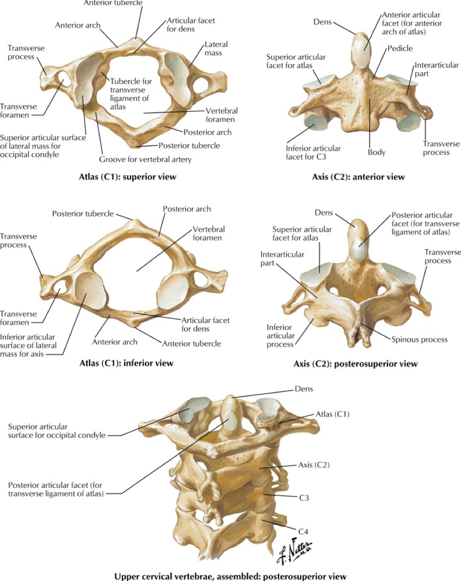
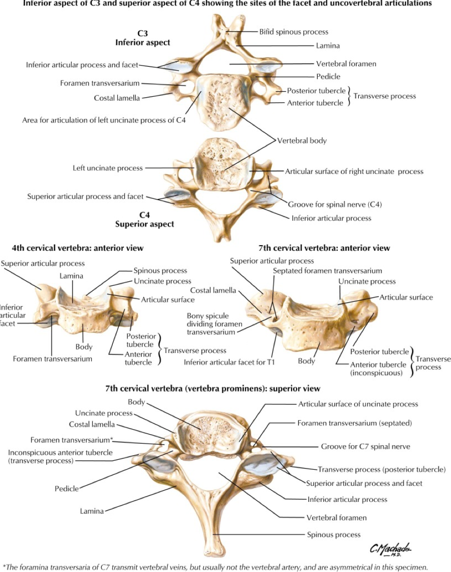
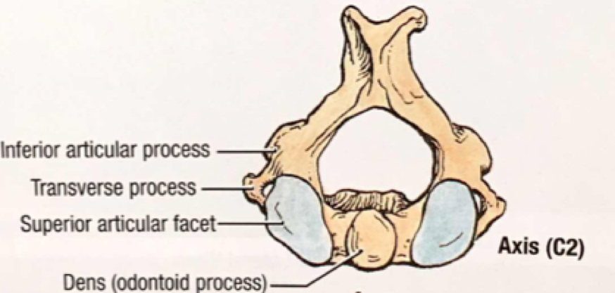
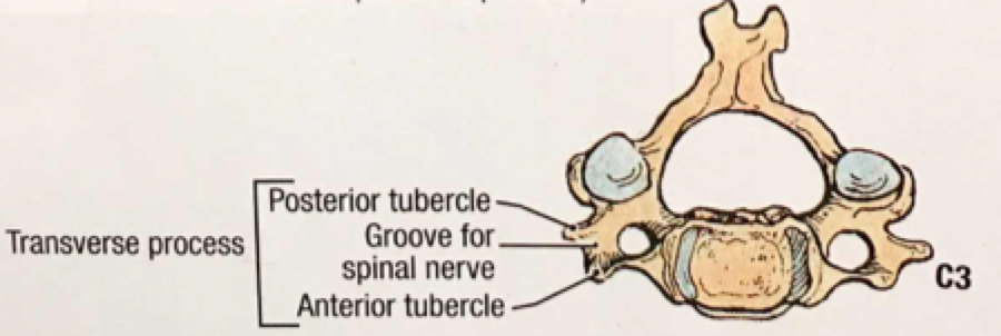
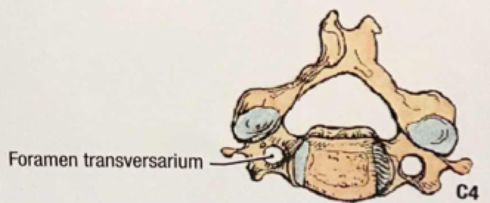
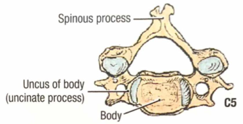
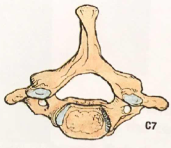
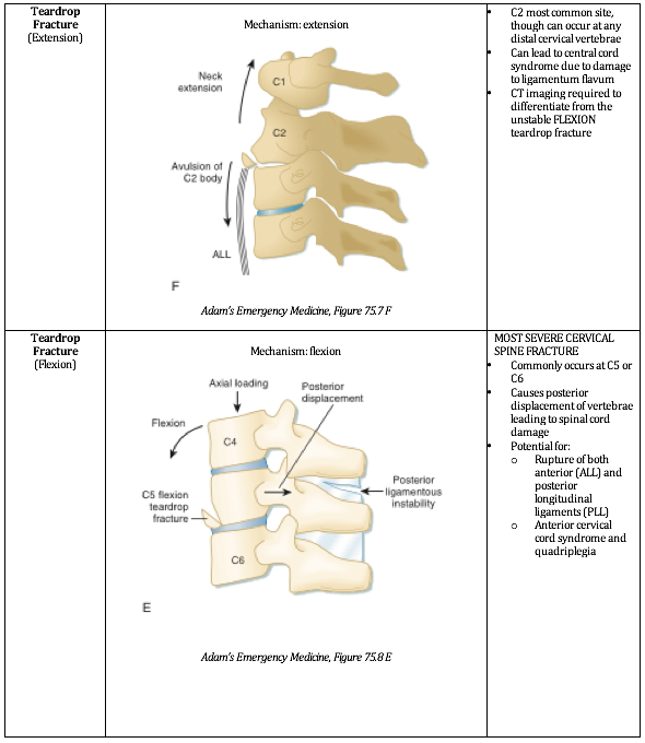
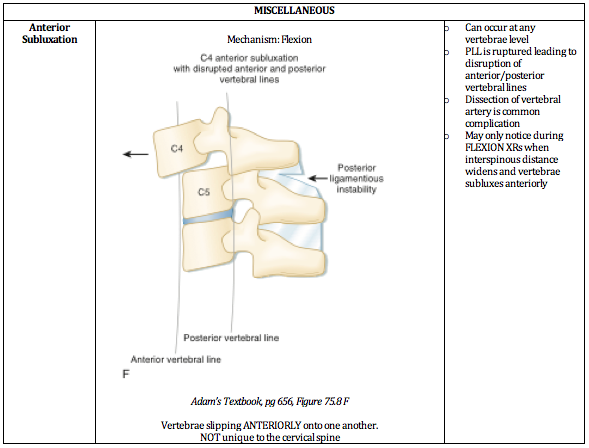
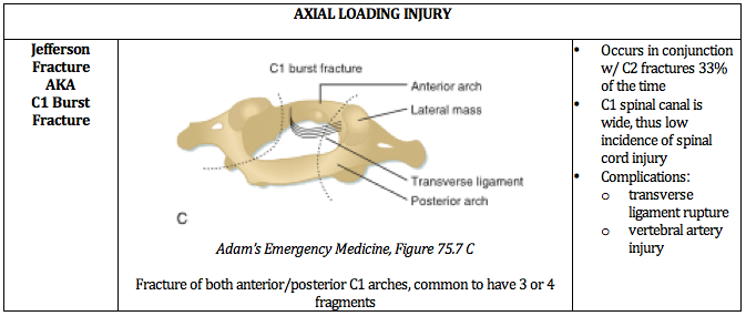
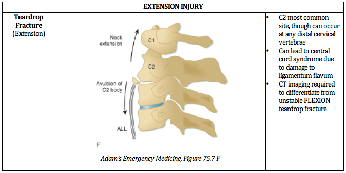
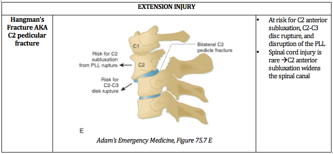
1. Unstable C-Spine Fractures

The top spot goes to a very nice succinct review by recent grads Sarah Sanders and Alison Marshall featuring beautiful images and a commentary by NLFH faculty Steve Hodges.


 How to cite this post

[Peer-Reviewed, Web Publication]  Hajjar M, Patel N, Karalius V, Ko J, (2019, December 30 ). The Top NUEM Blog Posts of 2019.  [NUEM Blog. Expert Commentary by Trueger S ]. Retrieved from http://www.nuemblog.com/blog/top-ten-2019.


Other posts you might enjoy…


C-spine Clearance with Negative CT: Are We There Yet?

Screen Shot 2019-05-07 at 10.46.34 AM.png

Written by: M. Terese Whipple, MD (NUEM PGY-3) Edited by: Quentin Reuter, MD (NUEM ‘18) Expert commentary by: Matthew Levine, MD


We have excellent decision rules for clinically clearing cervical spine injury in low risk patients without imaging. However, a frustrating situation arises when a CT of their c-spine is obtained and negative, but they are having persistent midline pain. What do we do then? Are we forced to order an MR of the c-spine even when they have no neurological deficits and our gestalt tells us there is no clinically significant injury? MR often means admission, worsening of already overwhelming ED crowding, and unhappy patients when they cannot remove the c-collar for at least several more hours. Recent data and recommendations suggest that this may not be the case; a negative CT may be enough to rule out clinically significant injury. This blog post will explore some of the historical and recent data on the subject of cervical spine clearance after CT scan alone.  


There has been great historical debate over the best management for patients with persistent midline pain after negative CT, however that evidence is beyond the scope of this post. Current common practice and the recommendation of the American College of Radiology leads down the path of cervical spine MR when this situation arises [1]. Due to the cumbersome logistics of MR, much work has been done to determine if MR truly adds value to the patient’s workup. Is MR catching clinically significant injury missed by CT that changes clinical management? The majority of studies have concluded that the answer to that question is no.  

In 2015 the Eastern Association for the Surgery of Trauma (EAST) sought to tackle this question by reviewing all studies to date examining C-spine evaluation in obtunded patients [2]. They evaluated 11 studies with a total of 1718 obtunded patients who underwent C-Spine imaging with CT. None were ultimately found to have unstable fractures or unstable ligamentous injury missed by CT. There was a 9% incidence of stable injuries missed on CT and found on follow up MR, flex-x, upright XR, or clinical follow up. They found a cumulative 100% NPV for unstable C-Spine injury with CT and 91% NPV for stable injury. They did rate the quality of evidence as low for various reasons, including non-comparable imaging protocols, inconsistently reported and variable outcomes, publication bias, and an overall inability to perform a meta-analysis with the data.  However, they rated the data from which they derived the NPV as moderate quality as the NPV was consistently 100% throughout all of the trials. Based on their analysis they provided the following recommendation for obtunded blunt trauma patients:

“We conditionally recommend cervical collar removal after negative high-quality c-spine CT scan results alone.”

 They went on to further clarify,   

“It should be acknowledged that cervical collar removal can result in neurological change and even paralysis, although this may be underreported in the literature. However we cannot continue indiscriminate two-stage sequential screening for C-spine injuries if the injury rate is near 0% for the first test and the second adjunctive test results in false positives and inconsistent treatment plans.”

But the real question that is more pertinent to us as EM physicians (obtunded MR’s are usually dealt with upstairs), is:  if we can remove the c-collars of obtunded patients after negative CT, why couldn’t that be extrapolated to awake patients?  Well, they commented on that too:   

“Therefore, if collars are to be removed in a high risk obtunded population […] cervical collar removal can be logically argued for any population-obtunded or not.” [2]

 They finally call for multicenter prospective research on the subject, again citing the low quality of evidence that they used for their recommendation. That call was answered in 2017 by the Western Trauma Association. The group completed a multi institution trial with 10,000 patients who were getting a CT for evaluation of cervical spine injury prospectively enrolled at 17 centers [3]. They found only 3 CT scans that missed clinically significant injury (.03%). All of those patients had focal neurological abnormalities on exam. There was no clinically significant injury missed by CT and exam combined. CT scan alone had an NPV of 99.97%, and an NPV of 100% when combined with clinical exam. Therefore, they proposed this diagnostic algorithm:

algorithm.png


Most trials have found similar results, with a few exceptions. Two trials prior to the publication of the Western Trauma Association (WTA) paper found that CT missed a few clinically significant injuries in patients with no neurological symptoms. Both trials enrolled significantly fewer patients than the WTA paper, and only enrolled patients with negative CT who would be evaluated with MR, meaning they couldn’t comment on the overall sensitivity of CT in unstable c-spine injury. The ReConect trial in 2016 found 5 of 767 patients (.6%) with injuries requiring surgical intervention that were missed on CT [4].  Another study with similar methods published in Annals of Emergency Medicine in 2011 evaluated those who had a negative CT but were MR’ed for persistent midline C-Spine tenderness [5]. They found that out of 178 patients, 5 had injury requiring operative management that was missed on CT but found on subsequent MR (2.8%) [5].  The Annals paper is certainly an outlier, with a considerably higher rate of missed clinically significant injury than the remainder of the literature, with rates usually between 0-1% [6-18]. The authors believe this may be due to more stringent methodology.  For instance, they required MR to be performed within 48 hours when it is the most sensitive for edema, and only enrolled patients with midline tenderness rather than subjective pain [5].  While this may be true, the results have not been replicated in subsequent studies.


With the publication of the WTA paper, evidence certainly seems to be tipping in favor of CT clearance of cervical spine in neurologically intact patients. However, a few questions remain. In every study discussed here, MR resulted in discharge with hard collar in a portion of patients. Indications ranged from stable injury to persistent pain with no evidence of injury on MR. It is unclear whether hard collar placement makes a difference in the clinical course of these patients, if their stable injuries would have become unstable without it, or if it has any long term impact on outcomes such as chronic pain. This is an important question not yet adequately addressed in the literature.  The majority of these trials were also completed at trauma centers with radiologists well trained in reading c-spine imaging and high quality CT scanners. It could be difficult to generalize this data to centers with older scanners or whose radiology departments are not as expert in trauma radiology.

Incredibly high quality and reproducible evidence is required to change practice when high stakes, such as potentially missed cervical spine injury, are involved. So far we have multiple trials showing an NPV of close to 100% when CT and good neurological exam are combined, and the conditional recommendation by the EAST group. Time will tell if recommendations in the future remove the “conditional” portion as CT technology continues to improve, further studies with stringent methodologies are conducted, and the results of the WTA paper are hopefully replicated.


Expert Commentary

Thank you Dr Whipple for that really practical review of a real-life common clinical question we face all the time: Can we remove the collar?  Some important takeaways are:

  1. There is a robust and growing body of evidence that removing the collar after a negative high-quality CT is safe if the patient is neurologically intact.

  2. This practice is endorsed by two major trauma organizations, EAST and WTA. 

The endorsement by respected major trauma societies is important in translating evidence into practice.  It seems like all that is left at this point for widespread implementation is overcoming culture.  This would likely require addressing the outlier studies listed by Dr Whipple to win over those still skeptical.  Part of overcoming culture would involve buy-in from neurosurgical societies.  What do neurosurgical societies say regarding clearing these patients?  There are many instances in which a patient is discharged with recommendations from the neurosurgeon to wear a hard collar despite a negative CT and MRI.  On the surface this seems like defensive medicine and impractical for the patient.  Is the patient really going to comply with this until follow up?  Is this collar really protecting them and preventing further injury which, after negative CT and MRI and with a normal neuro exam, seems exceedingly unlikely?  Does evidence support this practice?

In the end, decision rules should be used when you want evidence to support your clinical decisions, such as removing the C collar after negative imaging in a neurologically intact patient.  Do not use decision rules, however, to overturn or replace sound clinical judgement.  If there is something about a case that makes you still feel like you could be missing an outlier injury by removing the collar, listen to that voice inside of you. It is that sound clinical judgement that will guide you through your career, not decision rules.

Matt_Levine-33 (1).png
 

Matthew Levine, MD

Northwestern Medicine, Assistant Professor of Emergency Medicine


How To Cite This Post

[Peer-Reviewed, Web Publication] Whipple T, Reuter Q. (2019, May 13). C-spine clearance with negative CT: Are we there yet? [NUEM Blog. Expert Commentary by Levine M]. Retrieved from http://www.nuemblog.com/blog/cspine-clearance-ct


Other Posts You May Enjoy


References:

  1. American College of Radiology. ACR appropriateness criteria on suspected spine trauma. Available at: http://www.acr.org.

  2. Patel MB, et al. Cervical spine collar clearance in the obtunded adult blunt trauma patient: A systematic review and practice management guideline from the Eastern Association for the Surgery of Trauma. J Acute Care Trauma Surgery. 789(2): 432-441.

  3. Inaba, K et al. Cervical Spine Clearance: A Prospective Western Trauma Association Multi-Institutional Trial. J Trauma Acute Care Surg. 2016 Dec: 81(6): 1122-1130.doi: 10.1097/TA.0000000000001194

  4. Maung A, et al. Cervical spine MRI in patients with negative CT: A prospective, multicenter study of the Research Consortium of New England Centers for Trauma (ReCONECT). J Trauma Acute Care Surg. 82 (2): 263-269.

  5. Ackland HM, et al. Cervical Spine Magnetic Resonance Imaging in Alert, Neurologically Intact Trauma Patients With Persistent Midline Tenderness and Negative Computed Tomography Results. Ann of Em Med. 2011 Dec. 58 (6): 521-530.

  6. Chew B, et al. Cervical spine clearance in the traumatically injured patient: is multidector CT scanning sufficient alone? J Neurosurg Spine. 2013. 19: 576-581

  7. Bush L, et al. Evaluation of cervical spine clearance by computed tomographic scan alone in intoxicated patients with blunt trauma. JAMA Surg. 2016; 151 (9): 807-813

  8. D’Alise  et al. Magnetic resonance imaging for the evaluation of the cervical spine in the comatose or obtunded trauma patient. J Neurosurgery  (Spine 1) 1999; 91:54-59.

  9. Resnick S, et al. Clinical relevance of magnetic resonance imaging in cervical spine clearance: a prospective study. JAMA Surg. 2014; 149 (9): 934-9.

  10. Menaker J, Philp A, Boswell S, Scalea TM. Computed tomography alone for cervical spine clearance in the unreliable patient--are we there yet? J Trauma. 2008; 64(4):898–903.

  11. Chew BG, Swartz C, Quigley MR, Altman DT, Daffner RH, Wilberger JE. Cervical spine clearance in the traumatically injured patient: is multidetector CT scanning sufficient alone? Clinical article. J Neurosurg Spine. 2013; 19(5):576–81.

  12. Como JJ, Leukhardt WH, Anderson JS, Wilczewski PA, Samia H, Claridge JA. Computed tomography alone may clear the cervical spine in obtunded blunt trauma patients: a prospective evaluation of a revised protocol. J Trauma. 2011; 70(2):345–9. discussion 9-51.

  13. Khanna P, Chau C, Dublin A, Kim K, Wisner D. The value of cervical magnetic resonance imaging in the evaluation of the obtunded or comatose patient with cervical trauma, no other abnormal neurological findings, and a normal cervical computed tomography. J Trauma Acute Care Surg. 2012; 72(3):699–702.

  14. Schuster R, Waxman K, Sanchez B, Becerra S, Chung R, Conner S, Jones T. Magnetic resonance imaging is not needed to clear cervical spines in blunt trauma patients with normal computed tomographic results and no motor deficits. Arch Surg. 2005; 140(8):762–6.

  15. Anekstein Y, Jeroukhimov I, Bar-Ziv Y, Shalmon E, Cohen N, Mirovsky Y, Masharawi Y. The use of dynamic CT surview for cervical spine clearance in comatose trauma patients: a pilot prospective study. Injury. 2008; 39(3):339–46.

  16. Brohi K, Healy M, Fotheringham T, Chan O, Aylwin C, Whitley S, Walsh M. Helical computed tomographic scanning for the evaluation of the cervical spine in the unconscious, intubated trauma patient. J Trauma. 2005; 58(5):897–901.

  17. Harris TJ, Blackmore CC, Mirza SK, Jurkovich GJ. Clearing the cervical spine in obtunded patients. Spine (Phila Pa 1976). 2008; 33(14):1547–1553.

  18. Steigelman M, Lopez P, Dent D, Myers J, Corneille M, Stewart R, Cohn S. Screening cervical spine MRI after normal cervical spine CT scans in patients in whom cervical spine injury cannot be excluded by physical examination. Am J Surg. 2008; 196(6):857–862.

Posted on May 13, 2019 and filed under Trauma.

Unstable Cervical Spine Fractures

Screen Shot 2019-01-02 at 5.45.38 PM.png

Written by: Sarah Sanders, MD (NUEM PGY-4) Edited by: Alison Marshall, MD (NUEM Alum ‘17) Expert commentary by: Steve Hodges, MD


Fractures of the cervical spine are injuries that must be approached with caution. Some are stable, some are unstable, and mismanagement can lead to life-altering sequelae. Remembering which fractures fit into which category is imperative for optimum emergency department care.

A quick review of cervical spine anatomy is a helpful starting point:

All the cervical spine anatomy images are credited to Netter, FH Atlas of Human Anatomy, Sixth Edition.

1.png
2.png

All the cervical spine anatomy diagrams are credited to Agur, AMR & Dalley, AF of Grant’s Atlas of Anatomy: Twelfth Edition.

The common mnemonic “Jefferson Bit Off A Hangman’s Thumb,” is used to remember the unstable fractures, which will be reviewed in this post.

Screen Shot 2019-01-05 at 10.59.28 PM.png
Screen Shot 2019-01-05 at 11.05.37 PM.png
Screen Shot 2019-01-05 at 11.09.01 PM.png
Screen Shot 2019-01-05 at 11.10.04 PM.png
Screen Shot 2019-01-05 at 11.10.49 PM.png

Ultimately, understanding the mechanism of injury is crucial in identifying and accurately managing these injuries. The below PV cards are organized by mechanism and tailored down to be an on-shift reference.

In conclusion, cervical spine injuries required a high index of suspicion and caution by the emergency medicine physician as their variability and potential for neurological impairment is high. Hopefully this review can provide you with additional insight and ease of memory when the next Level 1 trauma rolls through the door.  


Expert Commentary

Thanks for this insightful post.  You've done a really nice job in laying out the most salient points.  It is important to have an understanding of the anatomy and this is a great review.  Knowing the factors that put people a high risk for injury is also paramount.   Certain chronic disease states and anatomic variances, as you note, do put select patient populations at risk for specific injury.  The mechanism of injury is something we always talk about, but when it comes to neck trauma we need to really pay attention to all available history including paramedic reports, or even cell phone video, to get the best possible picture or the mechanism of injury.   I can not stress enough the importance of a detailed neurological exam including sensation(s) and reflexes.  Any asymmetric finding should raise your level of suspicion for severe injury.   Moving to advanced imaging is especially important if there is a complaint of a focal neurological deficit; be that transient, subjective or blatantly obvious. 

Athletes or motorcyclist who have suspected cervical spine injury, who have protective shoulder pads and/or helmets pose a unique challenge.  Eventually the protective devices are going to need to be removed.   There are many opinions on how best to do this and whether an x-ray should be done before attempting the removal of protective gear.   From personal experience; it can be difficult to remove protective gear;  I recommend getting "all hands on deck" and using an methodical slow organized approach.  More recent thought is that cervical spine imaging should incorporate procedures for removal of equipment before initial radiographic evaluation.[1]  Once the gear is removed a c-collar should then be applied and you can proceed with imaging.

Recommendations for imaging the cervical spine for trauma has changed quit a lot over the last several years.  The National Emergency X-Radiography Utilization Study (NEXUS) and the Canadian C-Spine Rule (CCR) have been validated and have allowed our practice to advance such that we can effectively practice clinical medicine.  However, a word of caution on using these criteria with patients who could be impaired.  Sometimes the mild dementia, delirium or subtle drug, alcohol intoxication can lead us astray when we rely solely on these criteria.   The cross table lateral films and specifically flexion/extension views have fallen out of favor.  Most patients without focal neurological complaint or deficit are imaged with plain CT.  If your patient has a focal neurological complaint or deficit, a suspected ligamentous or disc injury an MRI should be done.  Depending on the exam and risk factors I would consider either a CTA or and MRA to evaluate for vascular injury.  

You asked about c-collars specifically.  What is available to you will be somewhat hospital - vendor specific.  I prefer the Aspen or the Miami collar, they are very similar in function overall and superior to the pre-hospital EMS ones.  When a patient has to be transported to another facility; make sure that the patient has full immobilization with back board and head-side blocks with the collar and head secured to the side blocks.    An immobilized patient that requires intubation can make an easy air way difficult and a difficult airway terrifying.  Make sure you have all your equipment prepared, including a surgical method, before you intubate.  The person holding c spine immobilization needs to knows their role.....don't let go and don't move.  This is the time when a video assisted intubation should be used.  Use either the intubating bronchoscope or a video laryngoscope.   This article talks a bit about managing airway in cervical spine injury and is a nice reference.[2]

Closing thoughts: maintain a high level of suspicion for injury in the setting of a focal neurological deficit, immobilize early immobilize often and don't be shy about intubating before transferring. 

  1. https://doi.org/10.1067/mem.2001.116333  Annals of Emergency Medicine; Baldwin et. al., "Football protective gear and cervical spine imaging" July 2001 Volume 38, Issue 1, Pages 26–30

  2. 10.4103/2229-5151.128013    International Journal of Critical Illness and Injury Science; Austin et. al., "Airway management in cervical spine injury" Jan-Mar; 4(1): 50–56

hodges.png
 

Steven W. Hodges, MD, FACEP

Assistant Medical Director

Northwestern Lake Forest Hospital


How To Cite This Post

[Peer-Reviewed, Web Publication] Sanders S, Marshall A (2019, January 21). Unstable Cervical Spine Fractures [NUEM Blog. Expert Commentary by Hodges S]. Retrieved from http://www.nuemblog.com/blog/cervical-spine-fractures.


Other Posts You May Enjoy


References

  1. Adams, J. Lin, M. Mahadevan, SV. “Spine Trauma and Spinal Cord Injury.” Section VIII, Chapter 75. Emergency Medicine: Clinical Essentials. Second Edition. P652 - 657.  

  2. Agur, AMR; Dalley AF. Grant’s Atlas of Anatomy. Twelfth Edition. Chapter 4: Back. 2009.

  3. Bergenheim, AT, Forssell, A. “Vertical Odontoid Fracture. Case Report.” Journal of Neurosurgery. Vol 74 (4) p665-667. 1991.

  4. Netter, F. Atlas of Human Anatomy. Section Head & Neck. Sixth Edition. 2014.

  5. Wheeless, CR. “Cervical Spine.” Wheeless Textbook of Orthopedics by Duke University. April 26 2016. 2 Jan 2017. http://www.wheelessonline.com/ortho/cervical_spine 

Posted on January 21, 2019 and filed under Trauma.