Posts tagged #c-spine clearance

C-Spine Intubation

Written by: Daniel Levine, MD (NUEM ‘24) Edited by: Zach Schmitz (NUEM ‘21)  Expert Commentary by: Matt Levine, MD

Written by: Daniel Levine, MD (NUEM ‘24) Edited by: Zach Schmitz (NUEM ‘21)
Expert Commentary by: Matt Levine, MD


The “Evidence” Behind Manual In-Line Stabilization During Intubation of Trauma Patients

Background

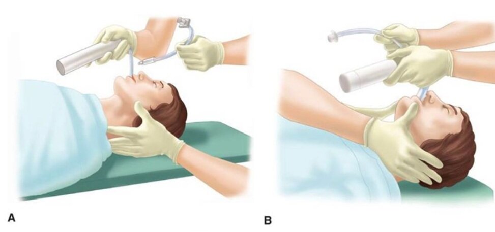
Even in the absence of frank head and neck trauma that may cause bleeding or distortions in usual anatomy, trauma patients present challenging airways because of cervical spine precautions. Standard-of-care technique according to EAST (Eastern Association for the Surgery of Trauma), West (Western Trauma Association), and ATLS (Advanced Trauma Life Support) guidelines for intubating acute trauma patients with known or potential cervical spine injury involves manual in-line stabilization (MILS). (1,2) This is a two-person technique whereby one provider performs laryngoscopy while another holds the patient’s neck in place.  The two most common techniques for this procedure are depicted below, one in which the stabilizer crouches down at the head of the bed (A), and the other where the stabilizer approaches from the side of the bed (B). (3)

(photo from Strange and Schafermeyer's Pediatric Emergency Medicine, 4th edition) (3)

(photo from Strange and Schafermeyer's Pediatric Emergency Medicine, 4th edition) (3)

Evidence

Like many practices in medicine, MILS has never been studied in randomized controlled trials, and the practice stems more from weak data and expert opinion. (4) The practice of spinal stabilization began during the 1970s after a retrospective review published in 1979 of 300 patients with acute cervical injuries who presented to Johns Hopkins hospital between 1950 and 1972. Although the main focus was on the effects of laminectomy and steroids, the review also found that 11 of the 300 patients developed neurologic deficits after reaching the hospital. Of the 11 patients, 7 developed these deficits “after neck immobilization was not provided”, with no clear comment as to whether immobilization was not provided during intubation or during some other process of the patient’s care. (5) These observations led to concerns that mobilization of the neck during intubation may worsen spinal cord injury, so manual in-line stabilization became standard of care in the 1980s.

Existing data for spinal stabilization comes from trials of cadaveric models, case series, and uninjured patients. Data from cadavers with post-mortem surgically created cervical spine injuries have shown mixed results on the effects of the amount of measured movement at the injured site with versus without MILS. For example, a 1993 study by Donaldson et al. found higher degrees of subluxation and angulation at C5-C6 during orotracheal intubation without MILS compared to with stabilization in five cadaveric specimens with injuries created in that area. (6) On the other hand, a 2001 Lennarson et al. study on cadavers found MILS significantly increased subluxation in C4-C5 during the same movements. (7) While it is somewhat counterintuitive that performing MILS might be associated with increased cervical motion, this may be explained by the laryngoscopist’s need to apply greater force with the laryngoscope in order to obtain an adequate view. This is what Santoni et al. (2009) found in a matched control study of 9 patients undergoing elective surgery. The patients in this study underwent two sequential laryngoscopies and oral intubations with a Macintosh 3 blade. Pressure transducers attached to the end of the blades detected higher maximum pressures at best glottic view with MILS compared to without. (8)

What is more clear in the literature on MILS than its effect on cervical motion is that it impairs glottic visualization and subsequent first pass intubation success. In the aforementioned Donaldson study on cadavers, MILS was shown to have a negative impact on Cormack-Lehane (CL) grade. (6) Similarly, in the aforementioned Santoni et al. study of 9 patients who underwent two sequential intubations with and without MILS, glottic visualization was worse in 6 patients with MILS, and intubation failure occurred in 2 of these 6 patients compared to no intubation failures among these patients when the intubation was performed without MILS. Thiboutot et al. (2008) performed a randomized controlled trial that further demonstrated this effect. In their study, 200 elective surgical patients were randomized to receive MILS or no MILS, and the primary endpoint was rate of failed intubation at 30 seconds with a Mac 3 blade. The rate of failed intubation was half in the MILS group (50%, 47/94), significantly higher compared to the control group (5.7%, 6/105). When they released manual in-line stabilization, they were able to intubate all patients. Secondary outcomes of rate of CL grade 3-4 as well as mean latency to successful intubation were also both significantly higher in the MILS group. (9) Additionally, these data were from patients undergoing elective surgery being intubated in the controlled OR setting by anesthesiologists. It is likely that the rate of failed intubation would be even higher in the chaotic emergency department environment with an acutely injured trauma patient. While 30 seconds is a somewhat arbitrary cutoff for a failed intubation, and it is quite possible many of the patients in the MILS group who “failed” may have been successfully intubated if a longer cut-off time were chosen, hypoxia caused by failed or delayed intubation is associated with poor outcome in central nervous system injury. (10)

Conclusion

In an ideal world, a large-scale randomized controlled trial of trauma patients studying the effects of MILS on mortality and important functional neurologic outcomes would help elucidate the utility of this commonly accepted practice. However realistically, completing such a study has significant obstacles. Cervical spine injuries are relatively rare (4% of trauma injured patients)4 and only a small fraction of those cases involve unstable injuries with potentially salvageable cord function. Thus, a study with sufficient power to detect any meaningful difference in outcomes would take many thousands of patients, many trauma centers, and many years to complete. Perhaps an even larger hurdle is the ethical and medicolegal hurdle of randomizing patients to not getting MILS and possibly putting them at risk of quadriplegia. (4) So what’s a clinician to do when faced with the common scenario of having to intubate a trauma patient? I personally like the approach that Dr. Reuben Strayer discusses in his video “Advanced Airway Management for the Emergency Physician” (link below). (11) To summarize his strategy:

Screen Shot 2021-10-17 at 10.22.17 AM.png

*The exception: in the rare situation where the patient has a highly suspected (e.g. obvious bony deformity, focal neurologic deficit) or known cervical spine injury, Dr. Strayer recommends lowering the threshold to perform a cricothyroidotomy. Additionally, he recommends considering an awake intubation approach in these patients. 


Another consideration is intubating using a hyper-angulated video GlideScope, which has been shown to have improved CL views and high rates of intubation success in c-spine immobilized patients. (12) That said, occasionally equipment availability or a bloody airway may preclude the use of video laryngoscopy in the trauma setting.

References

  1. Mayglothling J, Duane TM, Gibbs M, McCunn M, Legome E, Eastman AL, Whelan J, Shah KH; Eastern Association for the Surgery of Trauma. Emergency tracheal intubation immediately following traumatic injury: an Eastern Association for the Surgery of Trauma practice management guideline. J Trauma Acute Care Surg. 2012 Nov;73(5 Suppl 4).

  2. Brown CVR, Inaba K, Shatz DV, Moore EE, Ciesla D, Sava JA, Alam HB, Brasel K, Vercruysse G, Sperry JL, Rizzo AG, Martin M. Western Trauma Association critical decisions in trauma: airway management in adult trauma patients. Trauma Surg Acute Care Open. 2020 Oct 9;5(1)

  3. Leonard, J et al. "Strange and Schafermeyer's Pediatric Emergency Medicine, 4th edition." Chapter 24: Cervical Spine Injury. https://doctorlib.info/pediatric/schafermeyers-pediatric-emergency-medicine/24.html, accessed 5/7/21. 

  4. Manoach S, Paladino L. Manual in-line stabilization for acute airway management of suspected cervical spine injury: historical review and current questions. Ann Emerg Med. 2007 Sep;50(3):236-45. 

  5. Bohlman HH. Acute fractures and dislocations of the cervical spine. An analysis of three hundred hospitalized patients and review of the literature. J Bone Joint Surg Am. 1979;61:1119- 1142. 

  6. Donaldson WF 3rd, Towers JD, Doctor A, et al. A methodology to evaluate motion of the unstable spine during intubation techniques. Spine. 1993;18:2020-2023 
 

  7. Lennarson PJ, Smith DW, Sawin PD, Todd MM, Sato Y, Traynelis VC. Cervical spinal motion during intubation: efficacy of stabilization maneuvers in the setting of complete segmental instability. J Neurosurg. 2001 Apr;94(2 Suppl):265-70.

  8. Santoni BG, Hindman BJ, Puttlitz CM, Weeks JB, Johnson N, Maktabi MA, Todd MM. Manual in-line stabilization increases pressures applied by the laryngoscope blade during direct laryngoscopy and orotracheal intubation. Anesthesiology. 2009 Jan;110(1):24-31.

  9. Thiboutot, F et al. Effect of manual in-line stabilization of the C-spine on the rate of difficult orotracheal intubation by direct laryngoscopy; a randomized controlled trial. Can J Anaesth. 2009 Jun;56(6):412-8.  

  10. Chesnut RM, Marshall LF, Klauber MR, Blunt BA, Baldwin N, Eisenberg HM, Jane JA, Marmarou A, Foulkes MA. The role of secondary brain injury in determining outcome from severe head injury. J Trauma. 1993 Feb;34(2):216-22. 

  11. “Advanced Airway Management for the Emergency Physician”, uploaded by Reuben Strayer, https://vimeo.com/12440392

  12. Bathory I, Frascarolo P, Kern C, Schoettker P. Evaluation of the GlideScope for tracheal intubation in patients with cervical spine immobilisation by a semi-rigid collar. Anaesthesia. 2009 Dec;64(12):1337-41. 


Expert Commentary

So once again a review of a significant body of literature leaves a clinical question unanswered, leaving the practitioner to either follow dogma or make one’s own conclusions.  Like most of our medical decision making, this is a risk/benefit analysis.  So let’s go through the process.

Some background context to keep in mind:

Most cervical spine injury occurs from the initial traumatic event (primary neurologic injury).  Secondary neurologic injury is a cascade of events at the cellular level that worsen primary injury and is exacerbated by hypoxia and hypercarbia, which are frequent events in difficult/prolonged intubations.  These must be minimized when the brain or c spine are injured!  

The movements of the cervical spine that occur during ED care pale in magnitude to the cervical spine motion that caused the primary injury to occur.  These likely contribute less to neurologic outcome than secondary neurologic injury from other events during ED care like hypotension, hypoxia, and hypocarbia.

It’s too difficult to intubate with a collar on.  It must be carefully and temporarily removed.  As Dr. Levine taught us, MILS impairs glottic visualization and first pass intubation success.  Dr. Levine also taught us that we don’t know whether the injured cervical spine actually moves less or more with MILS during intubation attempts.

The synthesis:

These factors all lead me to agree with Dr. Strayer’s approach.  It is reasonable to minimize cervical spine motion as much as possible, but not at the expense of adequate glottic visualization. Maybe MILS helps minimize motion during intubation.  But abandon MILS when glottic visualization is suboptimal because MILS can be contributing to this, leading to hypoxia, hypercarbia, and secondary neurologic injury.  Practice MILS only until it is possibly prolonging airway success, because now it is more likely to be harming than helping.

Even more future questions remain.  Much of the prior literature is based on use of traditional orotracheal intubation techniques.  How much of that knowledge applies to the now widespread use of fiberoptic video intubations (i.e. Glidescope), which may have better first pass success rates and less neck motion?  Do we even need to perform MILS for these intubations?  Or can we reliably rapidly intubate with MILS and the Glidecope – so we can have our cake and eat it too?

Matthew Levine, MD

Associate Professor of Emergency Medicine

Department of Emergency Medicine

Northwestern Memorial Hospital


How To Cite This Post:

[Peer-Reviewed, Web Publication] Levine, D. Schmitz, Z. (2021, Oct 18). C-Spine. [NUEM Blog. Expert Commentary by Levine, M]. Retrieved from http://www.nuemblog.com/blog/cervical-spine-intubation


Other Posts You May Enjoy

Posted on October 18, 2021 and filed under Trauma.

Clearing C-Spine in Intoxicated Blunt Trauma Patients

Written by: Jason Chodakowski, MD (NUEM PGY-4) Edited by: Duncan Wilson, MD (NUEM ‘18) Expert commentary by: Matt Levine, MD

Written by: Jason Chodakowski, MD (NUEM PGY-4) Edited by: Duncan Wilson, MD (NUEM ‘18) Expert commentary by: Matt Levine, MD


Saturday night in the ED.  A 28 year old man presents after a low speed motor vehicle accident.  Police report that he was seen swerving in the road before rear ending a parked car at approximately 25 mph.  He presents to the ED without visible signs of trauma. His trauma exam reveals no cervical spine tenderness, but he is heavily intoxicated with a GCS of 13.  Head CT and cervical spine CT are negative and he is currently sleeping in the hallway, periodically waking up to remove his cervical collar. You have very low suspicion that he has a significant cervical spine injury, but you ask yourself, can I clear his cervical spine given his level of intoxication?

Evaluating C-Spine Injuries

The Eastern Association for the Surgery of Trauma (EAST) Practice Management Guidelines Committee recommends the following approach to the care of patients with suspected cervical spine injuries: [1]

  • In awake, alert patients with trauma without neurologic deficit or distracting injury who have no neck pain or tenderness with full range of motion of the cervical spine, imaging is not necessary and the cervical collar may be removed.

  • All other patients in whom cervical spine injury is suspected should have radiographic evaluation, preferably with cervical spine CT imaging.

    • In patients with negative CT imaging but persistent neck pain, the patient may have a cervical ligamentous injury.  Three treatment options exist:

      • Continue the cervical collar

      • Cervical collar may be removed after negative MRI

      • Cervical collar may be removed after negative and adequate flexion/extension plain films.

The Canadian C-Spine and National Emergency X-Radiography Utilization Study (NEXUS) criteria are two widely used, prospectively validated decision rules that can be used by clinicians to clinically rule out clinically significant cervical spine injury, thereby obviating the need for imaging.

Canadian C-Spine criteria [10]: If the patient has all of the below, then radiography is not necessary:

  1. No High Risk Factors: Age >/=65; Dangerous Mechanism, paresthesias in extremities

  2. AND has presence of at least one low risk factor: simple rear-end MVC, sitting position in ED, ambulatory at any time, delayed onset of neck pain, and absence of midline c spine tenderness

  3. AND able to range neck actively (i.e. rotate neck 45 degrees left and right)

National Emergency X-Radiography Utilization Study (NEXUS) criteria [9]: If the patient meets all of the below criteria, no radiology is required.

  1. No posterior midline cervical-spine tenderness

  2. No evidence of intoxication

  3. A normal level of alertness

  4. No focal neurologic deficit

  5. No painful distracting injuries

C-Spine Clearance in Intoxicated Patients

Intoxicated patients are an important population to consider in the setting of suspected cervical spine injury: not only do they make up nearly half of all blunt and penetrating trauma patients [2], but intoxication and reduced level of consciousness disqualify the use of the above decision-rules, thereby necessitating CT imaging. CT is insensitive for ligamentous injuries and current practice dictates that after a negative CT c-spine these patients (and obtunded patients generally) are left in a c-collar until they can be reassessed unaltered or have additional imaging performed, usually MRI. 

A wealth of gradually accumulating data challenges the need to keep obtunded patients (and therefore plausibly intoxicated patients) in prolonged immobilization or to obtain MRI after a single negative CT c-spine, notably: 

  • Smith et al [3]: meta-analysis, 16785 obtunded trauma patients 

    • 99.9% Sn and 99.9% Sp for CSI; NPV 100%

  • Panczykowski et al [4]: meta-analysis, 14327 obtunded or intubated patients

    • 99.9% Sn and 99.9% Sp for unstable cervical spine injury

  • Patel et a [5]: systematic review, 1718 obtunded blunt trauma patients

    • NPV 100% for unstable CSI, 91% for any stable CSI

  • Raza et al [6]: meta-analysis, 1850 obtunded blunt trauma patients

    • 93.7% Sn and 99%.7% Sp; NPV 99.7%

  • Hogan et al [7]: retrospective review, 1400 blunt trauma patients

    • NPV 98.9% for ligamentous injury; 100% for unstable CSI

EAST Practice Management Guidelines reflect these findings, conditionally recommending c-collar removal after a negative high-quality CT c-spine alone. [5]

Most recently, a prospective observational study of intoxicated patients with blunt trauma was published by Bush et al [8] in JAMA Surgery in 2016. The authors followed 1696 adult blunt trauma patients who underwent 2mm-thickness, three-view CT c-spine, finding that among intoxicated patients (alcohol or other drugs) a single negative CT c-spine alone had a NPV of 99.2% for all cervical spine injuries and 99.8% for unstable cervical spine injuries. Of the 632 intoxicated patients, only 1 had an unstable ligamentous injury that was missed on CT and later identified on MRI.  This patient had quadriplegia on initial evaluation. The incidence and types of CSI were similar between intoxicated and sober groups. 

Where Do We Go From Here?

Given the high incidence of intoxication in blunt trauma patients who are collared and require c-spine clearance, it is worth considering whether an otherwise neurologically intact intoxicated patient with a negative high-quality CT c-spine requires prolonged immobilization. This is of particular importance in patients that become combative and demand removal of their cervical collar.  In such cases, ED physicians may be forced to sedate the patient in order to keep the cervical collar on or obtain an MRI, which may place the patient at risk. While the data is admittedly limited, it does demonstrate that the incidence of clinically significant c-spine injury in the setting of a negative CT scan is very low, with some authors stating it approaches zero. Given this, it may be justifiable to remove an intoxicated patient’s cervical collar in the setting of a reassuring clinical exam and negative CT scan in settings when the risk of keeping the patient in a cervical collar until sober is deemed to outweigh the risks of missed cervical spine injury.


Expert Commentary

Everything we recommend in medicine is a risk-benefit analysis.  If there is extremely little to benefit, then do not recommend. If the risk exceeds the benefit, then do not recommend.  Keep this in mind when considering various cervical collar scenarios, and the concept of being risk-averse vs being risk-neurotic.

Most of us think we are risk averse, but no one thinks they are risk neurotic.  However, many witnessed practices regarding the use of cervical collars are exactly that.  For instance, a patient that had an MVC yesterday presents with neck pain after waking up this morning.  They have been moving all over, showered, dressed, etc. There is some midline tenderness so now they must lie flat and still and wear a collar. They are not allowed to walk, use the toilet, or move themselves onto a CT table even though they got themselves in and out of the car this morning.  This is risk neurosis. This patient has gained nothing from wearing this collar. Furthermore, we have inconvenienced ourselves by having to now logroll this patient for imaging studies, not to mention the bedpan for the negative pregnancy test. Why are we doing this to ourselves and our patients?  Risk neurosis.

Do not confuse this with the MVC patient who had immediate neck pain, was removed from the car by EMS and immediately placed in a collar.  That patient has not moved around yet and declared themselves low enough risk yet. It is reasonable to handle them with care until the doctor can assess, and possibly image before considering collar removal.  Risk averse.

Back to the intoxicated patient demanding collar removal.  My risk-benefit calculator which is continuously churning in my head considers the two options:  

  1. Sedate and restrain a neurologically intact patient without signs of spine injury, despite not meeting strict clearance criteria due to intoxication.  This puts him at risk for violent behavior, over sedation, aspiration, prolonged ED length of stay, etc. Even if the patient is hiding a fracture, is the struggle to restrain him protecting his spine or putting it at risk?

  2. Do not make him wear the collar but make him stay in the ED until he can be clinically reassessed (when sober).  Even if he has a c spine fracture, how likely is deterioration during this time? 

Which is riskier for the patient’s spine, the restraint to force a collar on or the relatively peaceful collarless period?  My risk-benefit calculator tells me the peaceful collarless period is safest.

So remember to ask yourself when faced with a cervical collar scenario what are the risks and benefits of applying this collar?  Is there any real benefit? And then ask yourself the truly difficult but introspective question: Am I being risk averse or am I being risk-neurotic?

Matt_Levine-33.png
 

Matt Levine, MD

Assistant Professor of Emergency Medicine

Northwestern Feinberg School of Medicine


Citations

  1. Como, John J., et al. "Practice management guidelines for identification of cervical spine injuries following trauma: update from the eastern association for the surgery of trauma practice management guidelines committee." Journal of Trauma and Acute Care Surgery 67.3 (2009): 651-659.

  2. Rivara, Frederick P., et al. "The magnitude of acute and chronic alcohol abuse in trauma patients." Archives of Surgery 128.8 (1993): 907-913.

  3. Smith, Jackie S. "A synthesis of research examining timely removal of cervical collars in the obtunded trauma patient with negative computed tomography: an evidence-based review." Journal of Trauma Nursing 21.2 (2014): 63-67.

  4. Panczykowski, David M., Nestor D. Tomycz, and David O. Okonkwo. "Comparative effectiveness of using computed tomography alone to exclude cervical spine injuries in obtunded or intubated patients: meta-analysis of 14,327 patients with blunt trauma: A review." Journal of neurosurgery 115.3 (2011): 541-549.

  5. Patel, Mayur B., et al. "Cervical spine collar clearance in the obtunded adult blunt trauma patient: a systematic review and practice management guideline from the Eastern Association for the Surgery of Trauma." The journal of trauma and acute care surgery 78.2 (2015): 430.

  6. Raza, Mushahid, et al. "Safe cervical spine clearance in adult obtunded blunt trauma patients on the basis of a normal multidetector CT scan—a meta-analysis and cohort study." Injury 44.11 (2013): 1589-1595.

  7. Hogan, Gerard J., et al. "Exclusion of Unstable Cervical Spine Injury in Obtunded Patients with Blunt Trauma: Is MR Imaging Needed when Multi–Detector Row CT Findings Are Normal? 1." Radiology 237.1 (2005): 106-113.

  8. Bush, Lisa, et al. "Evaluation of cervical spine clearance by computed tomographic scan alone in intoxicated patients with blunt trauma." JAMA surgery 151.9 (2016): 807-813.

  9. Hoffman, J.R., et. al. “Validity of a set of clinical criteria to rule out injury to the cervical spine in patients with blunt trauma. National Emergency X-Radiography Utilization Study Group.” NEJM. 2000. 343(2):94-99.

  10. Stiell, IG, et. al. “The Canadian C-Spine rule for Radiography in Alert and Stable Patients.” JAMA. 2001. 286(15):1841-8.


How To Cite This Post

[Peer-Reviewed, Web Publication] Chodakowski J, Wilson D. (2019, Sept 16). Clearing C-Spine in Intoxicated Blunt Trauma Patients. [NUEM Blog. Expert Commentary by Levine M]. Retrieved from http://www.nuemblog.com/blog/cspine-clearance-etoh.


Other Posts You May Enjoy

Posted on September 16, 2019 and filed under Trauma.

C-spine Clearance with Negative CT: Are We There Yet?

Screen Shot 2019-05-07 at 10.46.34 AM.png

Written by: M. Terese Whipple, MD (NUEM PGY-3) Edited by: Quentin Reuter, MD (NUEM ‘18) Expert commentary by: Matthew Levine, MD


We have excellent decision rules for clinically clearing cervical spine injury in low risk patients without imaging. However, a frustrating situation arises when a CT of their c-spine is obtained and negative, but they are having persistent midline pain. What do we do then? Are we forced to order an MR of the c-spine even when they have no neurological deficits and our gestalt tells us there is no clinically significant injury? MR often means admission, worsening of already overwhelming ED crowding, and unhappy patients when they cannot remove the c-collar for at least several more hours. Recent data and recommendations suggest that this may not be the case; a negative CT may be enough to rule out clinically significant injury. This blog post will explore some of the historical and recent data on the subject of cervical spine clearance after CT scan alone.  


There has been great historical debate over the best management for patients with persistent midline pain after negative CT, however that evidence is beyond the scope of this post. Current common practice and the recommendation of the American College of Radiology leads down the path of cervical spine MR when this situation arises [1]. Due to the cumbersome logistics of MR, much work has been done to determine if MR truly adds value to the patient’s workup. Is MR catching clinically significant injury missed by CT that changes clinical management? The majority of studies have concluded that the answer to that question is no.  

In 2015 the Eastern Association for the Surgery of Trauma (EAST) sought to tackle this question by reviewing all studies to date examining C-spine evaluation in obtunded patients [2]. They evaluated 11 studies with a total of 1718 obtunded patients who underwent C-Spine imaging with CT. None were ultimately found to have unstable fractures or unstable ligamentous injury missed by CT. There was a 9% incidence of stable injuries missed on CT and found on follow up MR, flex-x, upright XR, or clinical follow up. They found a cumulative 100% NPV for unstable C-Spine injury with CT and 91% NPV for stable injury. They did rate the quality of evidence as low for various reasons, including non-comparable imaging protocols, inconsistently reported and variable outcomes, publication bias, and an overall inability to perform a meta-analysis with the data.  However, they rated the data from which they derived the NPV as moderate quality as the NPV was consistently 100% throughout all of the trials. Based on their analysis they provided the following recommendation for obtunded blunt trauma patients:

“We conditionally recommend cervical collar removal after negative high-quality c-spine CT scan results alone.”

 They went on to further clarify,   

“It should be acknowledged that cervical collar removal can result in neurological change and even paralysis, although this may be underreported in the literature. However we cannot continue indiscriminate two-stage sequential screening for C-spine injuries if the injury rate is near 0% for the first test and the second adjunctive test results in false positives and inconsistent treatment plans.”

But the real question that is more pertinent to us as EM physicians (obtunded MR’s are usually dealt with upstairs), is:  if we can remove the c-collars of obtunded patients after negative CT, why couldn’t that be extrapolated to awake patients?  Well, they commented on that too:   

“Therefore, if collars are to be removed in a high risk obtunded population […] cervical collar removal can be logically argued for any population-obtunded or not.” [2]

 They finally call for multicenter prospective research on the subject, again citing the low quality of evidence that they used for their recommendation. That call was answered in 2017 by the Western Trauma Association. The group completed a multi institution trial with 10,000 patients who were getting a CT for evaluation of cervical spine injury prospectively enrolled at 17 centers [3]. They found only 3 CT scans that missed clinically significant injury (.03%). All of those patients had focal neurological abnormalities on exam. There was no clinically significant injury missed by CT and exam combined. CT scan alone had an NPV of 99.97%, and an NPV of 100% when combined with clinical exam. Therefore, they proposed this diagnostic algorithm:

algorithm.png


Most trials have found similar results, with a few exceptions. Two trials prior to the publication of the Western Trauma Association (WTA) paper found that CT missed a few clinically significant injuries in patients with no neurological symptoms. Both trials enrolled significantly fewer patients than the WTA paper, and only enrolled patients with negative CT who would be evaluated with MR, meaning they couldn’t comment on the overall sensitivity of CT in unstable c-spine injury. The ReConect trial in 2016 found 5 of 767 patients (.6%) with injuries requiring surgical intervention that were missed on CT [4].  Another study with similar methods published in Annals of Emergency Medicine in 2011 evaluated those who had a negative CT but were MR’ed for persistent midline C-Spine tenderness [5]. They found that out of 178 patients, 5 had injury requiring operative management that was missed on CT but found on subsequent MR (2.8%) [5].  The Annals paper is certainly an outlier, with a considerably higher rate of missed clinically significant injury than the remainder of the literature, with rates usually between 0-1% [6-18]. The authors believe this may be due to more stringent methodology.  For instance, they required MR to be performed within 48 hours when it is the most sensitive for edema, and only enrolled patients with midline tenderness rather than subjective pain [5].  While this may be true, the results have not been replicated in subsequent studies.


With the publication of the WTA paper, evidence certainly seems to be tipping in favor of CT clearance of cervical spine in neurologically intact patients. However, a few questions remain. In every study discussed here, MR resulted in discharge with hard collar in a portion of patients. Indications ranged from stable injury to persistent pain with no evidence of injury on MR. It is unclear whether hard collar placement makes a difference in the clinical course of these patients, if their stable injuries would have become unstable without it, or if it has any long term impact on outcomes such as chronic pain. This is an important question not yet adequately addressed in the literature.  The majority of these trials were also completed at trauma centers with radiologists well trained in reading c-spine imaging and high quality CT scanners. It could be difficult to generalize this data to centers with older scanners or whose radiology departments are not as expert in trauma radiology.

Incredibly high quality and reproducible evidence is required to change practice when high stakes, such as potentially missed cervical spine injury, are involved. So far we have multiple trials showing an NPV of close to 100% when CT and good neurological exam are combined, and the conditional recommendation by the EAST group. Time will tell if recommendations in the future remove the “conditional” portion as CT technology continues to improve, further studies with stringent methodologies are conducted, and the results of the WTA paper are hopefully replicated.


Expert Commentary

Thank you Dr Whipple for that really practical review of a real-life common clinical question we face all the time: Can we remove the collar?  Some important takeaways are:

  1. There is a robust and growing body of evidence that removing the collar after a negative high-quality CT is safe if the patient is neurologically intact.

  2. This practice is endorsed by two major trauma organizations, EAST and WTA. 

The endorsement by respected major trauma societies is important in translating evidence into practice.  It seems like all that is left at this point for widespread implementation is overcoming culture.  This would likely require addressing the outlier studies listed by Dr Whipple to win over those still skeptical.  Part of overcoming culture would involve buy-in from neurosurgical societies.  What do neurosurgical societies say regarding clearing these patients?  There are many instances in which a patient is discharged with recommendations from the neurosurgeon to wear a hard collar despite a negative CT and MRI.  On the surface this seems like defensive medicine and impractical for the patient.  Is the patient really going to comply with this until follow up?  Is this collar really protecting them and preventing further injury which, after negative CT and MRI and with a normal neuro exam, seems exceedingly unlikely?  Does evidence support this practice?

In the end, decision rules should be used when you want evidence to support your clinical decisions, such as removing the C collar after negative imaging in a neurologically intact patient.  Do not use decision rules, however, to overturn or replace sound clinical judgement.  If there is something about a case that makes you still feel like you could be missing an outlier injury by removing the collar, listen to that voice inside of you. It is that sound clinical judgement that will guide you through your career, not decision rules.

Matt_Levine-33 (1).png
 

Matthew Levine, MD

Northwestern Medicine, Assistant Professor of Emergency Medicine


How To Cite This Post

[Peer-Reviewed, Web Publication] Whipple T, Reuter Q. (2019, May 13). C-spine clearance with negative CT: Are we there yet? [NUEM Blog. Expert Commentary by Levine M]. Retrieved from http://www.nuemblog.com/blog/cspine-clearance-ct


Other Posts You May Enjoy


References:

  1. American College of Radiology. ACR appropriateness criteria on suspected spine trauma. Available at: http://www.acr.org.

  2. Patel MB, et al. Cervical spine collar clearance in the obtunded adult blunt trauma patient: A systematic review and practice management guideline from the Eastern Association for the Surgery of Trauma. J Acute Care Trauma Surgery. 789(2): 432-441.

  3. Inaba, K et al. Cervical Spine Clearance: A Prospective Western Trauma Association Multi-Institutional Trial. J Trauma Acute Care Surg. 2016 Dec: 81(6): 1122-1130.doi: 10.1097/TA.0000000000001194

  4. Maung A, et al. Cervical spine MRI in patients with negative CT: A prospective, multicenter study of the Research Consortium of New England Centers for Trauma (ReCONECT). J Trauma Acute Care Surg. 82 (2): 263-269.

  5. Ackland HM, et al. Cervical Spine Magnetic Resonance Imaging in Alert, Neurologically Intact Trauma Patients With Persistent Midline Tenderness and Negative Computed Tomography Results. Ann of Em Med. 2011 Dec. 58 (6): 521-530.

  6. Chew B, et al. Cervical spine clearance in the traumatically injured patient: is multidector CT scanning sufficient alone? J Neurosurg Spine. 2013. 19: 576-581

  7. Bush L, et al. Evaluation of cervical spine clearance by computed tomographic scan alone in intoxicated patients with blunt trauma. JAMA Surg. 2016; 151 (9): 807-813

  8. D’Alise  et al. Magnetic resonance imaging for the evaluation of the cervical spine in the comatose or obtunded trauma patient. J Neurosurgery  (Spine 1) 1999; 91:54-59.

  9. Resnick S, et al. Clinical relevance of magnetic resonance imaging in cervical spine clearance: a prospective study. JAMA Surg. 2014; 149 (9): 934-9.

  10. Menaker J, Philp A, Boswell S, Scalea TM. Computed tomography alone for cervical spine clearance in the unreliable patient--are we there yet? J Trauma. 2008; 64(4):898–903.

  11. Chew BG, Swartz C, Quigley MR, Altman DT, Daffner RH, Wilberger JE. Cervical spine clearance in the traumatically injured patient: is multidetector CT scanning sufficient alone? Clinical article. J Neurosurg Spine. 2013; 19(5):576–81.

  12. Como JJ, Leukhardt WH, Anderson JS, Wilczewski PA, Samia H, Claridge JA. Computed tomography alone may clear the cervical spine in obtunded blunt trauma patients: a prospective evaluation of a revised protocol. J Trauma. 2011; 70(2):345–9. discussion 9-51.

  13. Khanna P, Chau C, Dublin A, Kim K, Wisner D. The value of cervical magnetic resonance imaging in the evaluation of the obtunded or comatose patient with cervical trauma, no other abnormal neurological findings, and a normal cervical computed tomography. J Trauma Acute Care Surg. 2012; 72(3):699–702.

  14. Schuster R, Waxman K, Sanchez B, Becerra S, Chung R, Conner S, Jones T. Magnetic resonance imaging is not needed to clear cervical spines in blunt trauma patients with normal computed tomographic results and no motor deficits. Arch Surg. 2005; 140(8):762–6.

  15. Anekstein Y, Jeroukhimov I, Bar-Ziv Y, Shalmon E, Cohen N, Mirovsky Y, Masharawi Y. The use of dynamic CT surview for cervical spine clearance in comatose trauma patients: a pilot prospective study. Injury. 2008; 39(3):339–46.

  16. Brohi K, Healy M, Fotheringham T, Chan O, Aylwin C, Whitley S, Walsh M. Helical computed tomographic scanning for the evaluation of the cervical spine in the unconscious, intubated trauma patient. J Trauma. 2005; 58(5):897–901.

  17. Harris TJ, Blackmore CC, Mirza SK, Jurkovich GJ. Clearing the cervical spine in obtunded patients. Spine (Phila Pa 1976). 2008; 33(14):1547–1553.

  18. Steigelman M, Lopez P, Dent D, Myers J, Corneille M, Stewart R, Cohn S. Screening cervical spine MRI after normal cervical spine CT scans in patients in whom cervical spine injury cannot be excluded by physical examination. Am J Surg. 2008; 196(6):857–862.

Posted on May 13, 2019 and filed under Trauma.