Posts tagged #abdominal pain

Bariatric Emergencies

Written by: Maurice Hajjar, MD, MPH (NUEM ‘22) Edited by: Philip Jackson(NUEM ‘20) Expert Commentary by: Gabby Ahlzadeh, MD

Written by: Maurice Hajjar, MD, MPH (NUEM ‘22) Edited by: Philip Jackson(NUEM ‘20) Expert Commentary by: Gabby Ahlzadeh, MD


bariatric1.png
bariatric2.png
bariatric3.png

Expert Commentary

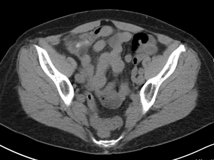
Thanks for this great post. Being familiar with the anatomy of these various procedures is essential to understanding the complications and why you cannot be reassured by a benign abdominal examination. 

With laparoscopic band procedures, complications are more common early on and are more related to band erosion or migration. Migration of the lap-band is best evaluated with an upper GI series with Gastrografin. Another important question to ask patients is if their lap band has been inflated recently. This is typically done in a progressive manner, where normal saline is instilled within the subcutaneous port. This can also be a source of obstruction, and if emergent, the band can be deflated by aspirating fluid from the port. This should be done under the guidance of a surgeon, if possible. 

Asking about dietary indiscretions can also sometimes clue you in to why a person is having abdominal pain or nausea. Especially immediately post-op, these patients have specific dietary guidelines and are often limited to liquids or pureed foods. Dumping syndrome can also occur in this patient population, more common with gastric bypass surgery, due to rapid gastric emptying. Typically, these patients have bloating, sweating, facial flushing, diarrhea, nausea, early satiety about 30 to 60 minutes after a meal. Dumping syndrome is typically diagnosed clinically, though laboratory testing should be performed to rule out electrolyte derangements. Dumping syndrome is typically treated with dietary modifications and should be discussed with the patient’s surgical team. 

As these procedures become more common and advanced, some are also performed endoscopically, which comes with its own set of complications. Another newer procedure is the intragastric balloon, which is a saline filled silicone balloon that is inflated in the stomach. In the first few days after placement, patients may experience abdominal pain, nausea, and vomiting. Other complications include balloon rupture, bowel obstruction, gastric outlet obstruction, gastric ulcer, pancreatitis, nonalcoholic steatohepatitis and cholecystitis.  

Overall, management of these patients in the emergency department should be done in consultation with the surgeon.

Gabrielle Ahlzadeh, MD.PNG

Gabrielle Ahlzadeh, MD

Clinical Assistant Professor of Emergency Medicine

University of Southern California


How To Cite This Post:

[Peer-Reviewed, Web Publication] Hajjar, M. Jackson, P. (2021, Apr 26). Bariatric Emergencies. [NUEM Blog. Expert Commentary by Ahlzadeh, G]. Retrieved from http://www.nuemblog.com/blog/bariatric-emergencies


Other Posts You May Enjoy

Posted on April 26, 2021 and filed under Gastrointestinal.

Pelvic Inflammatory Disease

Written by: Niki Patel, MD, MD (NUEM ‘22) Edited by: Luke Neill, MD  (NUEM ‘20) Expert Commentary by: Gabby Ahlzadeh, MD

Written by: Niki Patel, MD, MD (NUEM ‘22) Edited by: Luke Neill, MD (NUEM ‘20) Expert Commentary by: Gabby Ahlzadeh, MD


PID1.PNG
PID2.PNG
PID3.PNG
PID4.PNG
PID5.PNG
PID6.PNG
PID7.PNG
PID8.PNG
PID9.PNG
PID10.PNG
PID11.PNG
PID12.PNG
PID13.PNG
PID14.PNG
PID15.PNG

Expert Commentary

Thanks for this clear and succinct post. The differential diagnosis of lower abdominal and pelvic pain is extremely broad in both premenopausal and post-menopausal women. This is when the sexual history becomes important. A question we often overlook as part of the sexual history is asking about dyspareunia, which may help differentiate gynecological from intra-abdominal causes of abdominal pain, specifically in the case of PID. 

Patients with PID are frequently misdiagnosed with a urinary tract infection because they may have urinary symptoms, but the urinalysis often shows sterile pyuria, which should raise your suspicion for PID. 

And while the utility of the pelvic exam is constantly scrutinized and questioned in patients with vaginal bleeding, it is impossible to diagnose PID without it. Having said that, the clinical diagnosis is only 65-90% specific so even minimal symptoms with no other explanation warrant antibiotic therapy to reduce further complications. 

Underdiagnosis is even more significant in the adolescent patient population, who are at highest risk for developing PID. Over 70% of PID diagnoses among adolescents are made in the ED, with approximately 200,000 adolescents diagnosed annually. If the patient is accompanied by a family member or friend, having them step out to better elicit a sexual history is essential. HIV and syphilis testing should also be considered while these patients are in the ED. 

Ensuring follow-up for these patients within 48-72 hours is essential and must be emphasized. Patients should understand the complications of PID and the importance of antibiotic compliance prior to discharge, especially in younger patients. 

Gabrielle Ahlzadeh, MD.PNG

Gabrielle Ahlzadeh, MD

Clinical Assistant Professor of Emergency Medicine

University of Southern California


How To Cite This Post:

[Peer-Reviewed, Web Publication] Patel, N. Neill, L. (2021, Apr 5). Pelvic Inflammatory Disease. [NUEM Blog. Expert Commentary by Ahlzadeh, G]. Retrieved from http://www.nuemblog.com/blog/pelvic-inflammatory-disease


Other Posts You May Enjoy

SonoPro Tips and Tricks for Acute Cholecystitis

Written by: John Li, MD (NUEM ‘24) Edited by: Amanda Randolph (NUEM ‘21) Expert Commentary by: John Bailitz, MD & Mike Macias, MD

Written by: John Li, MD (NUEM ‘24) Edited by: Amanda Randolph (NUEM ‘21) Expert Commentary by: John Bailitz, MD & Mike Macias, MD


SonoPro Tips and Tricks

Welcome to the NUEM Sono Pro Tips and Tricks Series where Sono Experts team up to take you scanning from good to great for a problem or procedure!

For those new to the probe, we recommend first reviewing the basics in the incredible FOAMed Introduction to Bedside Ultrasound Book and 5 Minute Sono. Once you’ve got the basics beat, then read on to learn how to start scanning like a Pro!

Point of care right upper quadrant ultrasound has been shown to be a highly sensitive (82-91%), specific (66-95%), cost effective and efficient modality for emergency medicine physicians to quickly and effectively identify biliary pathology [1-5]. But despite its widespread utility, right upper quadrant ultrasound can often be a technically difficult study for the beginner sonographer, as there are multiple factors that can influence its ease of acquisition ranging from patient body habitus to bowel gas shadowing, and sonographer experience has been shown to influence its efficacy [1, 6-7].

Beyond the classic patient with right upper quadrant pain, what other scenarios do Sono-Pros use right upper quadrant ultrasound?

  1. Epigastric abdominal pain being “diagnosed” and even over treated as GERD. Pick up the probe in the symptomatic patient taking their PPI, EGD negative, or already treated for H. pylori

  2. Unexplained right shoulder or back pain. 

  3. Colicky pain in the right flank but no urinary findings of nephrolithiasis. 

  4. My gallstones are back! But my gallbladder is gone. Look for choledocholithiasis.

  5. Chronically ill elderly or immunosuppressed patients with unexplained fever or sepsis. 

SonoPro Tips - How to scan like a Pro

Always Start Smart: To Fail to Prepare is to Prepare to Fail whether in ED POCUS or ED Thoracotomy.

  1. Start with the patient in either the left lateral decubitus position or supine with the bed at approximately 30 degrees.

  2. Let the patient know “I’ll be asking you throughout this brief exam to take medium to deep breaths and hold for 5 sec, then automatically breathe out.” 

Still not not getting great views? 

  1. Scan between the ribs to use the liver as an acoustic window and avoid bowel gas. Switch to a small footprint phased array probe if needed. 

    • Not sure which intercostal space to use? Try about 7 centimeters to the right of the patient’s xiphoid process!

  2. Ask the patient to position their arms above their head to open the intercostal space. 

  3. Ask the patients to bend their knees to relax the abdominal muscles.

  4. In young, thin patients, the gallbladder may be more anterior and superior-- if you are scanning subcostally, try flattening out the probe even more!

Even a Small Pain in the Neck can be a Big Problem!

  1. Don’t forget the neck. There is a reason the gallbladder was so nicely distended and  easy to find. Be sure to scan carefully in two orthogonal planes to pick up subtle stones in the neck of the gallbladder!

    • If there is a lot of nearby bowel gas, tell your tech to look for these stones if your surgeons require a confirmatory comprehensive radiology ultrasound before operating. 

In this GIF, you can see a long-axis view of the gallbladder. When you are initially looking at the body and the fundus of the gallbladder, there are no clear shadowing stones. However, as the sonographer fans to the neck of the gallbladder, they can visualize multiple stones, which are casting shadows posteriorly. Image courtesy of the POCUS Atlas.

SonoPro Tips - Pro Pick Ups!

  1. Is that a stone or is that something else in the gallbladder? Roll the patient and see if the “stone” moves! 

    • If the stone in the fundus or body moves, then it’s more likely a mobile stone. 

    • If it doesn’t move, then consider a polyp or a malignancy. Polyps or malignancies generally are non-shadowing while stones are shadowing!

    • Impacted, “non-mobile” Neck Stone = Big Problem and likely to progress to acute cholecystitis. 

  2. What’s causing that shadow?

    • Stones shadow posteriorly. 

    • Edges shadow on the sides. Edge artifact results when ultrasound beams scatter passing by a smooth-walled structure, creating an anechoic stripe that could be confused with true shadowing!

  3. What if the entire gallbladder is casting a shadow?

    • Think about a gallbladder FULL of stones! This will cause only the most anterior stones to show up on ultrasound.

Here, on the right side of the screen you see a cross section of the gallbladder that has a large stone in it-- this is casting a shadow so you do not see the posterior wall of the gallbladder at all. This is called the wall echo sign-- where you will only see the most anterior surface of the stone. Image courtesy of the POCUS Atlas.

4. What are some of those pesky mimics of acute cholecystitis?

  • Think about hepatic pathologies! Acute hepatitis can cause a clinical Murphy’s sign. You can also have patients who present similarly when they have a congestive hepatopathy from their CHF. Even cirrhotic patients can present with a tender RUQ!

Here, you can see a dilated gallbladder with a thickened anterior wall and a small amount of pericholecystic fluid, all of which are consistent with acute cholecystitis. Image courtesy of the POCUS Atlas.

In this still image, you can see a thickened gallbladder wall (although be sure to measure the anterior wall, as the posterior wall can be thickened due to posterior acoustic enhancement!) and a small amount of pericholecystic fluid.  Image courtesy of the POCUS Atlas.

Here, you can see a dilated gallbladder with an obstructing stone in the neck of the gallbladder. Image courtesy of the POCUS Atlas.

SonoPro Tips - What the Pro’s Do Next!

Infographic courtesy of Justin Seltzer, MD

  1. If you see nonshadowing masses in the gallbladder:

    • Measure it! If the polyp is >1cm, then there’s a ~50% chance that this could be malignant, so be sure to refer these patients for additional imaging and close follow up. 

  2. What if you’re hoping to be really thorough and get a beautiful image of the CBD, but despite your best efforts, you cannot find it?

    • Draw some LFTs! A number of our emergency medicine colleagues, including Becker et. al and Lahham et. al, have done studies on this and it has been shown to be very unlikely that the CBD will be pathologically dilated in the setting of normal LFTs. On the flip side, if the LFTs appear cholestatic in nature, that’s another indication for a right upper quadrant ultrasound! [9-10]

SonoPro Tips - Where to Learn More

Do you want to see more pathologic images that you may see when you are doing a right upper quadrant ultrasound? Be sure to check out The Pocus Atlas by our expert editor Dr. Macias! It’s a great resource that also shows some of the rarer etiologies of gallbladder pathology, such as emphysematous cholecystitis or choledocholithiasis.

If you’re interested in looking at some of the evidence behind the right upper quadrant ultrasound, be sure to check out the evidence atlas here as well!


Expert Commentary

Thank you to NWEM1 John Li for bringing this great idea for a NUEM Blog Series to life. And another thanks to NUEM Blog Founder Mike Macias for his help on both content and graphics!

This new series is intended to push your Sono skills from just good, to really great. We will not rehash the basics. There are already abundant great resources available that we are truly thankful for and utilize everyday. But instead, we will share SonoPro Tips to help you more quickly master challenging POCUS applications and procedures. 

And there is no place better to start than Acute Cholecystitis. This is a great differentiator between the average and the expert clinician sonographer. As John outlines, start smart by expanding your indications and positioning your patient properly from the get go. Then breath, not you, the patient. Breath and hold again and again to bring the gallbladder and even difficult to discern pathology into clear view. Go beyond getting stones, and work to pick up, and explain other pathologies, as well as the bile ducts when needed.  

Thanks again John and Mike! Looking forward to the next post in this new series...

John-Bailitz.png

John Bailitz, MD

Vice Chair for Academics, Department of Emergency Medicine

Professor of Emergency Medicine, Feinberg School of Medicine

Northwestern Memorial Hospital

michael macias.PNG

Michael Macias, MD

Global Ultrasound Director, Emergent Medical Associates

Clinical Ultrasound Director, SoCal MEC Residency Programs


How To Cite This Post:

[Peer-Reviewed, Web Publication] Li, J. Randolph, A. (20201 Mar 22). SonoPro Tips and Tricks for Acute Cholecystitis. [NUEM Blog. Expert Commentary by Bailitz, J. Macias, M]. Retrieved from http://www.nuemblog.com/blog/sonopro-tips-and-tricks-for-acute-cholecystitis


Other Posts You May Enjoy

References

  1. Jain A, Mehta N, Secko M, Schechter J, Papanagnou D, Pandya S, Sinert R. History, Physical Examination, Laboratory Testing, and Emergency Department Ultrasonography for the Diagnosis of Acute Cholecystitis. Acad Emerg Med. 2017 Mar;24(3):281-297. doi: 10.1111/acem.13132. PMID: 27862628.

  2. Miller, Adam H., et al. “ED Ultrasound in Hepatobiliary Disease.” The Journal of Emergency Medicine, vol. 30, no. 1, 2006, pp. 69–74., doi:10.1016/j.jemermed.2005.03.017. 

  3. Shekarchi B, Hejripour Rafsanjani SZ, Shekar Riz Fomani N, Chahardoli M. Emergency Department Bedside Ultrasonography for Diagnosis of Acute Cholecystitis; a Diagnostic Accuracy Study. Emerg (Tehran). 2018;6(1):e11. Epub 2018 Jan 20. PMID: 29503836; PMCID: PMC5827043.

  4. American College of Emergency Physicians: Emergency Ultrasound Imaging Criteria Compendium. Oct. 2014, www.acep.org/globalassets/new-pdfs/policy-statements/emergency-ultrasound-imaging-criteria-compendium.pdf. 

  5. Hilsden R, Leeper R, Koichopolos J, et al. Point-of-care biliary ultrasound in the emergency department (BUSED): implications for surgical referral and emergency department wait times. Trauma Surg Acute Care Open. 2018;3(1):e000164. Published 2018 Jul 30. doi:10.1136/tsaco-2018-000164

  6. Ma, John, et al. Ma and Mateer's Emergency Ultrasound. McGraw-Hill Education, 2020. 

  7. Mallin, Mike, and Matthew Dawson. Introduction to Bedside Ultrasound: Volume 2. Emergency Ultrasound Solutions, 2013. 

  8. Macias, Michael. TPA, www.thepocusatlas.com/. 

  9. Becker BA, Chin E, Mervis E, Anderson CL, Oshita MH, Fox JC. Emergency biliary sonography: utility of common bile duct measurement in the diagnosis of cholecystitis and choledocholithiasis. J Emerg Med. 2014 Jan;46(1):54-60. doi: 10.1016/j.jemermed.2013.03.024. Epub 2013 Oct 11. PMID: 24126067.

  10. Lahham S, Becker BA, Gari A, Bunch S, Alvarado M, Anderson CL, Viquez E, Spann SC, Fox JC. Utility of common bile duct measurement in ED point of care ultrasound: A prospective study. Am J Emerg Med. 2018 Jun;36(6):962-966. doi: 10.1016/j.ajem.2017.10.064. Epub 2017 Nov 20. PMID: 29162442.

Posted on March 22, 2021 and filed under Ultrasound.

A Practical Approach to Abdominal Imaging

a practical approach to abdominal imaging.png

Written by: Zach Schmitz MD (PGY-3) Edited by: David Kaltman, MD (PGY-4) Expert commentary by: Samir Abboud, MD


I often find myself in a gray zone when it comes to imaging abdominal pain. Any third year medical student worth their salt can tell you to get the RUQ ultrasound for the fat, fertile, forty year-old female with RUQ abdominal pain, fever, positive Murphy’s sign, and leukocytosis. However, my patients don’t usually fit the textbook, and I’m often thinking about what I might miss or see with test X vs test Y. Below, I’ll touch on a few common dilemmas where the optimal choice of imaging modality isn’t immediately clear by focusing on what you actually gain or lose by ordering one imaging test over another.


Scenario 1: Stone or Appendicitis?

Case: 62 year old female with HTN and HLD presents with RLQ pain. The pain woke her this morning and has been intermittent all day, occurring exclusively when she urinates. It is sharp, non-radiating, and increasing in intensity. She never had a pain like this and can now barely sit still. She has thrown up a few times over the past few hours. Vitals are stable and she is afebrile. She appears uncomfortable with RLQ tenderness but no rebound or guarding. Labs show slight leukocytosis, and urine has no blood.

If I suspect stone over appendicitis, will a CT without contrast miss appendicitis?

  • CT, MR, and US are well studied in their ability to detect and accurately diagnose appendicitis.[1] 

    • CT with IV contrast is 96-100% sensitive and 91-100% specific. Per the American College of Radiology’s (ACR) appropriateness system, this is the most appropriate initial test for suspected appendicitis in adults.[2]

    • MR is 96% sensitive and 96% specific.[3]

    • Ultrasound has a wide range of data, with sensitivity ranging from 21-95.7% and specificity of 71-97%.[2]

  • CT without oral or IV contrast is nearly as useful for diagnosing appendicitis

    • A meta-analysis by Xiong et al included seven original studies investigating a total of 845 patients.[4]

      • Pooled sensitivity - 0.90 (95% CI: 0.86-0.92)

      • Pooled specificity - 0.94 (95% CI: 0.92-0.97)

      • Pooled positive likelihood ratio - 12.90 (95% CI: 4.80-34.67)

      • Pooled negative likelihood ratio - 0.09 (95% CI: 0.04-0.20)

Will a contrast enhanced CT for appendicitis ruin my chance to catch a kidney stone?

  • Non-contrast CT is the emergency standard in diagnosing nephrolithiasis with good reason - it is 97% sensitive and 95% specific.[5]

  • Will contrast ruin the ability to detect a stone?

    • This makes theoretical sense as stones and contrast are both hyper-intense on CT.

    • Sensitivity is decreased for small stones with contrast enhanced studies.

    • However, for stones > 3mm, sensitivity remains 95%.[5]

    • Only about 5% of stones that small ultimately require intervention.

Takeaways: You sacrifice a bit with a non-contrast study looking for appendicitis and a contrast enhanced study looking for stone, but both still work well. The American Urology Association recommends consultation for stones > 10mm.[6] Urology would also need to be involved with signs of sepsis, abscess, deterioration in renal function, intractable symptoms, or a transplant/solitary kidney. It seems I am very likely to see a stone requiring something other than watchful waiting on a CT with contrast. It is worse to miss an appendicitis than a 2mm stone, so contrast might make more sense if it’s close. 


Scenario 2: RUQ Ultrasound after Negative CT San

Case: 84 year old male with a history of prostate cancer and hypertension presents from a nursing home with 4 days of diffuse abdominal pain. He has had no vomiting or bowel movements over this time. No urinary symptoms. He is hemodynamically stable, and his abdomen is diffusely tender (maybe worse in the RUQ) and distended but overall not terribly impressive. You order a CT for possible obstruction and it just shows a large stool burden. The gallbladder was visualized and looked normal. 

If a CT is negative, should I get a RUQ US to look for cholecystitis?

  • RUQ Ultrasound

    • Per ACR, this is the most appropriate initial study for RUQ pain and suspected biliary disease.[7]

    • A 2012 meta analysis showed a sensitivity of 81% (95% CI 75-87%) and specificity of 88% for acute cholecystitis.[8]

    • It has the advantage of being dynamic, with a sonographic Murphy sign independently showing an 86% sensitivity and 35% specificity, positive predictive value of 43%, and negative predictive value of 82%.[9] 

  • Computed Tomography (CT) 

    • The same 2012 meta analysis only had one study with CT, but noted a sensitivity of 94% with fairly broad confidence intervals (95% CI 73-99) and a specificity of only 59%.[8]

    • ACR notes CT’s NPV for acute cholecystitis approaches 90%.[7]

    • A 2015 study looked at 101 patients who went to the OR and got both a CT and US. For acute cholecystitis, the sensitivities for CT and US were 92% and 79% respectively. For cholilithiasis, sensitivities for CT and US were 60% and 89% respectively.[10,11]

    • ACR states it is “usually appropriate” to proceed with CT for RUQ pain and suspected biliary disease with a negative or equivocal ultrasound.[7]

    • Although it lacks a sonographic murphy’s sign equivalent, its advantage is to help in operative planning and seeing complications, such as perforation or gangrene.

  • MRI has a sensitivity of 85% and a specificity of 81%. It is also considered “usually appropriate” by ACR if ultrasound is negative or equivocal[7]

  • Cholescintigraphy is the best imaging, showing 97% sensitivity and 90% specificity for acute cholecystitis. It is also the most appropriate study if you suspect acalculous cholecystitis.[7]

Takeaways: There are a few interesting points from this set of data. First, CT seems to have at least as good of ability to pick up cholecystitis compared to ultrasound. However, it is much worse in detecting gallstones themselves, which may be very relevant to a patient with abdominal pain. Second, the sensitivity of both RUQUS or CT isn’t really that great and we are probably missing a few episodes of cholecystitis. If there is a very high index of suspicion but negative imaging, it may be worthwhile to pursue additional workup. Overall, if the CT shows a normal gallbladder, and you are not worried about intractable biliary colic, the ultrasound probably won’t add much. 


Scenario 3: Female Pelvic Pain

Case: 33 year old female with a history of chlamydia infection presenting with right sided abdominal pain. The pain has gradually been getting worse for 1 day. She has had a few episodes of vomiting. There is some white vaginal discharge she always has. On exam, she is tachycardic, normotensive, and febrile to 101.5. She has RLQ tenderness with voluntary guarding. On pelvic exam, there is some white vaginal discharge, CMT, R adnexal tenderness that seems less intense than her RLQ tenderness, and no masses noted.

If this patient had a normal appendix and ovaries after a contrast enhanced CT for appendicitis, how useful is an additional transvaginal ultrasound to rule out gynecologic pathologies?

For ovarian torsion:

  • A retrospective study of 834 patients showed the NPV of a contrast enhanced CT of the pelvis for ovarian torsion is 100%.[12]

  • A prospective study of 199 patients showed doppler ultrasound has a sensitivity and specificity for torsion of 100 and 97%.[13]

For Tubo-Ovarian Abscess (TOA):

  • CT is thought to be between 78 and 100% sensitive.[14]

  • 2011 literature review gives a broad range of sensitivity and specificity for US in TOA with a sensitivity of 56-93% and specificity from 89-98%.[15]

Takeaways: ACR appropriates rates ultrasound as the most appropriate test for female pelvic pain.[14] However, it also rates CT with contrast as more appropriate for suspected appendicitis.[2] This patient raises concerns for both, and a CT was done first. CT is good for finding intra abdominal and pelvic abscess. It is more difficult to assess how useful ultrasound is for TOA, as many studies in the literature review were either before year 2000 or used a transabdominal approach. Overall, if someone has a CT scan for appendicitis that shows normal ovaries, the transvaginal ultrasound seems to add little for either torsion or TOA.


One potential dangerous conclusion from this set of data is that we should just CT everyone up front. While CT shows good sensitivities for many of the pathologies in question, simply ordering a CT first ignores the many good reasons - such as cost, radiation dose, speed, improved specificity and comparable sensitivity, resource utilization, sonographic murphy sign - RUQUS and pelvic ultrasound are the most appropriate initial tests for suspect biliary and pelvic pathology. That said, it a patient has an entirely normal CT that was already performed for other indicated reasons, the use of additional imaging may be unnecessary and should be considered carefully. Overall, the question of exactly what imaging test to order when ruling out common, emergent, abdominal pathologies is often a difficult one with shades of gray. By having a better understanding of exactly what type of information we are getting and missing from each test we order, emergency physicians can more quickly, safely, and accurately diagnose and treat our patients.


Expert Commentary

This is a thoughtful, well-reasoned approach to optimizing the imaging strategy in challenging, atypical clinical scenarios. To add a few nuances to some of the points raised:

When considering a contrast-enhanced versus non-contrast CT (both IV and PO) in the clinically ambiguous scenario, it is important to consider your patient’s body habitus. Figure 1 includes representative images from a non-contrast enhanced CT of a patient with a BMI above 25. You can clearly see the inflammatory stranding in the right lower quadrant mesenteric fat (Figure 1a) and portions of an appendicolith (Figure 1 b), in this patient who ultimately proved to have appendicitis. The natural contrast provided by the patient’s mesenteric fat in this scenario helps us work around the absence of IV contrast.

Figure 1a

Figure 1a

Figure 1b

Figure 1b

Figure 2 includes representative images from a contrast enhanced CT of a very thin patient, with a relative paucity of intra-abdominal fat. In this patient, the relative absence of natural contrast would greatly reduce our chances to diagnose appendicitis (or even identify the appendix) in the absence of IV contrast. PO contrast is additionally likely to be most helpful in very thin patients [Alabousi 2015].

Figure 2

Figure 2


The author asks (and answers) a very insightful question with regards to identifying kidney stones on contrast enhanced CT. A few points to add:

Assuming the contrast enhanced study is obtained prior to the excretory phase of imaging (and most routine studies are) ureteral stones should still be largely visible - the stones that will generally be more difficult to identify will be the non-obstructing stones still within the collecting system. Additionally, while there is indeed a small sacrifice in sensitivity for small stones with contrast enhanced studies, the identification of secondary complications is much improved.

Consider Figure 3, which demonstrates a 2 mm stone in the proximal left ureter identified on a contrast enhanced study. Notice the slightly delayed nephrogram on the left relative to the right, which could indicate a component of obstructive uropathy. Similarly, identification of such complications as pyonephrosis, pyelonephritis, and perinephric abscess is much improved with contrast enhanced images. For this reason, I would suggest that in the clinically ambiguous scenario, erring on the side of the contrast enhanced study would be wise.

Figure 3

Figure 3


It is important to note that the CT scanner installed in our emergency department is a dual-energy machine. Many of our other departmental scanners are dual-energy as well. With these scanners, we are able to apply algorithms to deconstruct the elemental composition of stones and provide more information than simply size and location - i.e. uric acid or non-uric acid stone - if requested. We can additionally generate virtual non-contrast images from the contrast-enhanced images, without exposing our patients to additional radiation. While it is tempting to think that we could recapture some of the sensitivity for renal stones using these virtual non-contrast images, this has unfortunately not been borne out in the literature at this time [Vrtiska 2010], though remains an area of continued investigation as imaging technology is further improved.

The advantages of dual-energy imaging are not only limited to the kidneys. With regards to the evaluation of biliary colic, virtual monochromatic images can be generated with resulting increased conspicuity of gallstones, even those that appear isodense to bile on the conventional images [Ratanaprasatporn 2018].

In general, if you find yourself with a high degree of suspicion for any disease process and discordant imaging findings, I would encourage you to call your radiologist. The additional clinical information exchanged during such a call may direct what additional data sets should be generated and what additional imaging studies may be of most benefit. Last, but certainly not least, that “second look” armed with additional clinical information can pick up on subtle findings that are, in isolation, entirely non-specific, but in a certain clinical scenario could clinch the diagnosis you are seeking.

References:

Alabousi A et al. Is Oral Contrast Necessary for Multidetector Computed Tomography Imaging of Patients With Acute Abdominal Pain? Canadian Association of Radiologists Journal. 2015;66(4): 318 - 322

Ratanaprasatporn L et al. Multimodality Imaging, including Dual-Energy CT, in the Evaluation of Gallbladder Disease. Radiographics 2018;38(1): 75-89

Vrtiska TJ et al. Genitourinary Applications of Dual-Energy CT. American Journal of Roentgenology. 2010;194: 1434-1442.

Abboud.png

Samir Abboud, MD

Assistant Professor of Radiology

Northwestern University


How To Cite This Post:

[Peer-Reviewed, Web Publication] Schmitz, Z. Kaltman, D. (2020, Feb 10). An Approach to Abdominal Imaging. [NUEM Blog. Expert Commentary by Abboud, S]. Retrieved from http://www.nuemblog.com/blog/abdominal-imaging.


Other Posts You Might Enjoy…


References

  1. Dahabreh IJ, Adam GP, Halladay CW, Steele DW, Daiello LA, Weiland LS, Zgodic A, Smith BT, Herliczek TW, Shah N, Trikalinos TA. Diagnosis of Right Lower Quadrant Pain and Suspected Acute Appendicitis. Comparative Effectiveness Review No. 157. (Prepared by the Brown Evidence-based Practice Center under Contract No. 290-2012-00012-I.) AHRQ Publication No. 15(16)-EHC025-EF. Rockville, MD: Agency for Healthcare Research and Quality; December 2015. www.effectivehealthcare.ahrq.gov/reports/final.cfm.

  2. American College of Radiology. ACR Appropriateness Criteria®: RLQ pain. Available at https://acsearch.acr.org/docs/69357/Narrative/ Accessed 5/10/19.

  3. Duke E, Kalb B, Arif-Tiwari H, et al. A Systematic Review and Meta-Analysis of Diagnostic Performance of MRI for Evaluation of Acute Appendicitis. AJR Am J Roentgenol 2016;206:508-17.

  4. Xiong B, Zhong B, Li Z, Zhou F, Hu R, Feng Z, Xu S, Chen F. Diagnostic Accuracy of Noncontrast CT in Detecting Acute Appendicitis: A Meta-analysis of Prospective Studies. Am Surg. 2015 Jun;81(6):626-9.

  5. Curhan G, Aronson M, Preminger G. Diagnosis and acute management of suspected nephrolithiasis in adults. UpToDate.com. April 30 2019. 

  6. Assimos D, Krambek A, Miller N et al. Surgical Management of Stones: AUA/Endourology Society Guideline (2016). https://www.auanet.org/guidelines/kidney-stones-surgical-management-guideline. Accessed 5/10/19.

  7. American College of Radiology. ACR Appropriateness Criteria®: RUQ pain. Available at https://acsearch.acr.org/docs/69474/Narrative/ .

  8. Kiewiet J.J., Leeuwenburgh M.M., Bipat S., et al: A systematic review and meta-analysis of diagnostic performance of imaging in acute cholecystitis. Radiology 2012; 264: pp. 708-720.

  9. Bree, Robert L. Further observations on the usefulness of the sonographic Murphy sign in the evaluation of suspected acute cholecystitis. Journal of Clinical Ultrasound. March/April 1995.

  10. Wertz JR1,2, Lopez JM3, Olson D4, Thompson WM1,2. Comparing the Diagnostic Accuracy of Ultrasound and CT in Evaluating Acute Cholecystitis. AJR Am J Roentgenol. 2018 Aug;211(2):W92-W97. doi: 10.2214/AJR.17.18884. Epub 2018 Apr 27.

  11. Fagenholz, P et al. Computed Tomography Is More Sensitive than Ultrasound for the Diagnosis of Acute Cholecystitis. Surg Infect (Larchmt). 2015 Oct;16(5):509-12. doi: 10.1089/sur.2015.102. Epub 2015 Sep 16. 

  12. Lam A1, Nayyar M2, Helmy M2, Houshyar R2, Marfori W2, Lall C2.Assessing the clinical utility of color Doppler ultrasound for ovarian torsion in the setting of a negative contrast-enhanced CT scan of the abdomen and pelvis. Abdom Imaging. 2015 Oct;40(8):3206-13. Doi: 10.1007/s00261-015-0535-4.

  13. Laufer, M. Ovarian and fallopian tube torsion. UpToDate. April 30 2019. https://www.uptodate.com/contents/ovarian-and-fallopian-tube-torsion?search=ovarian%20torsion&source=search_result&selectedTitle=1~70&usage_type=default&display_rank=1 .

  14. Beigi, R. Epidemiology, clinical manifestations, and diagnosis of tubo-ovarian abscess. UpToDate. April 30 2019. https://www.uptodate.com/contents/epidemiology-clinical-manifestations-and-diagnosis-of-tubo-ovarian-abscess?search=tuboovarian%20abscess&source=search_result&selectedTitle=2~24&usage_type=default&display_rank=2 .

  15. Lee DC1, Swaminathan AK. Sensitivity of ultrasound for the diagnosis of tubo-ovarian abscess: a case report and literature review. J Emerg Med. 2011 Feb;40(2):170-5. doi: 10.1016/j.jemermed.2010.02.033. Epub 2010 May 13.

  16. American College of Radiology. ACR Appropriateness Criteria®: Female Pelvic Pain. Available at https://acsearch.acr.org/docs/69503/Narrative/ .

Better than a shotgun approach to diagnosis: Ultrasound in Cholangitis

This week we discuss an interesting case and how bedside ultrasound can help you facilitate rapid diagnosis and disposition of patients presenting to the emergency department with right upper quadrant abdominal pain.